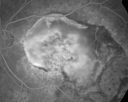
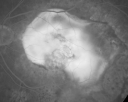
Scar - Large Macular Scar in Fellow Eye new Subretinal Fluid Good Eye
|
|
81-year-old woman has a macular scar in the left eye and then unfortunately developed a central retinal artery occlusion in the right eye. She responded nicely to her anterior chamber taps. She is on Cosopt and Travatan. She notices the right eye again declining the last few weeks.
VISUAL ACUITY: OD 20/100, OS 6/200. IOP: OD 12, OS 14. The right eye has a posterior chamber intraocular lens is in good position.
EXTENDED OPHTHALMOSCOPY:
OD: Vertical C/D ratio is 0.9. There is pigment epithelium thickening centrally.
OCT SCAN: The OCT scan of the right eye shows trace subfoveal fluid. Photos confirm clinical findings.
FLUORESCEIN ANGIOGRAPHY: Fluorescein angiography of the left eye shows a large staining macular scar. The left eye shows areas of stippled hyperfluorescence temporal to the fovea and inferior to the fovea, which are unchanged from an angiogram of December 8th.
IMPRESSION:
1. POSSIBLE CHOROIDAL NEOVASCULAR MEMBRANE – RIGHT EYE
2. RECENT CENTRAL RETINAL ARTERY OCCLUSION – RIGHT EYE
3. MACULAR SCAR – LEFT EYE
DISCUSSION: I explained to the patient that the right eye does have some fluid in the retina, but I do not see the angiogram being significantly different than the angiogram taken a few months ago and I suspect that some of this may be still healing from the central retinal artery occlusion. I suggested on the absence of more compelling problems that we leave the eye alone and I asked her to return for a check in three to four weeks, but I am going to have a fairly low threshold for instituting intravitreal Avastin injections in this eye, if the vision in the macula does not start to look better soon
|

Macular Scar Left Eye from Age-Related Macular Degenerationvaatamisi: 50981-year-old woman has a macular scar in the left eye and then unfortunately developed a central retinal artery occlusion in the right eye OD 20/100, OS 6/200.    
(0 hinnangut)
|
|

Macular Scar Left Eye from Age-Related Macular Degenerationvaatamisi: 37781-year-old woman has a macular scar in the left eye and then unfortunately developed a central retinal artery occlusion in the right eye OD 20/100, OS 6/200.    
(0 hinnangut)
|
|

Macular Scar Left Eye from Age-Related Macular Degenerationvaatamisi: 46181-year-old woman has a macular scar in the left eye and then unfortunately developed a central retinal artery occlusion in the right eye OD 20/100, OS 6/200.    
(0 hinnangut)
|
|

Macular Scar Left Eye from Age-Related Macular Degenerationvaatamisi: 49181-year-old woman has a macular scar in the left eye and then unfortunately developed a central retinal artery occlusion in the right eye OD 20/100, OS 6/200.    
(0 hinnangut)
|
|

Macular Scar Left Eye from Age-Related Macular Degenerationvaatamisi: 41281-year-old woman has a macular scar in the left eye and then unfortunately developed a central retinal artery occlusion in the right eye OD 20/100, OS 6/200.    
(0 hinnangut)
|
|

Macular Scar Left Eye from Age-Related Macular Degenerationvaatamisi: 37981-year-old woman has a macular scar in the left eye and then unfortunately developed a central retinal artery occlusion in the right eye OD 20/100, OS 6/200.    
(0 hinnangut)
|
|

Macular Scar Left Eye from Age-Related Macular Degenerationvaatamisi: 32881-year-old woman has a macular scar in the left eye and then unfortunately developed a central retinal artery occlusion in the right eye OD 20/100, OS 6/200.    
(0 hinnangut)
|
|

Macular Scar Left Eye from Age-Related Macular Degenerationvaatamisi: 34281-year-old woman has a macular scar in the left eye and then unfortunately developed a central retinal artery occlusion in the right eye OD 20/100, OS 6/200.    
(0 hinnangut)
|
|

Macular Scar Left Eye from Age-Related Macular Degenerationvaatamisi: 37981-year-old woman has a macular scar in the left eye and then unfortunately developed a central retinal artery occlusion in the right eye OD 20/100, OS 6/200.    
(0 hinnangut)
|
|

Macular Scar Left Eye from Age-Related Macular Degenerationvaatamisi: 38481-year-old woman has a macular scar in the left eye and then unfortunately developed a central retinal artery occlusion in the right eye OD 20/100, OS 6/200.    
(0 hinnangut)
|
|

Macular Scar Left Eye from Age-Related Macular Degenerationvaatamisi: 35081-year-old woman has a macular scar in the left eye and then unfortunately developed a central retinal artery occlusion in the right eye OD 20/100, OS 6/200.    
(0 hinnangut)
|
|

Macular Scar Left Eye from Age-Related Macular Degenerationvaatamisi: 37481-year-old woman has a macular scar in the left eye and then unfortunately developed a central retinal artery occlusion in the right eye OD 20/100, OS 6/200.    
(0 hinnangut)
|
|
|
|
81-year-old woman has a macular scar in the left eye and then unfortunately developed a central retinal artery occlusion in the right eye. She responded nicely to her anterior chamber taps. She is on Cosopt and Travatan. She notices the right eye again declining the last few weeks.
VISUAL ACUITY: OD 20/100, OS 6/200. IOP: OD 12, OS 14. The right eye has a posterior chamber intraocular lens is in good position.
EXTENDED OPHTHALMOSCOPY:
OD: Vertical C/D ratio is 0.9. There is pigment epithelium thickening centrally.
OCT SCAN: The OCT scan of the right eye shows trace subfoveal fluid. Photos confirm clinical findings.
FLUORESCEIN ANGIOGRAPHY: Fluorescein angiography of the left eye shows a large staining macular scar. The left eye shows areas of stippled hyperfluorescence temporal to the fovea and inferior to the fovea, which are unchanged from an angiogram of December 8th.
IMPRESSION:
1. POSSIBLE CHOROIDAL NEOVASCULAR MEMBRANE – RIGHT EYE
2. RECENT CENTRAL RETINAL ARTERY OCCLUSION – RIGHT EYE
3. MACULAR SCAR – LEFT EYE
DISCUSSION: I explained to the patient that the right eye does have some fluid in the retina, but I do not see the angiogram being significantly different than the angiogram taken a few months ago and I suspect that some of this may be still healing from the central retinal artery occlusion. I suggested on the absence of more compelling problems that we leave the eye alone and I asked her to return for a check in three to four weeks, but I am going to have a fairly low threshold for instituting intravitreal Avastin injections in this eye, if the vision in the macula does not start to look better soon