Enhanced S-Cone Syndrome - Goldman Favre (probably)
|
|
55-year-old woman was seen in the office on December 5, 2011. She has had difficulty with her vision for sometime. She always wore glasses since childhood, but while in college her vision was poor even with glasses and she sought evaluation for that. At various times in the past she has been diagnosed with Stargardt’s disease. She was told after she had an electroretinogram at USF sometime ago, that she had something with her blue cones. It was not normal and she recently is concerned, because she has a teen age son and she is worried about his possible future if he were to have eye problems, because he is thinking about going into medicine. She is in otherwise excellent health. She does have poor night vision, but her reading vision is pretty good.
VISUAL ACUITY: OD 20/40, OS 20/40. IOP: OD 15, OS 16. Color vision is 6 out of 12 correct in each either eye. There is 1+ nuclear sclerosis in both eyes.
EXTENDED OPHTHALMOSCOPY:
OD: Vertical C/D ratio is 0.3. There looks to be a vitreous separation, although I can’t be positive. There are 1+ vitreous cells. The nerve is slightly pale. The retinal vessels are definitely attenuated with attenuated retinal arterials in all quadrants extending out to the mid periphery. There is some mottling of the pigment under the fovea. There is no tri-radiate yellow pigment spots nor is there significant peripheral pigment degeneration, but there is mild peripheral pigment degeneration. There are definitely no bone spicules.
OS: Vertical C/D ratio is 0.3. There is 1+ vitreous cells. There is a partial vitreous separation. There is no retinoschisis in the periphery. There are no tri radiate yellow spots in the macula. There is pigment mottling of the macula and the peripheral retina and again, there is no evidence of any bone spicules and there is no retinoschisis in the periphery.
SPECTRALIS-SD-OCT SCAN: The OCT scan does show foveal schisis in each eye with what looks like possible fluid deep to the retina, but there are some areas splitting in the right eye. There is dense retinal atrophy in each eye. There is thinning of all layers of the retina.
FUNDUS AUTO FLUORESCENCE: The image in the right eye shows hyper auto fluorescence throughout the posterior pole with some loss in the macular pigment and generally just brightness. In the left eye there is also hyper auto fluorescence of the posterior pole. We did a peripheral auto fluorescencent shot in the right eye, which does show some speckled hypo auto fluorescence suggesting peripheral loss of retinal integrity.
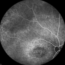
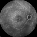
FLUORESCEIN ANGIOGRAPHY: Fluorescein angiography shows speckled hyperfluorescence in the macula of the right eye, a little bit of a bull’s eye type pattern. There is some loss of the macular pigment and then throughout the periphery there is increased hyperfluorescence. The retinal vessels are attenuated, but nothing leaks. The fluorescein angiogram of the left eye is similar and it is symmetric to the other eye and shows hyperfluorescence ringing the macula.
INDOCYANINE GREEN ANGIOGRAPHY: The indocyanine green angiogram doesn’t add anything. It shows normal, accept for some of the transmission from the lack of pigment of the pigment epithelium. Photos confirm clinical findings.
IMPRESSION:
1. PROBABLE GOLDMAN-FAVRE/ENHANCED S-CONE SYNDROME
DISCUSSION: I explained to the patient the fact that she had on ERG in the past showing a blue cone abnormality and her fundus appearance, including the angiogram and the clinical appearance is all consistent with enhanced S-cone syndrome, I think she probably has that, but the diagnostic tests for that would be an electroretinogram and I think given that she is still without a firm diagnosis of enhanced S-cone. It sounds like she was having her tests done just about the time when enhanced S-cone syndrome was being described. I think if she were to go back for a test now, the testing could be more definitive now that the parameters of the disease have been delineated. I have given her some places to go.
There is no rush. In the meantime I am fairly confident what she has is autosomal recessive. The risk of her son having it is very low. I told her if her son wants to come in for an exam, we could always do a fundus auto fluorescence and OCT. We don’t need to do any injections. The genetic locus for enhanced S-cone syndrome has been isolated. I have arranged for her to be tested for a defect in the NR2E3 gene. I told her she does not need to return here unless you or she note further problems.
|

Fundus Photograph - Enhanced S Cone Syndrome - Goldmann Favre - 1056 views55-year-old woman while in college her vision was poor even with glasses and she sought evaluation for that. She was told after she had an electroretinogram at USF 15 years ago, that she had something with her blue cones. She does have poor night vision, but her reading vision is pretty good.
VISUAL ACUITY: OD 20/40, OS 20/40    
(0 votes)
|
|

Fundus Photograph - Enhanced S Cone Syndrome - Goldmann Favre - 932 views55-year-old woman while in college her vision was poor even with glasses and she sought evaluation for that. She was told after she had an electroretinogram at USF 15 years ago, that she had something with her blue cones. She does have poor night vision, but her reading vision is pretty good.
VISUAL ACUITY: OD 20/40, OS 20/40    
(0 votes)
|
|

Fundus Photograph - Enhanced S Cone Syndrome - Goldmann Favre - 719 views55-year-old woman while in college her vision was poor even with glasses and she sought evaluation for that. She was told after she had an electroretinogram at USF 15 years ago, that she had something with her blue cones. She does have poor night vision, but her reading vision is pretty good.
VISUAL ACUITY: OD 20/40, OS 20/40    
(0 votes)
|
|

Fundus Autofluorescence - Enhanced S Cone Syndrome - Goldmann Favre - 854 views55-year-old woman while in college her vision was poor even with glasses and she sought evaluation for that. She was told after she had an electroretinogram at USF 15 years ago, that she had something with her blue cones. She does have poor night vision, but her reading vision is pretty good.
VISUAL ACUITY: OD 20/40, OS 20/40    
(0 votes)
|
|

Fundus Autofluorescence - Enhanced S Cone Syndrome - Goldmann Favre - 684 views55-year-old woman while in college her vision was poor even with glasses and she sought evaluation for that. She was told after she had an electroretinogram at USF 15 years ago, that she had something with her blue cones. She does have poor night vision, but her reading vision is pretty good.
VISUAL ACUITY: OD 20/40, OS 20/40    
(0 votes)
|
|

Fundus Autofluorescence - Enhanced S Cone Syndrome - Goldmann Favre - 636 views55-year-old woman while in college her vision was poor even with glasses and she sought evaluation for that. She was told after she had an electroretinogram at USF 15 years ago, that she had something with her blue cones. She does have poor night vision, but her reading vision is pretty good.
VISUAL ACUITY: OD 20/40, OS 20/40    
(0 votes)
|
|

Fundus Autofluorescence - Enhanced S Cone Syndrome - Goldmann Favre - 525 views55-year-old woman while in college her vision was poor even with glasses and she sought evaluation for that. She was told after she had an electroretinogram at USF 15 years ago, that she had something with her blue cones. She does have poor night vision, but her reading vision is pretty good.
VISUAL ACUITY: OD 20/40, OS 20/40    
(0 votes)
|
|

InfraRed Image - Enhanced S Cone Syndrome - Goldmann Favre - 581 views55-year-old woman while in college her vision was poor even with glasses and she sought evaluation for that. She was told after she had an electroretinogram at USF 15 years ago, that she had something with her blue cones. She does have poor night vision, but her reading vision is pretty good.
VISUAL ACUITY: OD 20/40, OS 20/40    
(0 votes)
|
|

OCT Line Scan - Enhanced S Cone Syndrome - Goldmann Favre - 551 views55-year-old woman while in college her vision was poor even with glasses and she sought evaluation for that. She was told after she had an electroretinogram at USF 15 years ago, that she had something with her blue cones. She does have poor night vision, but her reading vision is pretty good.
VISUAL ACUITY: OD 20/40, OS 20/40    
(0 votes)
|
|

OCT Line Scan - Enhanced S Cone Syndrome - Goldmann Favre - 536 views55-year-old woman while in college her vision was poor even with glasses and she sought evaluation for that. She was told after she had an electroretinogram at USF 15 years ago, that she had something with her blue cones. She does have poor night vision, but her reading vision is pretty good.
VISUAL ACUITY: OD 20/40, OS 20/40    
(0 votes)
|
|

OCT Line Scan - Enhanced S Cone Syndrome - Goldmann Favre - 611 views55-year-old woman while in college her vision was poor even with glasses and she sought evaluation for that. She was told after she had an electroretinogram at USF 15 years ago, that she had something with her blue cones. She does have poor night vision, but her reading vision is pretty good.
VISUAL ACUITY: OD 20/40, OS 20/40    
(0 votes)
|
|

Fluorescein Angiogram - Enhanced S Cone Syndrome - Goldmann Favre - 610 views55-year-old woman while in college her vision was poor even with glasses and she sought evaluation for that. She was told after she had an electroretinogram at USF 15 years ago, that she had something with her blue cones. She does have poor night vision, but her reading vision is pretty good.
VISUAL ACUITY: OD 20/40, OS 20/40    
(0 votes)
|
|

Fluorescein Angiogram - Enhanced S Cone Syndrome - Goldmann Favre - 528 views55-year-old woman while in college her vision was poor even with glasses and she sought evaluation for that. She was told after she had an electroretinogram at USF 15 years ago, that she had something with her blue cones. She does have poor night vision, but her reading vision is pretty good.
VISUAL ACUITY: OD 20/40, OS 20/40    
(0 votes)
|
|

Fluorescein Angiogram - Enhanced S Cone Syndrome - Goldmann Favre - 458 views55-year-old woman while in college her vision was poor even with glasses and she sought evaluation for that. She was told after she had an electroretinogram at USF 15 years ago, that she had something with her blue cones. She does have poor night vision, but her reading vision is pretty good.
VISUAL ACUITY: OD 20/40, OS 20/40    
(0 votes)
|
|

Fluorescein Angiogram - Enhanced S Cone Syndrome - Goldmann Favre - 529 views55-year-old woman while in college her vision was poor even with glasses and she sought evaluation for that. She was told after she had an electroretinogram at USF 15 years ago, that she had something with her blue cones. She does have poor night vision, but her reading vision is pretty good.
VISUAL ACUITY: OD 20/40, OS 20/40    
(0 votes)
|
|

Fluorescein Angiogram - Enhanced S Cone Syndrome - Goldmann Favre - 398 views55-year-old woman while in college her vision was poor even with glasses and she sought evaluation for that. She was told after she had an electroretinogram at USF 15 years ago, that she had something with her blue cones. She does have poor night vision, but her reading vision is pretty good.
VISUAL ACUITY: OD 20/40, OS 20/40    
(0 votes)
|
|

Fluorescein Angiogram - Enhanced S Cone Syndrome - Goldmann Favre - 463 views55-year-old woman while in college her vision was poor even with glasses and she sought evaluation for that. She was told after she had an electroretinogram at USF 15 years ago, that she had something with her blue cones. She does have poor night vision, but her reading vision is pretty good.
VISUAL ACUITY: OD 20/40, OS 20/40    
(0 votes)
|
|

Fluorescein Angiogram - Enhanced S Cone Syndrome - Goldmann Favre - 414 views55-year-old woman while in college her vision was poor even with glasses and she sought evaluation for that. She was told after she had an electroretinogram at USF 15 years ago, that she had something with her blue cones. She does have poor night vision, but her reading vision is pretty good.
VISUAL ACUITY: OD 20/40, OS 20/40    
(0 votes)
|
|

Fluorescein Angiogram - Enhanced S Cone Syndrome - Goldmann Favre - 521 views55-year-old woman while in college her vision was poor even with glasses and she sought evaluation for that. She was told after she had an electroretinogram at USF 15 years ago, that she had something with her blue cones. She does have poor night vision, but her reading vision is pretty good.
VISUAL ACUITY: OD 20/40, OS 20/40    
(0 votes)
|
|

Fluorescein Angiogram - Enhanced S Cone Syndrome - Goldmann Favre - 564 views55-year-old woman while in college her vision was poor even with glasses and she sought evaluation for that. She was told after she had an electroretinogram at USF 15 years ago, that she had something with her blue cones. She does have poor night vision, but her reading vision is pretty good.
VISUAL ACUITY: OD 20/40, OS 20/40    
(0 votes)
|
|

Fluorescein Angiogram - Enhanced S Cone Syndrome - Goldmann Favre - 468 views55-year-old woman while in college her vision was poor even with glasses and she sought evaluation for that. She was told after she had an electroretinogram at USF 15 years ago, that she had something with her blue cones. She does have poor night vision, but her reading vision is pretty good.
VISUAL ACUITY: OD 20/40, OS 20/40    
(0 votes)
|
|

Fluorescein Angiogram - Enhanced S Cone Syndrome - Goldmann Favre - 394 views55-year-old woman while in college her vision was poor even with glasses and she sought evaluation for that. She was told after she had an electroretinogram at USF 15 years ago, that she had something with her blue cones. She does have poor night vision, but her reading vision is pretty good.
VISUAL ACUITY: OD 20/40, OS 20/40    
(0 votes)
|
|

Fluorescein Angiogram - Enhanced S Cone Syndrome - Goldmann Favre - 498 views55-year-old woman while in college her vision was poor even with glasses and she sought evaluation for that. She was told after she had an electroretinogram at USF 15 years ago, that she had something with her blue cones. She does have poor night vision, but her reading vision is pretty good.
VISUAL ACUITY: OD 20/40, OS 20/40    
(0 votes)
|
|

Indocyanine Green Angiogram - Enhanced S Cone Syndrome - Goldmann Favre - 501 views55-year-old woman while in college her vision was poor even with glasses and she sought evaluation for that. She was told after she had an electroretinogram at USF 15 years ago, that she had something with her blue cones. She does have poor night vision, but her reading vision is pretty good.
VISUAL ACUITY: OD 20/40, OS 20/40    
(0 votes)
|
|

Indocyanine Green Angiogram - Enhanced S Cone Syndrome - Goldmann Favre - 526 views55-year-old woman while in college her vision was poor even with glasses and she sought evaluation for that. She was told after she had an electroretinogram at USF 15 years ago, that she had something with her blue cones. She does have poor night vision, but her reading vision is pretty good.
VISUAL ACUITY: OD 20/40, OS 20/40    
(1 votes)
|
|

Fundus Photograph - Enhanced S Cone Syndrome - Goldmann Favre - 580 views55-year-old woman while in college her vision was poor even with glasses and she sought evaluation for that. She was told after she had an electroretinogram at USF 15 years ago, that she had something with her blue cones. She does have poor night vision, but her reading vision is pretty good.
VISUAL ACUITY: OD 20/40, OS 20/40    
(0 votes)
|
|

Fundus Photograph - Enhanced S Cone Syndrome - Goldmann Favre - 505 views55-year-old woman while in college her vision was poor even with glasses and she sought evaluation for that. She was told after she had an electroretinogram at USF 15 years ago, that she had something with her blue cones. She does have poor night vision, but her reading vision is pretty good.
VISUAL ACUITY: OD 20/40, OS 20/40    
(0 votes)
|
|

Fundus Autofluorescence - Enhanced S Cone Syndrome - Goldmann Favre - 552 views55-year-old woman while in college her vision was poor even with glasses and she sought evaluation for that. She was told after she had an electroretinogram at USF 15 years ago, that she had something with her blue cones. She does have poor night vision, but her reading vision is pretty good.
VISUAL ACUITY: OD 20/40, OS 20/40    
(0 votes)
|
|

InfraRed Image - Enhanced S Cone Syndrome - Goldmann Favre - 505 views55-year-old woman while in college her vision was poor even with glasses and she sought evaluation for that. She was told after she had an electroretinogram at USF 15 years ago, that she had something with her blue cones. She does have poor night vision, but her reading vision is pretty good.
VISUAL ACUITY: OD 20/40, OS 20/40    
(0 votes)
|
|

OCT Line Scan - Enhanced S Cone Syndrome - Goldmann Favre - 514 views55-year-old woman while in college her vision was poor even with glasses and she sought evaluation for that. She was told after she had an electroretinogram at USF 15 years ago, that she had something with her blue cones. She does have poor night vision, but her reading vision is pretty good.
VISUAL ACUITY: OD 20/40, OS 20/40    
(0 votes)
|
|

OCT Line Scan - Enhanced S Cone Syndrome - Goldmann Favre - 546 views55-year-old woman while in college her vision was poor even with glasses and she sought evaluation for that. She was told after she had an electroretinogram at USF 15 years ago, that she had something with her blue cones. She does have poor night vision, but her reading vision is pretty good.
VISUAL ACUITY: OD 20/40, OS 20/40    
(0 votes)
|
|

OCT Line Scan - Enhanced S Cone Syndrome - Goldmann Favre - 506 views55-year-old woman while in college her vision was poor even with glasses and she sought evaluation for that. She was told after she had an electroretinogram at USF 15 years ago, that she had something with her blue cones. She does have poor night vision, but her reading vision is pretty good.
VISUAL ACUITY: OD 20/40, OS 20/40    
(0 votes)
|
|

Fluorescein Angiogram - Enhanced S Cone Syndrome - Goldmann Favre - 566 views55-year-old woman while in college her vision was poor even with glasses and she sought evaluation for that. She was told after she had an electroretinogram at USF 15 years ago, that she had something with her blue cones. She does have poor night vision, but her reading vision is pretty good.
VISUAL ACUITY: OD 20/40, OS 20/40    
(0 votes)
|
|

Fluorescein Angiogram - Enhanced S Cone Syndrome - Goldmann Favre - 502 views55-year-old woman while in college her vision was poor even with glasses and she sought evaluation for that. She was told after she had an electroretinogram at USF 15 years ago, that she had something with her blue cones. She does have poor night vision, but her reading vision is pretty good.
VISUAL ACUITY: OD 20/40, OS 20/40    
(0 votes)
|
|

Fluorescein Angiogram - Enhanced S Cone Syndrome - Goldmann Favre - 382 views55-year-old woman while in college her vision was poor even with glasses and she sought evaluation for that. She was told after she had an electroretinogram at USF 15 years ago, that she had something with her blue cones. She does have poor night vision, but her reading vision is pretty good.
VISUAL ACUITY: OD 20/40, OS 20/40    
(0 votes)
|
|

Fluorescein Angiogram - Enhanced S Cone Syndrome - Goldmann Favre - 440 views55-year-old woman while in college her vision was poor even with glasses and she sought evaluation for that. She was told after she had an electroretinogram at USF 15 years ago, that she had something with her blue cones. She does have poor night vision, but her reading vision is pretty good.
VISUAL ACUITY: OD 20/40, OS 20/40    
(0 votes)
|
|

Indocyanine Green Angiogram - Enhanced S Cone Syndrome - Goldmann Favre - 422 views55-year-old woman while in college her vision was poor even with glasses and she sought evaluation for that. She was told after she had an electroretinogram at USF 15 years ago, that she had something with her blue cones. She does have poor night vision, but her reading vision is pretty good.
VISUAL ACUITY: OD 20/40, OS 20/40    
(0 votes)
|
|

Indocyanine Green Angiogram - Enhanced S Cone Syndrome - Goldmann Favre - 448 views55-year-old woman while in college her vision was poor even with glasses and she sought evaluation for that. She was told after she had an electroretinogram at USF 15 years ago, that she had something with her blue cones. She does have poor night vision, but her reading vision is pretty good.
VISUAL ACUITY: OD 20/40, OS 20/40    
(0 votes)
|
|

Indocyanine Green Angiogram - Enhanced S Cone Syndrome - Goldmann Favre - 461 views55-year-old woman while in college her vision was poor even with glasses and she sought evaluation for that. She was told after she had an electroretinogram at USF 15 years ago, that she had something with her blue cones. She does have poor night vision, but her reading vision is pretty good.
VISUAL ACUITY: OD 20/40, OS 20/40    
(0 votes)
|
|

Indocyanine Green Angiogram - Enhanced S Cone Syndrome - Goldmann Favre - 586 views55-year-old woman while in college her vision was poor even with glasses and she sought evaluation for that. She was told after she had an electroretinogram at USF 15 years ago, that she had something with her blue cones. She does have poor night vision, but her reading vision is pretty good.
VISUAL ACUITY: OD 20/40, OS 20/40    
(0 votes)
|
|

OCT Video - Takes a LONG time to load - - Enhanced S Cone Syndrome - Goldmann Favre - 663 views55-year-old woman while in college her vision was poor even with glasses and she sought evaluation for that. She was told after she had an electroretinogram at USF 15 years ago, that she had something with her blue cones. She does have poor night vision, but her reading vision is pretty good.
VISUAL ACUITY: OD 20/40, OS 20/40    
(0 votes)
|
|

OCT Video - Takes a LONG time to load - Enhanced S Cone Syndrome - Goldmann Favre - 761 views55-year-old woman while in college her vision was poor even with glasses and she sought evaluation for that. She was told after she had an electroretinogram at USF 15 years ago, that she had something with her blue cones. She does have poor night vision, but her reading vision is pretty good.
VISUAL ACUITY: OD 20/40, OS 20/40    
(0 votes)
|
|
|
|
|
|
55-year-old woman was seen in the office on December 5, 2011. She has had difficulty with her vision for sometime. She always wore glasses since childhood, but while in college her vision was poor even with glasses and she sought evaluation for that. At various times in the past she has been diagnosed with Stargardt’s disease. She was told after she had an electroretinogram at USF sometime ago, that she had something with her blue cones. It was not normal and she recently is concerned, because she has a teen age son and she is worried about his possible future if he were to have eye problems, because he is thinking about going into medicine. She is in otherwise excellent health. She does have poor night vision, but her reading vision is pretty good.
VISUAL ACUITY: OD 20/40, OS 20/40. IOP: OD 15, OS 16. Color vision is 6 out of 12 correct in each either eye. There is 1+ nuclear sclerosis in both eyes.
EXTENDED OPHTHALMOSCOPY:
OD: Vertical C/D ratio is 0.3. There looks to be a vitreous separation, although I can’t be positive. There are 1+ vitreous cells. The nerve is slightly pale. The retinal vessels are definitely attenuated with attenuated retinal arterials in all quadrants extending out to the mid periphery. There is some mottling of the pigment under the fovea. There is no tri-radiate yellow pigment spots nor is there significant peripheral pigment degeneration, but there is mild peripheral pigment degeneration. There are definitely no bone spicules.
OS: Vertical C/D ratio is 0.3. There is 1+ vitreous cells. There is a partial vitreous separation. There is no retinoschisis in the periphery. There are no tri radiate yellow spots in the macula. There is pigment mottling of the macula and the peripheral retina and again, there is no evidence of any bone spicules and there is no retinoschisis in the periphery.
SPECTRALIS-SD-OCT SCAN: The OCT scan does show foveal schisis in each eye with what looks like possible fluid deep to the retina, but there are some areas splitting in the right eye. There is dense retinal atrophy in each eye. There is thinning of all layers of the retina.
FUNDUS AUTO FLUORESCENCE: The image in the right eye shows hyper auto fluorescence throughout the posterior pole with some loss in the macular pigment and generally just brightness. In the left eye there is also hyper auto fluorescence of the posterior pole. We did a peripheral auto fluorescencent shot in the right eye, which does show some speckled hypo auto fluorescence suggesting peripheral loss of retinal integrity.
FLUORESCEIN ANGIOGRAPHY: Fluorescein angiography shows speckled hyperfluorescence in the macula of the right eye, a little bit of a bull’s eye type pattern. There is some loss of the macular pigment and then throughout the periphery there is increased hyperfluorescence. The retinal vessels are attenuated, but nothing leaks. The fluorescein angiogram of the left eye is similar and it is symmetric to the other eye and shows hyperfluorescence ringing the macula.
INDOCYANINE GREEN ANGIOGRAPHY: The indocyanine green angiogram doesn’t add anything. It shows normal, accept for some of the transmission from the lack of pigment of the pigment epithelium. Photos confirm clinical findings.
IMPRESSION:
1. PROBABLE GOLDMAN-FAVRE/ENHANCED S-CONE SYNDROME
DISCUSSION: I explained to the patient the fact that she had on ERG in the past showing a blue cone abnormality and her fundus appearance, including the angiogram and the clinical appearance is all consistent with enhanced S-cone syndrome, I think she probably has that, but the diagnostic tests for that would be an electroretinogram and I think given that she is still without a firm diagnosis of enhanced S-cone. It sounds like she was having her tests done just about the time when enhanced S-cone syndrome was being described. I think if she were to go back for a test now, the testing could be more definitive now that the parameters of the disease have been delineated. I have given her some places to go.
There is no rush. In the meantime I am fairly confident what she has is autosomal recessive. The risk of her son having it is very low. I told her if her son wants to come in for an exam, we could always do a fundus auto fluorescence and OCT. We don’t need to do any injections. The genetic locus for enhanced S-cone syndrome has been isolated. I have arranged for her to be tested for a defect in the NR2E3 gene. I told her she does not need to return here unless you or she note further problems.