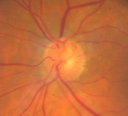
Unusual Swollen Optic Nerve
|
|
74-year-old woman was seen in the office on September 24, 2008. She has stage 4 non-small cell lung carcinoma and was treated for that starting in March of 2007. She has had decreasing vision in the left eye for at least the last two weeks, but in retrospect possibly the last month or two where she sees a blue spot in her vision in the left eye and the vision in that eye is not as good as the vision in the right eye previous to this. As far as she can recall the vision in the left eye was good. She has recently been cycling with chemotherapy for her lung cancer. She has been off the chemotherapy for about the last two weeks. She is now suffering some hearing loss possibly because of the chemotherapy. Her blood pressure has been fluctuating, but overall has been under reasonably good control.
VISUAL ACUITY: OD 20/30, OS 10/200. IOP: OD 16, OS 17.
SLIT LAMP EXAM: There is 2+ nuclear sclerosis in both eyes. We did not measure an afferent pupillary defect in the left eye, but I saw her dilated. She does see slight light desaturation in the left eye with the level in the right eye being 100 and the left eye being 80.
EXTENDED OPHTHALMOSCOPY:
OD: Vertical C/D ratio is 0.3. There is no posterior vitreous separation. The macula and periphery look healthy.
OS: Vertical C/D ratio is 0.1. There is 3+ optic nerve edema. There are dilated telangiectatic vessels 360 degrees around the optic nerve. The nerve does not look pale. There is macular edema. The retinal vessels outside of the macula look normal with no venous dilation.
Her blood pressure was 125/70.
OCT SCAN: The OCT scan of the right eye was normal. The OCT scan of the left eye shows increased retinal thickness from the macular edema. The central foveal thickness is 517 microns.
Photos confirm clinical findings.
ORBITAL ULTRASOUND: Shows no significant distension of the optic nerve or fluid around the optic nerve or masses around the optic nerve, but the ultrasound would only pick up fairly large masses.
IMPRESSION:
1. OPTIC NERVE EDEMA – LEFT EYE
2. MACULAR EDEMA – LEFT EYE
3. CONCOMITANT NON-SMALL CELL LUNG CANCER
DISCUSSION: I explained to the patient that the optic nerve swelling in the left eye could be from ischemia, possibly from a cancer-induced type of coagulable state. It is also possible she may have a paraneoplastic syndrome. There has been at least one case reported of optic nerve swelling associated with non-small cell lung carcinoma, which was thought to be paraneoplastic in nature. Finally, there is concern about compressive lesions affecting the optic nerve, especially given the relative absence of light desaturation in the left eye. I will talk to her doctor to make sure an MRI scan with and without contrast of the head and orbits is checked. She did have a brain metastasis from her lung cancer which had shrunk from 10 cm to 3 cm after radiation therapy, but to her knowledge, she has not had an MRI scan done in the recent past. Her radiation therapy was administered at the time of diagnosis in March of 2007.
One Year Later:
pleasant 74-year-old woman had an atypical neuroretinitis in the left eye and that eye is doing a little better lately. She noticed her vision fluctuating and wavy, overall though she is still not seeing very well out of the left eye.
VISUAL ACUITY: OS 20/400. IOP: 14. There is 1+ nuclear sclerosis.
EXTENDED OPHTHALMOSCOPY:
OS: Vertical C/D ratio is 0.0. There is some vascular remodeling. On the optic nerve there is a 300 micron yellow exudate under the fovea. The macula is now dry.
Patient REFUSED Fluorescein Angiogram
|

Atypical Neuroretinitis - ?Ischemic - ?Paraneoplastic - Optic Nerve Swelling and Foveal Edema and Exudate829 views74-year-old woman has stage 4 non-small cell lung carcinoma decreasing vision in the left eye for at least the last two weeks, VA 10/200. Trace APD OS. No pain    
(0 votes)
|
|

Atypical Neuroretinitis - ?Ischemic - ?Paraneoplastic - Line Scan OS Shows Nerve and Retinal Edema572 views74-year-old woman has stage 4 non-small cell lung carcinoma decreasing vision in the left eye for at least the last two weeks, VA 10/200. Trace APD OS. No pain    
(0 votes)
|
|

Atypical Neuroretinitis - ?Ischemic - ?Paraneoplastic - Optic Nerve Swelling597 views74-year-old woman has stage 4 non-small cell lung carcinoma decreasing vision in the left eye for at least the last two weeks, VA 10/200. Trace APD OS. No pain    
(0 votes)
|
|

Atypical Neuroretinitis - ?Ischemic - ?Paraneoplastic - Swelling of Optic Nerve Left Eye and Foveal Exudate720 views74-year-old woman has stage 4 non-small cell lung carcinoma decreasing vision in the left eye for at least the last two weeks, VA 10/200. Trace APD OS. No pain    
(0 votes)
|
|

Atypical Neuroretinitis - ?Ischemic - ?Paraneoplastic - Normal Right Eye464 views74-year-old woman has stage 4 non-small cell lung carcinoma decreasing vision in the left eye for at least the last two weeks, VA 10/200. Trace APD OS. No pain    
(0 votes)
|
|

Atypical Neuroretinitis - ?Ischemic - ?Paraneoplastic - Red Free Photo Shows Optic Nerve Telangiectasis509 views74-year-old woman has stage 4 non-small cell lung carcinoma decreasing vision in the left eye for at least the last two weeks, VA 10/200. Trace APD OS. No pain    
(0 votes)
|
|

Atypical Neuroretinitis - ?Ischemic - ?Paraneoplastic - Telangiectatic Vessels on Nerve After Edema Subsided645 viewsNo treatment was done and the nerve swelling subsided over about 6 months. This photo is taken one year after the ones showing swelling. There is still a trace APD.    
(0 votes)
|
|

Atypical Neuroretinitis - ?Ischemic - ?Paraneoplastic - Macular Exudate OS554 viewsNo treatment was done and the nerve swelling subsided over about 6 months. This photo is taken one year after the ones showing swelling. There is still a trace APD.    
(0 votes)
|
|

Atypical Neuroretinitis - ?Ischemic - ?Paraneoplastic - Macular Exudate Left Eye667 viewsNo treatment was done and the nerve swelling subsided over about 6 months. This photo is taken one year after the ones showing swelling. There is still a trace APD.    
(0 votes)
|
|

Atypical Neuroretinitis - ?Ischemic - ?Paraneoplastic Fundus Photo 1 year after initial episode VA 20/200574 viewsNo treatment was done and the nerve swelling subsided over about 6 months. This photo is taken one year after the ones showing swelling. There is still a trace APD.    
(0 votes)
|
|

Atypical Neuroretinitis - ?Ischemic - ?Paraneoplastic - Line Scan OS 1 year After Initial Episode488 views74-year-old woman has stage 4 non-small cell lung carcinoma decreasing vision in the left eye for at least the last two weeks, VA 10/200. Trace APD OS. No pain    
(0 votes)
|
|
|
|
|
74-year-old woman was seen in the office on September 24, 2008. She has stage 4 non-small cell lung carcinoma and was treated for that starting in March of 2007. She has had decreasing vision in the left eye for at least the last two weeks, but in retrospect possibly the last month or two where she sees a blue spot in her vision in the left eye and the vision in that eye is not as good as the vision in the right eye previous to this. As far as she can recall the vision in the left eye was good. She has recently been cycling with chemotherapy for her lung cancer. She has been off the chemotherapy for about the last two weeks. She is now suffering some hearing loss possibly because of the chemotherapy. Her blood pressure has been fluctuating, but overall has been under reasonably good control.
VISUAL ACUITY: OD 20/30, OS 10/200. IOP: OD 16, OS 17.
SLIT LAMP EXAM: There is 2+ nuclear sclerosis in both eyes. We did not measure an afferent pupillary defect in the left eye, but I saw her dilated. She does see slight light desaturation in the left eye with the level in the right eye being 100 and the left eye being 80.
EXTENDED OPHTHALMOSCOPY:
OD: Vertical C/D ratio is 0.3. There is no posterior vitreous separation. The macula and periphery look healthy.
OS: Vertical C/D ratio is 0.1. There is 3+ optic nerve edema. There are dilated telangiectatic vessels 360 degrees around the optic nerve. The nerve does not look pale. There is macular edema. The retinal vessels outside of the macula look normal with no venous dilation.
Her blood pressure was 125/70.
OCT SCAN: The OCT scan of the right eye was normal. The OCT scan of the left eye shows increased retinal thickness from the macular edema. The central foveal thickness is 517 microns.
Photos confirm clinical findings.
ORBITAL ULTRASOUND: Shows no significant distension of the optic nerve or fluid around the optic nerve or masses around the optic nerve, but the ultrasound would only pick up fairly large masses.
IMPRESSION:
1. OPTIC NERVE EDEMA – LEFT EYE
2. MACULAR EDEMA – LEFT EYE
3. CONCOMITANT NON-SMALL CELL LUNG CANCER
DISCUSSION: I explained to the patient that the optic nerve swelling in the left eye could be from ischemia, possibly from a cancer-induced type of coagulable state. It is also possible she may have a paraneoplastic syndrome. There has been at least one case reported of optic nerve swelling associated with non-small cell lung carcinoma, which was thought to be paraneoplastic in nature. Finally, there is concern about compressive lesions affecting the optic nerve, especially given the relative absence of light desaturation in the left eye. I will talk to her doctor to make sure an MRI scan with and without contrast of the head and orbits is checked. She did have a brain metastasis from her lung cancer which had shrunk from 10 cm to 3 cm after radiation therapy, but to her knowledge, she has not had an MRI scan done in the recent past. Her radiation therapy was administered at the time of diagnosis in March of 2007.
One Year Later:
pleasant 74-year-old woman had an atypical neuroretinitis in the left eye and that eye is doing a little better lately. She noticed her vision fluctuating and wavy, overall though she is still not seeing very well out of the left eye.
VISUAL ACUITY: OS 20/400. IOP: 14. There is 1+ nuclear sclerosis.
EXTENDED OPHTHALMOSCOPY:
OS: Vertical C/D ratio is 0.0. There is some vascular remodeling. On the optic nerve there is a 300 micron yellow exudate under the fovea. The macula is now dry.
Patient REFUSED Fluorescein Angiogram