Serous Retinal Detachment Both Eyes - Wet AMD - Possible Central Serous Retinopathy - 62 YO Male
|
|
62-year-old man was fine until about a year ago when he noticed decreasing vision in the right eye. There was fluid in the macula in both eyes; not in the center of the macula in the left but nasally and in the center of the macula in the right eye. He had one treatment with Avastin in the right eye on January 7th, which did not particularly help. He also had a trial of eplerenone at a dose of 50 mg PO Q day x 6 weeks without any effect either. . He does not take any steroids and is not taking any phosphodiesterase inhibitors and his blood pressure is reasonable.
VISUAL ACUITY: Vision OD is 20/100, OS is 20/20. IOP: OD 12, OS 13.
SLIT LAMP EXAM: There is 1+ nuclear sclerosis in both eyes.
EXTENDED OPHTHALMOSCOPY:
OD: Vertical C/D ratio is 0.2. There are pigment irregularities centrally.
OS: Vertical C/D ratio is 0.2. There is no posterior vitreous separation. There are some peripapillary pigment irregularities.
SPECTRALIS SD-OCT SCAN: The OCT scan of the right eye does show trace subretinal fluid overlying an area of thickening, which looks to be subretinal and possibly neovascular, in the center of the macula. The choroidal thickness under the center of the fovea is 400 microns. The left eye OCT scan shows a normal central foveal thickness in the retina. There is, however, serous macular detachment nasal to the fovea. In the superotemporal optic nerve, there does appear to be some subretinal areas which look to possibly be neovascular. The choroidal thickness under the center of the fovea in the left eye is 384 microns. (NOTE: Retinal thickness measured with Spectralis OCT is approximately 70 µm greater than that measured with Stratus OCT. This increased measurement corresponds to the inclusion of the outer segment-RPE-Bruch's membrane complex by Spectralis OCT.)
FUNDUS PHOTOGRAPHY – AUTOFLUORESCENCE: Autofluorescence image in the right eye shows hypoautofluorescence inferior to the fovea from chronic subretinal fluid. Centrally though, there is hypoautofluorescence, but it is not too dark in the center of the macula. In the left eye, the autofluorescence image is nearly normal with just a little bit hyperautofluorescence in the area of the pigment epithelial thickening adjacent to the optic nerve at about 1:00 o’clock, about a disc diameter off the nerve.
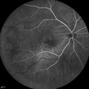
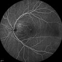
FLUORESCEIN ANGIOGRAPHY: FA of the right eye shows early hypofluorescence with fairly brisk hyperfluorescence inferior to the fovea, which looks to be from devitalization of the pigment epithelium. In the late frames, there is no pinpoint hyperfluorescence on the fluorescein angiogram, just sort of diffuse leakage as you would expect and an occult subfoveal choroidal neovascular membrane. The FA of the left eye shows
some hyperfluorescence adjacent to the optic nerve, but again nothing pinpoint and nothing extreme as you would expect in an occult subretinal choroidal neovascular membrane.
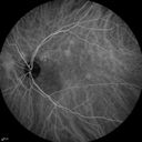
INDOCYANINE GREEN ANGIOGRAM: The right eye shows some hyperfluorescence at four minutes in the center of the macula, and then in the later images, there is a poorly defined area of hyperfluorescence centrally, measuring about 3mm² which does involve the center of the fovea. The indocyanine green angiogram of the left eye shows, within the first five minutes, some hyperfluorescence adjacent to the optic nerve just superotemporal to the nerve. In the later frames, that area does light up a little bit.
IMPRESSION:
1. SEROUS RETINAL DETACHMENT – BOTH EYES
2. POSSIBLE WET AGE-RELATED MACULAR DEGENERATION – BOTH EYES
3. POSSIBLE CENTRAL SEROUS RETINOPATHY – BOTH EYES
4. PIGMENT EPITHELIAL DETACHMENT – BOTH EYES
DISCUSSION: I explained to the patient given the subretinal deposits in both eyes and the minimal choroidal thickness on the indocyanine and fluorescein appearance of the retina, this looks a little more like wet macular degeneration than central serous retinopathy. I suggested, prior to contemplating photodynamic laser, he have a series of monthly intravitreal injections for six months in both eyes. I think that will probably dry up the left eye and may dry up the right eye a little bit. If Avastin and Lucentis do not work in the right eye, Eylea could also be tried. If all that fails, it would not be unreasonable to consider photodynamic laser.
|

Serous Macular Detachment Both Eyes - Wet AMD vs. CSR - 62 Year Old Man - Color Photo434 views    
(0 votes)
|
|

Serous Macular Detachment Both Eyes - Wet AMD vs. CSR - 62 Year Old Man - Color Photo407 views    
(0 votes)
|
|

Serous Macular Detachment Both Eyes - Wet AMD vs. CSR - 62 Year Old Man - Color Photo409 views    
(0 votes)
|
|

Serous Macular Detachment Both Eyes - Wet AMD vs. CSR - 62 Year Old Man - IR374 views    
(0 votes)
|
|

Serous Macular Detachment Both Eyes - Wet AMD vs. CSR - 62 Year Old Man - IR374 views    
(0 votes)
|
|

Serous Macular Detachment Both Eyes - Wet AMD vs. CSR - 62 Year Old Man - FAF409 views    
(0 votes)
|
|

Serous Macular Detachment Both Eyes - Wet AMD vs. CSR - 62 Year Old Man - FAF433 views    
(0 votes)
|
|

Serous Macular Detachment Both Eyes - Wet AMD vs. CSR - 62 Year Old Man - SD - OCT574 views    
(0 votes)
|
|

Serous Macular Detachment Both Eyes - Wet AMD vs. CSR - 62 Year Old Man - SD - OCT598 views    
(0 votes)
|
|

Serous Macular Detachment Both Eyes - Wet AMD vs. CSR - 62 Year Old Man - SD - OCT438 views    
(0 votes)
|
|

Serous Macular Detachment Both Eyes - Wet AMD vs. CSR - 62 Year Old Man - SD - OCT419 views    
(0 votes)
|
|

Serous Macular Detachment Both Eyes - Wet AMD vs. CSR - 62 Year Old Man - Fluorescein angiogram356 views    
(0 votes)
|
|

Serous Macular Detachment Both Eyes - Wet AMD vs. CSR - 62 Year Old Man - Fluorescein angiogram350 views    
(0 votes)
|
|

Serous Macular Detachment Both Eyes - Wet AMD vs. CSR - 62 Year Old Man - Fluorescein angiogram372 views    
(0 votes)
|
|

Serous Macular Detachment Both Eyes - Wet AMD vs. CSR - 62 Year Old Man - Fluorescein angiogram348 views    
(0 votes)
|
|

Serous Macular Detachment Both Eyes - Wet AMD vs. CSR - 62 Year Old Man - Fluorescein angiogram331 views    
(0 votes)
|
|

Serous Macular Detachment Both Eyes - Wet AMD vs. CSR - 62 Year Old Man - Fluorescein angiogram347 views    
(0 votes)
|
|

Serous Macular Detachment Both Eyes - Wet AMD vs. CSR - 62 Year Old Man - Fluorescein angiogram374 views    
(0 votes)
|
|

Serous Macular Detachment Both Eyes - Wet AMD vs. CSR - 62 Year Old Man ICG354 views    
(0 votes)
|
|

Serous Macular Detachment Both Eyes - Wet AMD vs. CSR - 62 Year Old Man ICG329 views    
(0 votes)
|
|

Serous Macular Detachment Both Eyes - Wet AMD vs. CSR - 62 Year Old Man ICG346 views    
(0 votes)
|
|

Serous Macular Detachment Both Eyes - Wet AMD vs. CSR - 62 Year Old Man ICG 355 views    
(0 votes)
|
|

Serous Macular Detachment Both Eyes - Wet AMD vs. CSR - 62 Year Old Man ICG311 views    
(0 votes)
|
|

Serous Macular Detachment Both Eyes - Wet AMD vs. CSR - 62 Year Old Man ICG320 views    
(0 votes)
|
|
|
|
62-year-old man was fine until about a year ago when he noticed decreasing vision in the right eye. There was fluid in the macula in both eyes; not in the center of the macula in the left but nasally and in the center of the macula in the right eye. He had one treatment with Avastin in the right eye on January 7th, which did not particularly help. He also had a trial of eplerenone at a dose of 50 mg PO Q day x 6 weeks without any effect either. . He does not take any steroids and is not taking any phosphodiesterase inhibitors and his blood pressure is reasonable.
VISUAL ACUITY: Vision OD is 20/100, OS is 20/20. IOP: OD 12, OS 13.
SLIT LAMP EXAM: There is 1+ nuclear sclerosis in both eyes.
EXTENDED OPHTHALMOSCOPY:
OD: Vertical C/D ratio is 0.2. There are pigment irregularities centrally.
OS: Vertical C/D ratio is 0.2. There is no posterior vitreous separation. There are some peripapillary pigment irregularities.
SPECTRALIS SD-OCT SCAN: The OCT scan of the right eye does show trace subretinal fluid overlying an area of thickening, which looks to be subretinal and possibly neovascular, in the center of the macula. The choroidal thickness under the center of the fovea is 400 microns. The left eye OCT scan shows a normal central foveal thickness in the retina. There is, however, serous macular detachment nasal to the fovea. In the superotemporal optic nerve, there does appear to be some subretinal areas which look to possibly be neovascular. The choroidal thickness under the center of the fovea in the left eye is 384 microns. (NOTE: Retinal thickness measured with Spectralis OCT is approximately 70 µm greater than that measured with Stratus OCT. This increased measurement corresponds to the inclusion of the outer segment-RPE-Bruch's membrane complex by Spectralis OCT.)
FUNDUS PHOTOGRAPHY – AUTOFLUORESCENCE: Autofluorescence image in the right eye shows hypoautofluorescence inferior to the fovea from chronic subretinal fluid. Centrally though, there is hypoautofluorescence, but it is not too dark in the center of the macula. In the left eye, the autofluorescence image is nearly normal with just a little bit hyperautofluorescence in the area of the pigment epithelial thickening adjacent to the optic nerve at about 1:00 o’clock, about a disc diameter off the nerve.
FLUORESCEIN ANGIOGRAPHY: FA of the right eye shows early hypofluorescence with fairly brisk hyperfluorescence inferior to the fovea, which looks to be from devitalization of the pigment epithelium. In the late frames, there is no pinpoint hyperfluorescence on the fluorescein angiogram, just sort of diffuse leakage as you would expect and an occult subfoveal choroidal neovascular membrane. The FA of the left eye shows
some hyperfluorescence adjacent to the optic nerve, but again nothing pinpoint and nothing extreme as you would expect in an occult subretinal choroidal neovascular membrane.
INDOCYANINE GREEN ANGIOGRAM: The right eye shows some hyperfluorescence at four minutes in the center of the macula, and then in the later images, there is a poorly defined area of hyperfluorescence centrally, measuring about 3mm² which does involve the center of the fovea. The indocyanine green angiogram of the left eye shows, within the first five minutes, some hyperfluorescence adjacent to the optic nerve just superotemporal to the nerve. In the later frames, that area does light up a little bit.
IMPRESSION:
1. SEROUS RETINAL DETACHMENT – BOTH EYES
2. POSSIBLE WET AGE-RELATED MACULAR DEGENERATION – BOTH EYES
3. POSSIBLE CENTRAL SEROUS RETINOPATHY – BOTH EYES
4. PIGMENT EPITHELIAL DETACHMENT – BOTH EYES
DISCUSSION: I explained to the patient given the subretinal deposits in both eyes and the minimal choroidal thickness on the indocyanine and fluorescein appearance of the retina, this looks a little more like wet macular degeneration than central serous retinopathy. I suggested, prior to contemplating photodynamic laser, he have a series of monthly intravitreal injections for six months in both eyes. I think that will probably dry up the left eye and may dry up the right eye a little bit. If Avastin and Lucentis do not work in the right eye, Eylea could also be tried. If all that fails, it would not be unreasonable to consider photodynamic laser.