Idiopathic Polypoidal Choroidal Vasculopathy (IPCV) and Nonexudative wet AMD in the right eye
|
|
69-year-old woman was seen in the office on April 3, 2013. She has noticed decreasing vision in the left eye. Starting a few years ago she was in Costa Rica. She is spending more time though here. Her mother lost vision from macular degeneration and was told some time ago that there is no treatments, so the patient presumed that there wasn’t any good treatment for her eye, but when she saw you, you suggested because of the bleeding in the left eye, she come here for an evaluation. Her right eye is a little hazier than it was last year, but not a lot.
VISUAL ACUITY: OD 20/50, OS 20/200. IOP: OD 14, OS 13.
SLIT EXAMINATION: There is 2+ nuclear sclerosis in both eyes.
EXTENDED OPHTHALMOSCOPY:
OD: Vertical C/D ratio is 0.2. There is no posterior vitreous separation. There is pigment epithelium thickening centrally.
OS: Vertical C/D ratio is 0.2. There is no posterior vitreous separation. There is pigment epithelium thickening centrally with subretinal hemorrhage inferotemporal to the fovea.
SPECTRALIS-SD-OCT SCAN: The OCT scan of the right eye shows the macula to be dry with pigment epithelium thickening. The left eye shows the macula to be wet with intraretinal and subretinal fluid.
FUNDUS PHOTOGRAPHY - AUTO FLUORESCENCE: The image in the right eye does show some hyper auto fluorescence centrally and the left eye has hypo auto fluorescence centrally and there was some hyper auto fluorescence inferotemporally.
COLOR PHOTOS: The color images in the right eye show the macular drusen. The left eye shows the hemorrhage inferotemporal to the fovea and the pigment epithelial detachment.
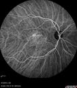
FLUORESCEIN ANGIOGRAPHY: Fluorescein angiography of the left eye shows early leakage with late staining of the entire neovascular complex, which fills much of the macula. There is hypofluorescence going to the blood inferotemporally. The right eye shows mostly staining of the drusen. There is however, some stippled hyperfluorescence centrally in the late frames.
INDOCYANINE GREEN ANGIOGRAPHY: The indocyanine green angiogram in the left eye shows early polypoidal choroidal vasculopathy with a clear lacy network with polyps temporal to the fovea. In the right eye in the late frames, the entire lesion stains. The right eye similarly has a staining lesion centrally suggesting an occult subfoveal choroidal neovascular membrane.
IMPRESSION:
1. WET AGE-RELATED MACULAR DEGENERATION – BOTH EYES
2. IDIOPATHIC POLYPOIDAL CHOROIDAL VASCULOPATHY – LEFT EYE
3. MACULAR HEMORRHAGE – LEFT EYE
4. INTRARETINAL AND SUBRETINAL FLUID – LEFT EYE
5. CATARACT
DISCUSSION: I explained to the patient the left eye does have bleeding and wet age-related macular degeneration and vision loss. With intravitreal Avastin there is a chance of possibly improving the vision.
I treated the left eye with intravitreal injection of Avastin (1.25 mg/0.05 ml) without any difficulty today. In the meantime the right eye on the indocyanine green angiogram is a suspicious lesion and that needs to be watched closely, if she does start to manifest vision problems, it may be worthwhile considering treatment in the right eye as well.
Finally, the left eye has polypoidal choroidal vasculopathy in the EVEREST study, recently suggested that in that situation there is a place for photodynamic laser, especially in patients who are not responsive to anti VEGF therapy.
|

Idiopathic Polypoidal Choroidal Vasculopathy - False Color644 views    
(0 votes)
|
|

Idiopathic Polypoidal Choroidal Vasculopathy - False Color527 views    
(0 votes)
|
|

Idiopathic Polypoidal Choroidal Vasculopathy - False Color399 views    
(0 votes)
|
|

Idiopathic Polypoidal Choroidal Vasculopathy - IR403 views    
(0 votes)
|
|

Idiopathic Polypoidal Choroidal Vasculopathy - IR572 views    
(0 votes)
|
|

Idiopathic Polypoidal Choroidal Vasculopathy - FAF474 views    
(0 votes)
|
|

Idiopathic Polypoidal Choroidal Vasculopathy - FAF424 views    
(0 votes)
|
|

Idiopathic Polypoidal Choroidal Vasculopathy - FAF417 views    
(0 votes)
|
|

Idiopathic Polypoidal Choroidal Vasculopathy - SD-OCT557 views    
(0 votes)
|
|

Idiopathic Polypoidal Choroidal Vasculopathy - SD-OCT557 views    
(0 votes)
|
|

Idiopathic Polypoidal Choroidal Vasculopathy - SD-OCT543 views    
(0 votes)
|
|

Idiopathic Polypoidal Choroidal Vasculopathy - SD-OCT614 views    
(0 votes)
|
|

Idiopathic Polypoidal Choroidal Vasculopathy - Fluorescein Angiogram680 views    
(0 votes)
|
|

Idiopathic Polypoidal Choroidal Vasculopathy - Fluorescein Angiogram464 views    
(0 votes)
|
|

Idiopathic Polypoidal Choroidal Vasculopathy - Fluorescein Angiogram504 views    
(0 votes)
|
|

Idiopathic Polypoidal Choroidal Vasculopathy - Fluorescein Angiogram459 views    
(0 votes)
|
|

Idiopathic Polypoidal Choroidal Vasculopathy - Fluorescein Angiogram548 views    
(0 votes)
|
|

Idiopathic Polypoidal Choroidal Vasculopathy - Fluorescein Angiogram434 views    
(0 votes)
|
|

Idiopathic Polypoidal Choroidal Vasculopathy - Fluorescein Angiogram468 views    
(0 votes)
|
|

Asymptomatic Occult Subfoveal CNVM in Good Eye Visible on ICG656 views    
(0 votes)
|
|

Asymptomatic Occult Subfoveal CNVM in Good Eye Visible on ICG486 views    
(0 votes)
|
|

Idiopathic Polypoidal Choroidal Vasculopathy - Indocyanine Green Angiogram - Polyp Visible Left Eye. Branching Network Both Eye648 views    
(0 votes)
|
|

Idiopathic Polypoidal Choroidal Vasculopathy - Indocyanine Green Angiogram - Polyp Visible Left Eye. Branching Network Both Eye613 views    
(0 votes)
|
|

Idiopathic Polypoidal Choroidal Vasculopathy - Indocyanine Green Angiogram - Polyp Visible Left Eye. Branching Network Both Eye598 views    
(0 votes)
|
|
|
|
69-year-old woman was seen in the office on April 3, 2013. She has noticed decreasing vision in the left eye. Starting a few years ago she was in Costa Rica. She is spending more time though here. Her mother lost vision from macular degeneration and was told some time ago that there is no treatments, so the patient presumed that there wasn’t any good treatment for her eye, but when she saw you, you suggested because of the bleeding in the left eye, she come here for an evaluation. Her right eye is a little hazier than it was last year, but not a lot.
VISUAL ACUITY: OD 20/50, OS 20/200. IOP: OD 14, OS 13.
SLIT EXAMINATION: There is 2+ nuclear sclerosis in both eyes.
EXTENDED OPHTHALMOSCOPY:
OD: Vertical C/D ratio is 0.2. There is no posterior vitreous separation. There is pigment epithelium thickening centrally.
OS: Vertical C/D ratio is 0.2. There is no posterior vitreous separation. There is pigment epithelium thickening centrally with subretinal hemorrhage inferotemporal to the fovea.
SPECTRALIS-SD-OCT SCAN: The OCT scan of the right eye shows the macula to be dry with pigment epithelium thickening. The left eye shows the macula to be wet with intraretinal and subretinal fluid.
FUNDUS PHOTOGRAPHY - AUTO FLUORESCENCE: The image in the right eye does show some hyper auto fluorescence centrally and the left eye has hypo auto fluorescence centrally and there was some hyper auto fluorescence inferotemporally.
COLOR PHOTOS: The color images in the right eye show the macular drusen. The left eye shows the hemorrhage inferotemporal to the fovea and the pigment epithelial detachment.
FLUORESCEIN ANGIOGRAPHY: Fluorescein angiography of the left eye shows early leakage with late staining of the entire neovascular complex, which fills much of the macula. There is hypofluorescence going to the blood inferotemporally. The right eye shows mostly staining of the drusen. There is however, some stippled hyperfluorescence centrally in the late frames.
INDOCYANINE GREEN ANGIOGRAPHY: The indocyanine green angiogram in the left eye shows early polypoidal choroidal vasculopathy with a clear lacy network with polyps temporal to the fovea. In the right eye in the late frames, the entire lesion stains. The right eye similarly has a staining lesion centrally suggesting an occult subfoveal choroidal neovascular membrane.
IMPRESSION:
1. WET AGE-RELATED MACULAR DEGENERATION – BOTH EYES
2. IDIOPATHIC POLYPOIDAL CHOROIDAL VASCULOPATHY – LEFT EYE
3. MACULAR HEMORRHAGE – LEFT EYE
4. INTRARETINAL AND SUBRETINAL FLUID – LEFT EYE
5. CATARACT
DISCUSSION: I explained to the patient the left eye does have bleeding and wet age-related macular degeneration and vision loss. With intravitreal Avastin there is a chance of possibly improving the vision.
I treated the left eye with intravitreal injection of Avastin (1.25 mg/0.05 ml) without any difficulty today. In the meantime the right eye on the indocyanine green angiogram is a suspicious lesion and that needs to be watched closely, if she does start to manifest vision problems, it may be worthwhile considering treatment in the right eye as well.
Finally, the left eye has polypoidal choroidal vasculopathy in the EVEREST study, recently suggested that in that situation there is a place for photodynamic laser, especially in patients who are not responsive to anti VEGF therapy.