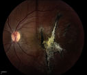
Image search results - "choroidal"
|

Choroidal Rupture and small Hemorrhage - Assault1112 views34 year old man. The patient reports he was assaulted by a couple of adolescents near downtown St. Petersburg about forty-eight hours ago. He reports the pain in the left eye has significantly improved, but his vision is a little fuzzy and he sees some floaters in the left eye. 20/125 OD, 20/20 OS. Pinhole improves in the right eye to 20/50. IOP: 15 OD, 13 OS.
SLIT LAMP EXAMINATION: Biomicroscopy reveals 3+ pupillary reaction OD, 2+ OS with no afferent pupillary defect. EXTENDED OPHTHALMOSCOPY: Extended funduscopy with Volk 90-diopter lens and fundus drawings of both eyes reveal a clear view with no posterior vitreous separation. C/D ratio is 0.2. There is no vitreous debris or hemorrhage. Large retinal vessels and macula are healthy. The retinal periphery, inspected with scleral depression for 360° shows no retinal tears, breaks, or detachments.
|
|

Choroidal Rupture and small Hemorrhage - Assault817 views34 year old man. The patient reports he was assaulted by a couple of adolescents near downtown St. Petersburg about forty-eight hours ago. He reports the pain in the left eye has significantly improved, but his vision is a little fuzzy and he sees some floaters in the left eye. 20/125 OD, 20/20 OS. Pinhole improves in the right eye to 20/50. IOP: 15 OD, 13 OS.
SLIT LAMP EXAMINATION: Biomicroscopy reveals 3+ pupillary reaction OD, 2+ OS with no afferent pupillary defect. EXTENDED OPHTHALMOSCOPY: Extended funduscopy with Volk 90-diopter lens and fundus drawings of both eyes reveal a clear view with no posterior vitreous separation. C/D ratio is 0.2. There is no vitreous debris or hemorrhage. Large retinal vessels and macula are healthy. The retinal periphery, inspected with scleral depression for 360° shows no retinal tears, breaks, or detachments.
|
|

Choroidal Rupture and small Hemorrhage - Assault670 views34 year old man. The patient reports he was assaulted by a couple of adolescents near downtown St. Petersburg about forty-eight hours ago. He reports the pain in the left eye has significantly improved, but his vision is a little fuzzy and he sees some floaters in the left eye. 20/125 OD, 20/20 OS. Pinhole improves in the right eye to 20/50. IOP: 15 OD, 13 OS.
SLIT LAMP EXAMINATION: Biomicroscopy reveals 3+ pupillary reaction OD, 2+ OS with no afferent pupillary defect. EXTENDED OPHTHALMOSCOPY: Extended funduscopy with Volk 90-diopter lens and fundus drawings of both eyes reveal a clear view with no posterior vitreous separation. C/D ratio is 0.2. There is no vitreous debris or hemorrhage. Large retinal vessels and macula are healthy. The retinal periphery, inspected with scleral depression for 360° shows no retinal tears, breaks, or detachments.
|
|

Choroidal Rupture and small Hemorrhage - Assault638 views34 year old man. The patient reports he was assaulted by a couple of adolescents near downtown St. Petersburg about forty-eight hours ago. He reports the pain in the left eye has significantly improved, but his vision is a little fuzzy and he sees some floaters in the left eye. 20/125 OD, 20/20 OS. Pinhole improves in the right eye to 20/50. IOP: 15 OD, 13 OS.
SLIT LAMP EXAMINATION: Biomicroscopy reveals 3+ pupillary reaction OD, 2+ OS with no afferent pupillary defect. EXTENDED OPHTHALMOSCOPY: Extended funduscopy with Volk 90-diopter lens and fundus drawings of both eyes reveal a clear view with no posterior vitreous separation. C/D ratio is 0.2. There is no vitreous debris or hemorrhage. Large retinal vessels and macula are healthy. The retinal periphery, inspected with scleral depression for 360° shows no retinal tears, breaks, or detachments.
|
|

Choroidal Rupture and small Hemorrhage - Assault840 views34 year old man. The patient reports he was assaulted by a couple of adolescents near downtown St. Petersburg about forty-eight hours ago. He reports the pain in the left eye has significantly improved, but his vision is a little fuzzy and he sees some floaters in the left eye. 20/125 OD, 20/20 OS. Pinhole improves in the right eye to 20/50. IOP: 15 OD, 13 OS.
SLIT LAMP EXAMINATION: Biomicroscopy reveals 3+ pupillary reaction OD, 2+ OS with no afferent pupillary defect. EXTENDED OPHTHALMOSCOPY: Extended funduscopy with Volk 90-diopter lens and fundus drawings of both eyes reveal a clear view with no posterior vitreous separation. C/D ratio is 0.2. There is no vitreous debris or hemorrhage. Large retinal vessels and macula are healthy. The retinal periphery, inspected with scleral depression for 360° shows no retinal tears, breaks, or detachments.
|
|

Choroidal Rupture and small Hemorrhage - Assault606 views34 year old man. The patient reports he was assaulted by a couple of adolescents near downtown St. Petersburg about forty-eight hours ago. He reports the pain in the left eye has significantly improved, but his vision is a little fuzzy and he sees some floaters in the left eye. 20/125 OD, 20/20 OS. Pinhole improves in the right eye to 20/50. IOP: 15 OD, 13 OS.
SLIT LAMP EXAMINATION: Biomicroscopy reveals 3+ pupillary reaction OD, 2+ OS with no afferent pupillary defect. EXTENDED OPHTHALMOSCOPY: Extended funduscopy with Volk 90-diopter lens and fundus drawings of both eyes reveal a clear view with no posterior vitreous separation. C/D ratio is 0.2. There is no vitreous debris or hemorrhage. Large retinal vessels and macula are healthy. The retinal periphery, inspected with scleral depression for 360° shows no retinal tears, breaks, or detachments.
|
|

Classic Subfoveal Choroidal Neovascular Membrane - 20/400 Vision - 6 - 12 months old lesion708 views78-year-old woman six months or a year ago decreasing vision in the right eye. OD 20/400. Fluorescein angiography shows a classic subfoveal choroidal neovascular membrane in the right eye 3 disc-diameters across, which has brisk leakage. The left eye shows staining drusen.
|
|

Classic Subfoveal Choroidal Neovascular Membrane - 20/400 Vision - 6 - 12 months old lesion611 views78-year-old woman six months or a year ago decreasing vision in the right eye. OD 20/400. Fluorescein angiography shows a classic subfoveal choroidal neovascular membrane in the right eye 3 disc-diameters across, which has brisk leakage. The left eye shows staining drusen.
|
|

Classic Subfoveal Choroidal Neovascular Membrane - 20/400 Vision - 6 - 12 months old lesion631 views78-year-old woman six months or a year ago decreasing vision in the right eye. OD 20/400. Fluorescein angiography shows a classic subfoveal choroidal neovascular membrane in the right eye 3 disc-diameters across, which has brisk leakage. The left eye shows staining drusen.
|
|

Classic Subfoveal Choroidal Neovascular Membrane - 20/400 Vision - 6 - 12 months old lesion581 views78-year-old woman six months or a year ago decreasing vision in the right eye. OD 20/400. Fluorescein angiography shows a classic subfoveal choroidal neovascular membrane in the right eye 3 disc-diameters across, which has brisk leakage. The left eye shows staining drusen.
|
|

Classic Subfoveal Choroidal Neovascular Membrane - 20/400 Vision - 6 - 12 months old lesion557 views78-year-old woman six months or a year ago decreasing vision in the right eye. OD 20/400. Fluorescein angiography shows a classic subfoveal choroidal neovascular membrane in the right eye 3 disc-diameters across, which has brisk leakage. The left eye shows staining drusen.
|
|

Classic Subfoveal Choroidal Neovascular Membrane - 20/400 Vision - 6 - 12 months old lesion536 views78-year-old woman six months or a year ago decreasing vision in the right eye. OD 20/400. Fluorescein angiography shows a classic subfoveal choroidal neovascular membrane in the right eye 3 disc-diameters across, which has brisk leakage. The left eye shows staining drusen.
|
|

Classic Subfoveal Choroidal Neovascular Membrane - 20/400 Vision - 6 - 12 months old lesion580 views78-year-old woman six months or a year ago decreasing vision in the right eye. OD 20/400. Fluorescein angiography shows a classic subfoveal choroidal neovascular membrane in the right eye 3 disc-diameters across, which has brisk leakage. The left eye shows staining drusen.
|
|

Steel Dart Perforating Injury - 15 year old - 3 weeks post-injury Vision 20/400879 views15-year-old injured in his left eye with a steel dart. The dart entered superiorly at the pars plana and his vision is clearing as the blood absorbs.
VISUAL ACUITY: OD 20/20, OS 20/400.
|
|

Steel Dart Perforating Injury - 15 year old - 3 weeks post-injury Vision 20/400489 views15-year-old injured in his left eye with a steel dart. The dart entered superiorly at the pars plana and his vision is clearing as the blood absorbs.
VISUAL ACUITY: OD 20/20, OS 20/400.
|
|

Steel Dart Perforating Injury - 15 year old - 3 weeks post-injury Vision 20/400467 views15-year-old injured in his left eye with a steel dart. The dart entered superiorly at the pars plana and his vision is clearing as the blood absorbs.
VISUAL ACUITY: OD 20/20, OS 20/400.
|
|

Steel Dart Perforating Injury - 15 year old - 3 weeks post-injury Vision 20/400509 views15-year-old injured in his left eye with a steel dart. The dart entered superiorly at the pars plana and his vision is clearing as the blood absorbs.
VISUAL ACUITY: OD 20/20, OS 20/400.
|
|

Steel Dart Perforating Injury - 15 year old - 3 weeks post-injury Vision 20/400508 views15-year-old injured in his left eye with a steel dart. The dart entered superiorly at the pars plana and his vision is clearing as the blood absorbs.
VISUAL ACUITY: OD 20/20, OS 20/400.
|
|

Steel Dart Perforating Injury - 15 year old - 3 weeks post-injury Vision 20/400481 views15-year-old injured in his left eye with a steel dart. The dart entered superiorly at the pars plana and his vision is clearing as the blood absorbs.
VISUAL ACUITY: OD 20/20, OS 20/400.
|
|

Steel Dart Perforating Injury - 15 year old - 3 weeks post-injury Vision 20/400468 views16-year-old had a dart injury to the left eye August 3rd of 2009. He has had a few surgeries since then, although he never did require a vitrectomy. His vision is improving remarkably.
VISUAL ACUITY: OS: Without correction 20/40
|
|

Perforating Steel Tip Dart Injury - 1 year later - no vitrectomy - 20/40 Vision 479 views16-year-old had a dart injury to the left eye one year ago. His vision is improving remarkably.
VISUAL ACUITY: OS: 20/40
|
|

Perforating Steel Tip Dart Injury - 1 year later - no vitrectomy - 20/40 Vision 415 views16-year-old had a dart injury to the left eye one year ago. His vision is improving remarkably.
VISUAL ACUITY: OS: 20/40
|
|

Perforating Steel Tip Dart Injury - 1 year later - no vitrectomy - 20/40 Vision 483 views16-year-old had a dart injury to the left eye one year ago. His vision is improving remarkably.
VISUAL ACUITY: OS: 20/40
|
|

Shotgun wound to face with Perforating Pellet - Final Vision 20/400 650 views18-year-old had a gunshot wound to the left eye. He had surgery on December 31st (1 week post injury) and February 4th and his vision has remarkably done well.
VISUAL ACUITY: Vision OS is 3/200.
|
|

Shotgun wound to face with Perforating Pellet - Final Vision 20/400 569 views18-year-old had a gunshot wound to the left eye. He had surgery on December 31st (1 week post injury) and February 4th and his vision has remarkably done well.
VISUAL ACUITY: Vision OS is 3/200.
|
|

Shotgun wound to face with Perforating Pellet - Final Vision 20/400 778 views18-year-old had a gunshot wound to the left eye. He had surgery on December 31st (1 week post injury) and February 4th and his vision has remarkably done well.
VISUAL ACUITY: Vision OS is 3/200.
|
|

Serous Choroidal Effusions2031 viewsSerous choroidal effusions following anterior segment surgery. Final visual outcome was 20/20.
|
|

Choroidal Folds1045 viewsChoroidal Folds imaged during angiography and the corresponding OCT
|
|

1406 viewsAdult male with trauma to the right eye and orbital floor fracture. Hemorrhage with Choroidal Rupture.
|
|

FAF choroidal rupture557 views
|
|

Montage FAF of Coroidal Rupture504 views
|
|

Montage of Choroidal Rupture (Color)714 views
|
|

Choroidal Malignant Melanoma - Montage455 viewsChoroidal malignant melanoma status post plaque radiotherapy OD in 1986, patient also has a submacular hemorrhage OD, and radiation retinopathy OD.
|
|

Choroidal Malignant Melanoma - early phase FA588 viewsearly phase of FA at 23 seconds (Choroidal malignant melanoma status post plaque radiotherapy OD in 1986, patient also has a submacular hemorrhage OD, and radiation retinopathy OD.)
|
|

Choroidal Malignant Melanoma - mid phase FA443 viewsmid-phase of FA at 5 minutes 10 seconds (Choroidal malignant melanoma status post plaque radiotherapy OD in 1986, patient also has a submacular hemorrhage OD, and radiation retinopathy OD.)
|
|

Choroidal Hemorrhage1385 viewsHemorrhagic Choroidal Effusion
|
|

Choroidal Rupture Secondary to Ocular Trauma345 viewsChoroidal Rupture Secondary to Ocular Trauma
|
|

Choroidal Folds649 viewsMontage OS of Choroidal Folds
|
|

Choroidal Folds secondarly to Dry ARMD703 viewsChoroidal Folds and Dry ARMD in OS
|
|

Choroidal Rupture Secondary to Ocular Trauma812 views
|
|

Melanoma574 views
|
|

Choroidal Rupture Secondary to Ocular Trauma665 views
|
|

Choriodal Rupture with Hemorrhage793 views
|
|

Choroidal Rupture with Subretinal Hemorrhage436 viewsThis patient was punched in the eye with a fist, resulting in a choroidal rupture.
|
|

CHOROIDAL MELANOMA Stereo Pair282 viewsThis patient was seen approximately five years ago with a suspicious choroidal nodule. The patient did not return for scheduled follow up appointments. When referred back to the practice, the nodule had erupted through Bruchs’ membrane to assume a brawny mushroom cloud configuration which is visible when the stereo images are merged.
|
|

choroidal melanoma224 viewsAsian man aged 37 choroidal melanoma in left eye
|
|

choroidal melanoma204 viewsAn Asian male, 37 years old, choroidal melanoma.
|
|

choroidal melanoma.156 viewsAn Asian male, 37 years old, choroidal melanoma.
|
|

choroidal melanoma144 viewsAn Asian male, 37 years old, choroidal melanoma.
|
|

choroidal melanoma.135 viewsAn Asian male, 37 years old, choroidal melanoma.
|
|
|
|
|
|