|
|

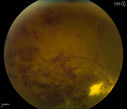
Choroidal Melanoma - Exudative Retinal Detachment 82 Year Old Man 333 views82-year-old man who had 3 falls 2 weeks ago. After the falls he started checking his vision and noticed there was a veil over the left eye, which he had seen for about two weeks in the superior visual field. OD 20/25, OS 20/40
(patient was medically never well enough for brachytherapy and died 6 months later from heart disease)
|
|

333 views
|
|

Vitreomacular traction with traction over retinal vessels in non-diabetic - Fluorescein leakage in areas of traction - Progressing vision loss left eye333 views80-year-old woman had vitrectomy surgery done in the right eye in 2007. Vision has been poor since despite a good anatomical result. Recently her vision in the left eye has been declining. The vision has declined in the left eye. Vision OD is 20/160, OS is 20/80
|
|

X-linked Juvenille Retinoschisis - Peripheral Retinal Vascular Anomalies - Vitreous Hemorrhage - 8 Year Old Boy333 views8-year-old child OD 20/40, OS 20/50
OD: Vertical C/D ratio is 0.2. There are foveal cysts. There is also a retinal elevation inferiorly and there are patchy peripheral retinal hemorrhages.
OS: Vertical C/D ratio is 0.2. There are foveal cysts. There is peripheral retinal issues with some hemorrhage in some areas of peripheral retinoschisis.
|
|

Acute Central Serous Retinopathy - CSR - Steroid Induced (probably) - Good Vision - ICG Affected Eye - Choroidal Leakage333 views42-year-old man was seen in the office on October 5, 2011. He had noticed starting in August after a course of antibiotic and steroids, that he developed new spots in his vision in the right eye. He may have had an episode like this sometime in the past. He did take steroids a few years ago and his vision did change at that time, but then returned.
VISUAL ACUITY: OD 20/32, OS 20/32. The OCT scan of the right eye confirms subretinal fluid.
|
|

Combined Artery and Vein Occlusion333 viewsPatient comes in with decreased vision in both eyes. VA 20/200-OD, 20/60-OS. Fundus exam shows great amount of macular edema due to artery and vein occlusions. There is some neovascularization on the optic nerve in the right eye. Patient was treated with Eylea Injection in the left eye today and will return for Eylea Injection in the right eye.
|
|

Central Serous Retinopathy 25 year old professional baseball player - Atypical - Choroidal Hypo-perfusion?333 views
|
|

Non-specific (uncharaterized / unknown) Macular Dystrophy 333 views59-year-old man has macular dystrophy in both eyes. He had some vision changes in medical school in the 1980s and saw Dr. Gass for that. He had some pigment epithelial irregularities in both eyes. He had strabismus as a child and had muscle surgery. He is color blind, along with many people in his family, and as far as he knows, it is congenital.
20/25, 20/30
|
|

Uveitis - Retinitis - Vasculitis - possibly Syphilitis333 viewsVisit 1: 12-06-12
74-year-old man 12-3-12 visit. 2 months ago noticed vision loss, first in the right eye than the left. He is getting tired easily. He can’t walk for a long distance. He has lost five pounds within the last month. He also had a severe itchy episode, but not quite a rash a few days ago, which went away after he took a shower. FTA-ABS is positive but a spinal tap, which was negative. OD 1/200, OS 20/63.
|
|

Vasculitis - Retinitis - Uveitis - Vision NLP right eye , 20/50 left eye333 views
|
|

Peripheral Retinal Degeneration333 viewsPatient comes in with double vision. VA was 20/20 in both eyes. Fundus exam shows Retinal Degenerative changes in both eyes. Offer to correct double vision with temporary Fresnel prism
|
|

Choroidal Neovascular Membrane - Multifocal Scars - PIC vs MFC vs OHS 333 views
|
|

Coat's Disease333 views47 year old african-american male, presented under the assumption he had Sarcoidosis but appears more like Coat's Disease
|
|

Vitreomacular traction - 20/50 vision and symptomatic with metamorphopsia333 views
|
|

333 views
|
|

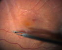
Vitrectomy with membrane peel333 viewsIntraoperative images showing the peeling of an epiretinal membrane - macular pucker - with a 25 gauge forcepts.
|
|

hydroxychloroquine (Plaquenil) Toxicity - Taiwanese 56 year old female (Asian)333 views56 year old female on 400 mg plaquenil for 22 years presents with vision loss. VA 20/50 both eyes. Plaquenil was stopped. The vision in the left eye continued to deteriorate to 3/200. 5'2" 127 lb (6.6 mg/kg body weight)
|
|

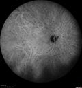
Birdshot Chorioretinitis - Fresh Presentation333 viewsUltawidefield ICG shows best the multifocal lesions. 61 year old woman with floaters for less than 6 months. HLA-A29 Positive
|
|

Birdshot Chorioretinitis - Fresh Presentation333 viewsUltawidefield ICG shows best the multifocal lesions. 61 year old woman with floaters for less than 6 months. HLA-A29 Positive
|
|

Cone Dystrophy - Autosomal Recessive333 views74 year old man with 20/25 vision OD and subtle bull's eye on FAF. Left eye is 20/200 with atrophy of the outer retina centrally
|
|

Asymptomatic wet AMD - Subfoveal CNM both eyes333 views82 year old woman - 20/25 OU. Images show occult subfoveal CNVM both eyes best seen on late ICG. The left eye also has probable polypoidal choroidal vasculopathy. No treatment was recommended on the first visit - follow-up at 1 month.
|
|

Recurrent MEWDS Left Eye - Teenage Girl333 viewsPatient presented in 2014 (first images) 2 months following vision loss left eye. Vision went from 20/60 - 20/20. OCT showed outer retinal irregularities which normalized. Then 3 years later in 2017 she had full blown MEWDS. She presented acutely and all the images are typical. Both episodes where in the same time of year, about 3 months after her flu shot for sports.
|
|

CRVO with Paracentral Acute Middle Maculopath - PAMM333 views56 year old woman with sudden vision loss, hemorrhages in all four quadrants. FA shows good retinal circulation. The OCT shows PAMM lesions in the affected eye. Vision did improve on second visit from 20/160 to 20/60 in about a month
|
|

Central Serous Retinopathy - Subretinal Fibrin333 views66 year old female with 20 year history of single episode of CSR. Now with vision loss for a month but improving - VA 20/63 OD; 20/32 OS
|
|

Central Serous Retinopathy - Subretinal Fibrin333 views66 year old female with 20 year history of single episode of CSR. Now with vision loss for a month but improving - VA 20/63 OD; 20/32 OS
|
|

Central Serous Retinopathy - Subretinal Fibrin333 views66 year old female with 20 year history of single episode of CSR. Now with vision loss for a month but improving - VA 20/63 OD; 20/32 OS
|
|

Proliferative Diabetic Retinopathy - NVD regressed with PRP laser333 views76 year old diabetic man - Presented one year ago with NVD in the left eye. This regressed with laser but then a year later worsened. Additional PRP was done and the NVD regressed again.
|
|

Neuroretinitis - NOT cat scratch - 5 year old333 viewsPresented with nerve swelling, serous RD and uveitis. Ultimately responded to steroids and methotrexate. ANA is positive. All other extensive testing negative including MRI worried about MS
|
|

Cystoid Macular Edema following Cataract surgery and vitrectomy for floaters333 views73 year old woman who had decreased vision 4 months following combined vitrectomy and cataract surgery. VA 20/63 improved in 1 month to 20/25 with ketorolac and prednisolone drops.
|
|

Cystoid Macular Edema following Cataract surgery and vitrectomy for floaters333 views73 year old woman who had decreased vision 4 months following combined vitrectomy and cataract surgery. VA 20/63 improved in 1 month to 20/25 with ketorolac and prednisolone drops.
|
|

Choroidal Osteoma 13 Year Old333 views13 year old with 6 months of vision loss right eye and vision of 20/40
|
|

Persistent subfoveal fluid following retinal detachment repair333 views62 year old man following pneumatic retinopexy for macula-off retinal detachment. Subfoveal fluid persisted for more than a year.
|
|

Choroidal thickening with subretinal fluid333 viewsPossible BDUMP - 77 year old man no history of cancer with vision loss on and off for a few months. He has dense cataract which affect the images.
|
|

Neuroretinitis and Multifocal Retinitis333 views62 year old female with vision loss in the left eye to 20/160. Positive Bartonellas IgG. Vision recovered in 2 months. She was treated with Oral Erythromycin BID for 2 weeks.
|
|

Multifocal Choroiditis Panuveitis with Subretinal Fibrosis Acute Phase333 views36 year old latin american female 3 weeks of vision loss and new headaches. MRI was normal. VA initially 20/32 OD; 20/20 OS Final vision was 20/63 OD; 20/20 OS
|
|

Macular Damage post Macular Hole Surgery333 viewsThere is a huge hole in the middle of the macula and several small holes in the posterior pole. The patient had 2 surgeries for an idiopathic stage II hole and then was offered a third procedure by the same surgeon and sought a second opinion (and was told to not have anything more done)
|
|

Coats' Disease -51 year old asymptomatic male333 views20/20 vision - had laser to non-perfusion because of proliferation.
|
|

Outer Retinal Tubulation333 views78 year old man chronic wet AMD - 20/200
|
|

Outer Retinal Tubulation333 views78 year old man chronic wet AMD - 20/200
|
|

Macroaneurysm - Resolved without treatment333 views87 year old female with vision loss OD. Initial FA showed no leakage so no treatment was done and the fluid absorbed over 4 months. Initial VA 20/100, Final VA 20/80
|
|

Angioid Streaks - CNVM OS with vision loss333 views48 year old soccer player. Prior trauma OS (with soccer ball). Vision loss for 3 weeks to 20/60 - with Lucentis vision improved to 20/30
|
|

CRVO 25 year old man - Heterozygous Factor V Leiden333 views20/100 initial vision. Improved to 20/30 with Lucentis which continued for 3 years, treat and extend, and stopped.
|
|

Pseudophakic Cystoid Macular Edema Both Eyes333 views81 year old man with vision loss about 4 months following ECCE. Did not respond to topical therapy but did fine with PST kenalog
|
|

Pseudophakic Cystoid Macular Edema Both Eyes333 views81 year old man with vision loss about 4 months following ECCE. Did not respond to topical therapy but did fine with PST kenalog
|
|

Optic Nerve Pit Maculopathy333 views10 year old child who had inner retinal fenestration (see youtube video on scohen125 channel) and did very well.
|
|

Vitreomacular Traction -> Macular Hole -> Aborted Macular hole333 viewsProgression of VMT in both eyes over time
|
|

Vitreomacular Traction -> Macular Hole -> Aborted Macular hole333 viewsProgression of VMT in both eyes over time
|
|

Sickle Retinopathy - Chronic retinal detachment OS - Proliferation OD333 views40 year old with known SC disease. Failed to return 4 years ago for treatment and lost ision in the left eye. Then returned for one visit and refused further treatment.
|
|

Sickle Retinopathy - Chronic retinal detachment OS - Proliferation OD333 views40 year old with known SC disease. Failed to return 4 years ago for treatment and lost ision in the left eye. Then returned for one visit and refused further treatment.
|
|

Outer retinal holes and cataract from YAG laser vitreolysis333 views44 year old man with 7 year history of multiple lasers for floaters. FAF and OCT show outer retinal holes and lens has focal defects
|
|

Severe PDR with preretinal fibrosis333 views71 year old female untreated for a year following injections because of hurricane Irma. VA 20/30 OD and 5/200 OS. Sudden vision loss 1 month ago left eye was likely an ischemic event. Right eye is being treated with injections and PRP laser.
|
|

Moderate PDR333 views43 year old man 20/32 vision in both eyes. Being started on Anti-VEGF injections which will be followed by PRP.
|
|

Melanocytoma - Lightly Pigmented333 views42 year old female with very mild visual field and vision loss in the left eye: 20/20 OU
|
|

Central Serous Chorioretinopathy - Moderate333 views49 year old man with episodes in 2011, 2017, and 2018 - now with some paracentral vision loss in the right eye and subretinal fluid.
|
|

Branch Retinal Artery Occlusion - No Plaque333 views74 year old man with 2 days of vision loss. Vision OS is 20/200 but improved to 20/25 in 1 month. He has dysfibrinogen anemia.
|
|

Branch Retinal Artery Occlusion - No Plaque333 views74 year old man with 2 days of vision loss. Vision OS is 20/200 but improved to 20/25 in 1 month. He has dysfibrinogen anemia.
|
|

Macroaneurysm - Macular Hemorrhage - Branch Retinal Aterial Occlusion333 views62 year old African American female with chronic hypertension and vision loss for 2 weeks. VA is 20/400
|
|

Multifocal Choroiditis and Subretinal Fibrosis with new Subretinal Neovascular Membrane333 views26 year old African American Girl Saw me at age 10 (16 years ago) with acute multifocal choroiditis and panuveitis which evolved into subretinal fibrosis. She was initially treated with 3 years of methotrexate. Vision ended up 20/20 in both eyes. Her eyes have been inactive since and she was lost to follow-up.
A week ago she noticed sudden decreased vision in the right eye. Within the past few weeks there are more floaters in the right eye.
VA OD: sc6’/200 PHNI
VA OS: sc20/16
|
|

Chronic BRAO with Hemorrhages - Sclerosed Vessel333 views95 year old female with blurred vision in both eyes in for a checkup. History of perpapillary CNVM in the left eye 2 years prior not treated. History of stroke with multiple emboli 1 year ago.
Vision 20/32 OD, 20/40 OS
|
|

Punctate Inner Choroidopathy (PIC) Mild333 views24 year old female with high myopia. Noticing some distortion on grid. Vision 20/20 OU. Was followed frequently for several months with no progression and no CNVM.
|
|

Macular Telangiectasis and Vision loss following vitrectomy with membrane peel333 views76 year old man with MacTel who had membrane peel in the right eye 10 years ago with permanent vision loss.
VA OD: sc20/200-4 and VA OS: sc20/25-1
|
|

PDR and DME and VH333 views57 year old diabetic man with vision loss in the left eye for several months. He has DME in the left eye and VH in the left eye and PDR in both eyes. He has been started in the left eye on Anti-VEGF therapy. VA on presentation was 20/25 OD and 20/200 OS
|
|

PDR and Vitreous Hemorrhage - High Risk Left Eye - Low Risk Right Eye333 views50 year old man with type I diabetes mellitus for 26 years. New Vitreous Hemorrhage in the left eye. Both eyes have NVE. Both also have foveal hypoplasia
|
|

Choroidal Neovascular Membrane following Prior Central Serous Chorioretinopathy - OCT-A333 views67 year old female with CSR in the left eye first 20 years ago. She was managed with out therapy and has had a few episodes since. Now ther eis distortion in the left eye for a few days. She had a steroid shot in her shoulder 5 months ago.
VA 20/20 OD, 20/50 OS- ICG and OCT-A confirm CNVM in the left eye
|
|

Eales Disease and fresh vitreous hemorrhage - 20 year old man333 views20 year old mane with fresh vitreous hemorrhage in the right eye. At age 15 he had a PPV and laser in the left eye and laser in the right eye. The vision is OD 20/80 PH 20/25, OS 20/25. The left eye has a mild cataract. He had prior testing for coagulopathies which was negative. Testing done for syphillis and TB was negative. Additional laser was done to prevent further bleeding in the right eye.
|
|

Stellate non-hereditary idiopathic foveomacular retinoschisis (SNIFR) and optic nerve drusen333 views84 year old man No visual complaints. Not diabetic, cataract surgery 8 years ago. Meds: Omeprazole, Tamsulosin (Flomax)
VA 20/32 OD, 20/20 OS
|
|

Fuch's Heterochromic Iridocyclitis - Iris Photos333 views71 year old man with inflammation in the left eye for over 50 years. The left eye got a tube shunt 15 years ago. Vision is 20/32 OD, 20/40 OS
|
|

Peripheral exudative hemorrhagic chorioretinopathy - PECHR - Subfoveal fluid333 views85 year old female with
Fluctuating vision in the right eye.
VA 20/200 OD, 20/32 OS
|
|

Peripapillary Lesion Hemangioma or CNVM Early FA332 views67-year-old man with a history of a peripapillary lesion in the right eye.
|
|

New BRVO Left Eye Old BRVO Right Eye 332 views90-year-old man decreased vision maybe one year since cataract surgery he is no longer able to read. OD 20/200, OS 20/200
|
|

Diabetic Macular Edema Left Eye - Pre Laser332 views82-year-old woman diabetic for many years, last eye exam 5 years ago with gradual vision loss. OD 20/60, OS 20/70. IOP: OD 16, OS 17.
|
|

Diabetic Macular Edema Left Eye - Pre Laser332 views82-year-old woman diabetic for many years, last eye exam 5 years ago with gradual vision loss. OD 20/60, OS 20/70. IOP: OD 16, OS 17.
|
|

Pistol to head in attempted suicide 3 Months Ago - Retinal Atrophy - Shockwave Retinopathy VA Right Eye - Light Perception - Left Eye - 20/400332 views66-year-old woman had a gunshot wound to the head on August 12, 2010, with the entry wound just on the right side, just about below her temple, toward the lower part of the temple and the exit wound on the left side just above her cheek, a little behind the eye. Since then she recovered and did not need any surgery. When that all cleared she realized her vision was poor in both eyes. She has seen a neuro-ophthalmologist who thought she had traumatic optic neuropathy. OD light perception, OS 20/400.
|
|

332 views
|
|

332 views
|
|

Choroidal Melanoma - Exudative Retinal Detachment 82 Year Old Man 332 views82-year-old man who had 3 falls 2 weeks ago. After the falls he started checking his vision and noticed there was a veil over the left eye, which he had seen for about two weeks in the superior visual field. OD 20/25, OS 20/40
(patient was medically never well enough for brachytherapy and died 6 months later from heart disease)
|
|

Proliferative Diabetic Retinopathy and Mild Vitreous Hemorrhage332 views58-year-old woman has diabetic retinopathy in both eyes with neovascularization of the optic nerve, worse in the right eye than the left eye. Vision OD is 20/25, OS is 20/20
|
|

Wet AMD controlled with Avastin 20/60 Vision332 views79-year-old woman has wet age-related macular degeneration in both eyes. The left eye had intravitreal injections of Avastin starting in May of 2009. Her vision remains good in the left eye and poor in the right eye.
VISUAL ACUITY: OD 8/200, OS 20/60.
|
|

Lumigan (Bimatoprost) Cystoid Macular Edema - Eye Better 1 month after Stopping Lumigan332 views 82-year-old man had cataract surgery in 1993. He has had glaucoma for seven or eight years for a long time. He is taking Lumigan only in the left eye. About a year ago he started in the right eye and then starting just about a few months ago he has noticed intermittent blurring in the vision in the right eye. Vision 20/25 OD, 20/20 OS
|
|

Diabetic Macular Edema - Mild332 views70 year old man with mild edema did well without treatment
|
|

Diabetic Macular Edema - Mild332 views70 year old man with mild edema did well without treatment
|
|

BRVO332 views50 yr old Female with a BRVO and ME in OS
|
|

Vasculitis - Retinitis - Uveitis - Vision NLP right eye , 20/50 left eye332 views
|
|

Retinal Capillary Hemangioma due to Von Hipple-Lindau Syndrome332 viewsA 47-year old female comes in for a second opinion for retinal capillary Hemangioma in the left eye. VA was HM (hand motion) in the left eye. She has had multiple procedures done for the left eye including, Retinal Detachment surgery, YAG Laser PI, YAG Laser Capsulotomy, and Cataract surgery.
|
|

Uveal Choroidal Melanoma332 viewsPatient comes in for evaluation on a Choroidal Melanoma in the right eye. VA was 20/25 in both eyes. The melanoma is in the temporal aspect of the right eye. It measured at 0.7mm elevated after doing a BSCAN Ultrasound.
|
|

Medium Choroidal Melanoma Left Eye 332 views
|
|

Proleferative Vitreoretinopathy - Panretinal Retinoschisis 332 views
|
|

332 views
|
|

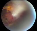
Free Floating Dislocated Lens in Vitreous332 viewsPatient comes in aphakic with dislocated lens floating to the back of the eye when laying down. Lens is laying up against the endothelium of the cornea when patient is right side up..
|
|

332 views
|
|

Cuticular Drusen - 26 year old female - Glomerulonephritis332 viewsVA 20/25 OU. Images are over 4 years. FA shows starry sky early images - This is because of loss of RPE over the drusen creating very small window defects.
|
|

Hairy Cell Leukemia - Retinal Hemorrhage and twig Branch Vein Occlusion332 views79 year old man He has had hairy cell leukemia since 2002. He is in remission. His last blood tests were 9/2018. He just moved down here and needs a new leukemia doctor. His vision is fine.
VA OD: Dcc20/25
VA OS: Dcc20/25
IOP: TP: OD:12 OS:12
|
|

Macular Laser Scars Left Eye - 5/200 Vision332 views83 year old woman who had extensive macular laser in the left eye about 15 years ago for DME. Vision is now 20/50 right eye and 3/200 left eye
|
|

Recurrent MEWDS Left Eye - Teenage Girl332 viewsPatient presented in 2014 (first images) 2 months following vision loss left eye. Vision went from 20/60 - 20/20. OCT showed outer retinal irregularities which normalized. Then 3 years later in 2017 she had full blown MEWDS. She presented acutely and all the images are typical. Both episodes where in the same time of year, about 3 months after her flu shot for sports.
|
|

CRVO with Paracentral Acute Middle Maculopath - PAMM332 views56 year old woman with sudden vision loss, hemorrhages in all four quadrants. FA shows good retinal circulation. The OCT shows PAMM lesions in the affected eye. Vision did improve on second visit from 20/160 to 20/60 in about a month
|
|

Proliferative Diabetic Retinopathy - NVD regressed with PRP laser332 views76 year old diabetic man - Presented one year ago with NVD in the left eye. This regressed with laser but then a year later worsened. Additional PRP was done and the NVD regressed again.
|
|

Cystoid Macular Edema following Cataract surgery and vitrectomy for floaters332 views73 year old woman who had decreased vision 4 months following combined vitrectomy and cataract surgery. VA 20/63 improved in 1 month to 20/25 with ketorolac and prednisolone drops.
|
|
| 16652 files on 167 page(s) |
 |
 |
 |
 |
 |
97 |  |
 |
 |
 |
|