|
|

Vitrectomy with membrane peel330 viewsIntraoperative images showing the peeling of an epiretinal membrane - macular pucker - with a 25 gauge forcepts.
|
|

Vitrectomy with membrane peel330 viewsIntraoperative images showing the peeling of an epiretinal membrane - macular pucker - with a 25 gauge forcepts.
|
|

330 views
|
|

Orbital Cellulitis - Optic nerve compression with ocular ischemia330 views81 year old woman with constricted eye movement, orbital cellulitis and severe vision loss in the right eye (LP). She has whitening of the retina and the OCT shows ischemic changes as well as subretinal fluid and blood. The vision in this eye declined to NLP and the eye remained ischemic despite control of her infection.
|
|

hydroxychloroquine (Plaquenil) Toxicity - Taiwanese 56 year old female (Asian)330 views56 year old female on 400 mg plaquenil for 22 years presents with vision loss. VA 20/50 both eyes. Plaquenil was stopped. The vision in the left eye continued to deteriorate to 3/200. 5'2" 127 lb (6.6 mg/kg body weight)
|
|

Macular Telangiectasia and dry AMD - Simulating wet AMD330 viewsThis patient was told different things by different retina specialists. Her case is complex because she has OCT findings of Macular Telangiectasis and dry AMD. This makes it look like she has wet AMD. Her vision is stable.
|
|

Bilateral Branch Retinal Artery Occlusion - Intraarterial (hollenhorst) plaques visible330 views67 year old with acute visual field loss in each eye. Vision is 20/25 right eye 20/32 left eye. Images show intra-arterial plaques in each eye. The right eye on the optic nerve and the left eye in a branch retinal artery.
|
|

Late Onset Retinal Degeneration (L-ORD)330 views55 year old with acute vision loss from a CNVM in the right eye. He responded to Lucentis therapy. His mother and her family has been confirmed genetically to have L-ORD and were part of the early reports.
|
|
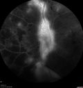
PDR NVD left eye and NVE right eye330 views41 year old diabetic woman with PDR in both eyes. High risk in the left eye. She had PRP, Avastin, and vitrectomy x 2 in the left eye with final vision of 20/25. The right eye had eventually PRP laser
|
|

Travatan Induced CME Left eye330 views63 year old woman 4 years post ECCE and 2 years post PPV for floaters. She developed CME in the left eye after 1 year of travatan. The travatan was stopped and the CME and serous retinal detachment resolved.
|
|

Asymptomatic wet AMD - Subfoveal CNM both eyes330 views82 year old woman - 20/25 OU. Images show occult subfoveal CNVM both eyes best seen on late ICG. The left eye also has probable polypoidal choroidal vasculopathy. No treatment was recommended on the first visit - follow-up at 1 month.
|
|

Recurrent MEWDS Left Eye - Teenage Girl330 viewsPatient presented in 2014 (first images) 2 months following vision loss left eye. Vision went from 20/60 - 20/20. OCT showed outer retinal irregularities which normalized. Then 3 years later in 2017 she had full blown MEWDS. She presented acutely and all the images are typical. Both episodes where in the same time of year, about 3 months after her flu shot for sports.
|
|

Multifocal Bilateral Central Serous Retinopathy in African American Male330 views67 year old african american male with : Blurred Vision OD. Duration of Problem: 4-6 weeks. VA 20/32 OD 20/20 OS
|
|

Chronic CSR with mild vision loss - Macular Atrophy Right Eye330 views55 year old man has not been to an eye doctor in years. VA 20/40 OD and 20/25 OS. OCT shows outer retinal atrophy and choroidal thickening. FAF shows guttering. FA shows no active lesions. ICG shows patchy mild leakage visible at about 5 minutes.
|
|

Persistent Subfoveal Fluid Following Retinal Detachment Repair330 viewsPatient had pneumatic retinopexy for macula off RD and subfoveal fluid persisted for 12 months but was gone at 18 months. IR images show pockets of fluid after the first week.
|
|

Retinal Capillary Hemangioma - Endophytic and Exophytic330 views46 year old female with normal vision - her father died of pancreatic cancer
|
|

Choroidal thickening with subretinal fluid330 viewsPossible BDUMP - 77 year old man no history of cancer with vision loss on and off for a few months. He has dense cataract which affect the images.
|
|

Multifocal Choroiditis Panuveitis with Subretinal Fibrosis Acute Phase330 views36 year old latin american female 3 weeks of vision loss and new headaches. MRI was normal. VA initially 20/32 OD; 20/20 OS Final vision was 20/63 OD; 20/20 OS
|
|

Multifocal Choroiditis Panuveitis with Subretinal Fibrosis Acute Phase330 views36 year old latin american female 3 weeks of vision loss and new headaches. MRI was normal. VA initially 20/32 OD; 20/20 OS Final vision was 20/63 OD; 20/20 OS
|
|

Choroidal Thickening330 views78 year old man followed for 4 months with choroidal thickening. Cursory systemic evaluation with no imaging shows no cancer. Vision is stable or improving.
|
|

Neuroretinitis - Negative cat scratch serology twice - Possible Behcets330 views65 year old man with no direct cat exposure and vision loss from neuroretinitis. His work up was positive for HLA B51. He had negative cat scratch titers twice. Vision dropped from 20/60 - 20/200 and then improved to 20/40 over 2 months
|
|

Macular Damage post Macular Hole Surgery330 viewsThere is a huge hole in the middle of the macula and several small holes in the posterior pole. The patient had 2 surgeries for an idiopathic stage II hole and then was offered a third procedure by the same surgeon and sought a second opinion (and was told to not have anything more done)
|
|

Coats' Disease -51 year old asymptomatic male330 views20/20 vision - had laser to non-perfusion because of proliferation.
|
|

Coats' Disease -51 year old asymptomatic male330 views20/20 vision - had laser to non-perfusion because of proliferation.
|
|

Nerve Fiber Defect from Optic Disk Drusen330 views31 year old male - asymptomatic
|
|

Outer Retinal Tubulation330 views78 year old man chronic wet AMD - 20/200
|
|

Choroideremia - Complete CHM gene deletion330 viewsVision loss since age 20 - now age 36 VA 20/160 (about) OU
|
|

Neuroretinitis Positive IgM Bartonella henselae330 views33 year old with vision loss. Her vision improved and she was treated with ciprofloxacen
|
|

Chronic Central Serous Chorioretinopathy330 views77 year old man was seen at uveitis clinic and tested positive to Anti-retinal antibody against 35 kDa and anti-optic nerve antibody. He did not have EDI OCT, FAF, or ICG. All testing was consistent with Chronic CSR. His immunosuppresives were stopped and he did fine for 3 years.
|
|

Non-exudative wet AMD right eye (treatment naive quiescent)330 views82 year old man. ICG shows CNVM in the right eye. This stayed quiet for 3 years and then started leaking and responded well to Avastin.
|
|

Ischemic CRVO OD and Arterial Macroaneurysm OS330 views83 year old female post PPV, Laser, Ahmed valve for CRVO with vitreous heme and NVG in the right eye. VA was 20/160 3 months following vitrectomy. Left eye has MA which was not treated and involuted post-heme
|
|

Stellate Non-heredtiary Idiopathic Foveomacular Retinoschisis (SNIFR)330 views77 year old man who is healthy and 6'6" tall with 20/40 vision and no complaints. OCT shows diffuse retinoschisis.
|
|

Macroaneurysm - Macular Hemorrhage - Branch Retinal Aterial Occlusion330 views62 year old African American female with chronic hypertension and vision loss for 2 weeks. VA is 20/400
|
|

Macroaneurysm - Macular Hemorrhage - Branch Retinal Aterial Occlusion330 views62 year old African American female with chronic hypertension and vision loss for 2 weeks. VA is 20/400
|
|

Concentric Geographic Atrophy330 views76 year old man Gradual vision loss
20/32 OD; 20/40 OS
No medicines, Non-smoker
Working and Driving
|
|
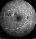
Chronic BRAO with Hemorrhages - Sclerosed Vessel330 views95 year old female with blurred vision in both eyes in for a checkup. History of perpapillary CNVM in the left eye 2 years prior not treated. History of stroke with multiple emboli 1 year ago.
Vision 20/32 OD, 20/40 OS
|
|

Subtle Branch Retinal Artery Occlusion330 views68 year old man with shadow in the left eye yesterday. IR and OCT show subtle retinal swelling. FA is normal
|
|

Adult Pseudovitelliform Macular Dystrophy330 views89 year old female states dark smudge on ceiling during night time came and went. Decreased vision in the left eye
VA OD: Dcc20/32
VA OS: Dcc20/63
|
|
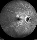
Peripheral Exudative Hemorrhagic Chorioretinopathy330 views93 year old female with shadow in side vision in the left eye. Referred for retinal detachment - Exudative lesion subsided in about 6 months without treatment.
Vision 20/63 OD and 20/100 OS (from dry AMD)
|
|

PDR and Vitreous Hemorrhage - High Risk Left Eye - Low Risk Right Eye330 views50 year old man with type I diabetes mellitus for 26 years. New Vitreous Hemorrhage in the left eye. Both eyes have NVE. Both also have foveal hypoplasia
|
|

Acquired Toxoplasmosis Retinitis Para Foveal - Following Venison Consumption330 views56 year old man who became very sick after eating venison at the end of December 2019. Presented 6 weeks later with a scotoma. Initial photos show presentation with 20/63 vision. Vision dropped to 5/200. All tests (including PCR of anterior chamber tap) were negative except serum toxoplasmosis IgG and IgM. Vision improved to 20/32 but the scotoma remained on oral trimetheprim/sulfa
|
|

Heavy Panretinal Photocoagulation for Proliferative Diabetic Retinopathy about 40 years ago330 views64 year old female - images from 2018 - heavy PRP about 30 years ago
VA 20/20 OD, 20/50 OS
|
|

Choroidal Folds - Nanophthalmous - Axial length 20330 views67 year old female Decreased Vision OU with choriodal folds. Axial length is 20. There is a CHRPE lesion in the left eye.
VA 20/25 OU. Has already had cataract and glaucoma surgery.
|
|

PDR OS with NVE329 views57-year-old man has diabetic retinopathy in both eyes.
Diabetic for 14 years with HgB A1C often over 10.
VISUAL ACUITY: OD 20/30, OS 20/40. PDR OS BDR OD
|
|

Diabetic Macular Edema Left Eye - Pre Laser329 views82-year-old woman diabetic for many years, last eye exam 5 years ago with gradual vision loss. OD 20/60, OS 20/70. IOP: OD 16, OS 17.
|
|

Proliferative Diabetic Retinopathy in patient with Moyamoya Disease329 views28-year-old womandiabetic since age nine, and she also has had multiple strokes. OD is 20/30, OS is 20/30.
|
|

329 views
|
|

Chronic Pseudophakic Cystoid Macular Edema - Haptic Migrated into Anterior Chamber Through Peripheral Iridotomy329 views57-year-old man with congenital nystagmus. He had cataract surgery done 30 years ago and has decreased vision left eye for 6 months. OD is 20/50, OS is 20/100
|
|

Retinal Pigment Epithelial Detachment with no Sub-Retinal Fluid...329 viewsA 38-year old male who comes in with blurred vision in the left eye. VA is 20/30. Notices a defect inferior of his central vision. Did an Fluorescence Angiogram to determine an RPE with no sub retinal fluid. Also OCT confirms. Patient was injected with Avastin.
|
|

Central Serous Retinopathy329 viewsPatient comes in with blurred vision. Patient also had a lung transplant. VA was 20/150 with pinhole correction to 20/60 in the right eye. Fundus exam in the right eye shows a great amount of sub retinal fluid over the macula. Will monitor to see if it goes away on its own, if not then will consider laser treatment.
|
|
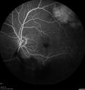
Vasculitis - Retinitis - Uveitis - Vision NLP right eye , 20/50 left eye329 views
|
|

No significant macular edema329 views
|
|

Serous Macular Detachment Both Eyes - Wet AMD vs. CSR - 62 Year Old Man ICG329 views
|
|

BRVO Inferiorly with Retinal Hemorrhages in the Left Eye329 viewsPatient comes in with complaint of "spot in vision", sort of a cloudy haze in the left eye. Patient's VA was 20/100-1 with no improvement with pinhole in the left eye. Fundus photos show multiple retinal hemorrhages scattered with a branch retinal vein occlusion inferiorily. Patient is also diabetic.
|
|

Hypertensive Retinopathy329 viewsPatient comes in complaining of spots in vision in both eyes. VA was 20/25 - right eye and 20/20- left eye. Fundus exam reveals little hemorrhages with cotton wool spots due to hypertension and anemia.
|
|

Proleferative Vitreoretinopathy - Panretinal Retinoschisis - VIDEO TAKES TIME TO LOAD329 views
|
|

Deep skin tunel remains after silicon buckle removed329 viewssuperior lid skin tunnel made by 2 years of erosion of the buckle
|
|

Optic Neuritis/Neuroretinitis329 viewsPt. states lost some vision in his left eye last week, was seen by eye MD and referred for eval. Also complains of pain in left eye with eye movement. VA was 20/400 in the left eye. Fundus photo and HD-OCT imaging show optic nerve swelling and fluid underneath the retina. Will consult with Neuro-Ophthalmologist for further evaluation.
|
|

Macular Pucker 20/40 vision - minimal symptoms for 1 year Right eye329 viewsFA shows leakage from traction from pucker in the right eye - cystoid macular edema
|
|

329 views
|
|

329 views
|
|

Branch Retinal Vein Occlusion329 viewsPatient's VA was 20/40, right eye. Fundus exam shows branch retinal vein occlusion nasally to the optic nerve in the right eye.
|
|

Toxoplasmosis Retinitis - Vitritis and Anerior Uveitis329 views
|
|

Pterygium and RK Scars329 viewsPatient comes in for SCL fitting. VA is 20/30. right eye and 20/70, left eye. Slit lamp photos show 8-RK Scars in both eyes. Pterygium, nasally, left eye.
|
|

New wet AMD OS with subretinal hemorrhage and recent vision loss, Scar OD329 views
|
|

Orbital Cellulitis - Optic nerve compression with ocular ischemia329 views81 year old woman with constricted eye movement, orbital cellulitis and severe vision loss in the right eye (LP). She has whitening of the retina and the OCT shows ischemic changes as well as subretinal fluid and blood. The vision in this eye declined to NLP and the eye remained ischemic despite control of her infection.
|
|

Branch Retinal Vein Occlusion329 views84 year old man - 2 months vision loss - VA 20/40 - 20/20 with one injection of Eylea
|
|

Late Onset Retinal Degeneration (L-ORD)329 views55 year old with acute vision loss from a CNVM in the right eye. He responded to Lucentis therapy. His mother and her family has been confirmed genetically to have L-ORD and were part of the early reports.
|
|

Recurrent MEWDS Left Eye - Teenage Girl329 viewsPatient presented in 2014 (first images) 2 months following vision loss left eye. Vision went from 20/60 - 20/20. OCT showed outer retinal irregularities which normalized. Then 3 years later in 2017 she had full blown MEWDS. She presented acutely and all the images are typical. Both episodes where in the same time of year, about 3 months after her flu shot for sports.
|
|

Central Serous Retinopathy - Subretinal Fibrin329 views66 year old female with 20 year history of single episode of CSR. Now with vision loss for a month but improving - VA 20/63 OD; 20/32 OS
|
|

Proliferative Diabetic Retinopathy - NVD regressed with PRP laser329 views76 year old diabetic man - Presented one year ago with NVD in the left eye. This regressed with laser but then a year later worsened. Additional PRP was done and the NVD regressed again.
|
|

329 views
|
|

Dry AMD - Confluent Drusen Sparing Center of Macula which has Atrophy329 views65 year old woman, VA 20/40 OD; 20/80 OS. The center of the macula has few or no drusen with predominantly non-geographic atrophy
|
|

Dry AMD - Confluent Drusen Sparing Center of Macula which has Atrophy329 views65 year old woman, VA 20/40 OD; 20/80 OS. The center of the macula has few or no drusen with predominantly non-geographic atrophy
|
|

Pattern Dystrophy - Adult Vitellifrom (Best)329 views71 year old female - lesions nasal to the fovea in both eyes. (20/40 OU)
|
|

Prefoveal Opercula and early Lamellar Macular Hole329 views73 year old man with 20/32 vision and pre-foveal opercula. The OCT shows very early lamellar hole with lamellar hole epiretinal proliferation
|
|

Enhanced S Cone Syndrome - Goldmann Favre - NR2E3 Mutation329 views82 year old man with poor vision for many years. VA HM OD, 5/200 OS. Diagnosed at age 12 with retinitis pigmentosa. Nystagmus.
|
|

329 views
|
|

329 views
|
|

Cardiac Catheterization induced Cilioretinal Artery Occlusion - Paracentral Acute Middle Maculopathy329 views69 year old female with sudden vision loss on completing cardiac catheterization. The SD OCT shows a PAMM lesion and the lesion looks gray on color imaging. There is delayed filling of the cilioretinal artery.
|
|

Multifocal Choroiditis Panuveitis - New Choroidal Neovascular Membrane Right Eye329 views74 year old female had vitrectomy elsewhere for possible ocular lymphoma - 5 years of shimmery vision changes then 3 weeks ago noticed vision loss right eye.
|
|

Multifocal Choroiditis Panuveitis - New Choroidal Neovascular Membrane Right Eye329 views74 year old female had vitrectomy elsewhere for possible ocular lymphoma - 5 years of shimmery vision changes then 3 weeks ago noticed vision loss right eye.
|
|

PAMM post CRAO329 views
|
|

Neuroretinitis and Multifocal Retinitis329 views62 year old female with vision loss in the left eye to 20/160. Positive Bartonellas IgG. Vision recovered in 2 months. She was treated with Oral Erythromycin BID for 2 weeks.
|
|

Neuroretinitis and Multifocal Retinitis329 views62 year old female with vision loss in the left eye to 20/160. Positive Bartonellas IgG. Vision recovered in 2 months. She was treated with Oral Erythromycin BID for 2 weeks.
|
|

Choroidal Thickening329 views78 year old man followed for 4 months with choroidal thickening. Cursory systemic evaluation with no imaging shows no cancer. Vision is stable or improving.
|
|

Chronic Central Serous Chorioretinopathy329 views77 year old man was seen at uveitis clinic and tested positive to Anti-retinal antibody against 35 kDa and anti-optic nerve antibody. He did not have EDI OCT, FAF, or ICG. All testing was consistent with Chronic CSR. His immunosuppresives were stopped and he did fine for 3 years.
|
|

Familial Exudative Vitreoretinopathy - FEVR - Stage 4b OD329 views10 year old child with poor vision OD from birth. The left eye had vascular remodelling in the temporal periphery with preretinal abnormalitlies seen on OCT. The patient never returned for a fluorescein angiogram. Left eye is either stage 1 or stage 2. no family history
|
|

Punctate Inner Choroidopathy - New CNVM right eye329 views42 year old female with recent vision loss in the right eye - Right eye is 20/80 - Vision improved to 20/16 with Ranabizumab injections over 3 months
|
|

White Retinal Artiole Left eye - Inferotemporal329 views31 year old female with migraines and headaches for the last 12-13 years. Sometimes she gets the visual symptoms with the migraine. When she gets the migraines the pain is on the left side of her head. She gets the problem a few times a month, sometimes more. They usually last 5-6 hours. She has not had a permanent vision change. When she gets a vision change there are spotty dots of blue neon lights in her vision. With her glasses her two eyes are about the same. VA 20/16 in Each Eye
|
|

Stage 2-3 Macular Hole - Closed with Vitrectomy and ILM peel with Brilliant blue329 views68 year old female was 20/100 with macular hole - VA improved to 20/25 post-op. C3F8 was used
|
|

Vitreous Hemorrhage with no evidence of PDR in the left eye329 views74 year old man with vision loss OS for about a week. The FA shows no PDR in the left eye but the right eye has very mild NVD. Diabetes for 40 years now on insulin.
|
|
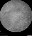
Stellate Non-heredtiary Idiopathic Foveomacular Retinoschisis (SNIFR)329 views77 year old man who is healthy and 6'6" tall with 20/40 vision and no complaints. OCT shows diffuse retinoschisis.
|
|

Melanocytoma - Lightly Pigmented329 views42 year old female with very mild visual field and vision loss in the left eye: 20/20 OU
|
|

Autoimmune Retinopathy329 views91 year old female with severe vision loss over the past 3 years. She has shimmery scotomas. The main findings are outer retinal atrophy evident on OCT and FAF. VA 20/80 OD and 20/125 OS
|
|

Posterior Scleritis - positive T-sign329 views62 year ols man was sick about 6 weeks ago and then 2 weeks ago the the left eye got red. He was treated for conjunctivitis and the eye did not get better. Topical steroids and oral non-steroidal anti-inflammatory medications did not help.
VA OD: Dcc20/25 NccJ5
VA OS: Dcc5'/200
The scleritis cleared within 2 weeks of starting high dose (80 mg) oral steroids. Blood tests were all negative.
|
|

Hyperpigmented Vasoproliferative Tumor on Buckle with New Serous Retinal Detachment 20 years post RD Repair329 views67 year old female RD repair OU 1999 then severe (20/400) macular pucker OD removed 3 months later (twin sister also had bilateral RD’s with PVR). Then dislocated IOL surgery right eye 9/2012. New vision loss in the right eye with serous retinal detachment. Argon laser to dark tumor led to resolution of subretinal fluid within a month.
|
|
| 16652 files on 167 page(s) |
 |
 |
 |
 |
 |
99 |  |
 |
 |
 |
|