Retina Gallery ~ Full Sized Retina Images

Library of Free, Non-Copyrighted Retina Images and Videos

Choose your language:
Branch Retinal Artery Occlusion with Hollenhorst Plaque
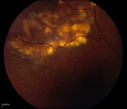
This patient was found to have retinal edema along the superotemporal arcade secondary to Branch Retinal Artery Occlusion. A Hollenhorst plaque is visible at the first bifurcation of central artery.
Keywords: Artery;occlusion;January;2015;Hollenhorst;plaque;edema;superior

Branch Retinal Artery Occlusion with Hollenhorst Plaque

This patient was found to have retinal edema along the superotemporal arcade secondary to Branch Retinal Artery Occlusion. A Hollenhorst plaque is visible at the first bifurcation of central artery.

DR1.jpg DR6.jpg BRAO_with_HHP.JPG Calcific_Nodules__.JPG Melanoma_Stereo_pair.JPG