Retina Gallery ~ Full Sized Retina Images

Library of Free, Non-Copyrighted Retina Images and Videos

Wähle Sprache:
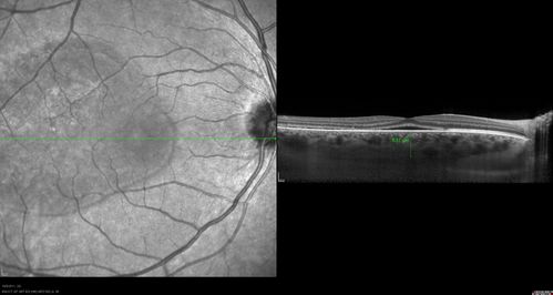
42-year-old man was seen in the office on October 5, 2011.  He had noticed starting in August after a course of antibiotic and steroids, that he developed new spots in his vision in the right eye.  He may have had an episode like this sometime in the past.  He did take steroids a few years ago and his vision did change at that time, but then returned.  

VISUAL ACUITY:  OD 20/32,  OS 20/32.  The OCT scan of the right eye confirms subretinal fluid.

42-year-old man was seen in the office on October 5, 2011. He had noticed starting in August after a course of antibiotic and steroids, that he developed new spots in his vision in the right eye. He may have had an episode like this sometime in the past. He did take steroids a few years ago and his vision did change at that time, but then returned.

VISUAL ACUITY: OD 20/32, OS 20/32. The OCT scan of the right eye confirms subretinal fluid.

pigmentedmass_082611_rily08.jpg xljr_gk11.jpg FA_R1.JPG juxtapapillaryCNVM_ReticularDisease_kawi_021113_29.jpg postpvrrd_schisis_anps_122012_022113_01.png