Retina Gallery ~ Full Sized Retina Images

Library of Free, Non-Copyrighted Retina Images and Videos

Choose your language:


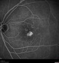
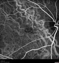
85 year old man with Large, confluent drusen may be accompanied by subretinal spaces that appear to be filled with fluid. This fluid might be from poor absorption of subretinal fluid or possibly from mechanical strain where the drusen elevate the retina. The patient was in for a yearly check and you saw a problem in the right and better eye. His left eye does not see very well but he does well enough to drive. The right, better eye, looks like it may have wet macular degeneration. VA OD: sc20/25 NscJ10 VA OS: sc20/50 NscJ10-1

AMD_drusen_fluid1_rosi_112816_01.jpg
Confluent Soft Drusen with Subetinal Fluid - Right Eye831 viewsAsymptomatic 85 year old man. OCT shows pocket of subfoveal fluid in the center of the macula. This could be mechanical from the drusen preventing the retina from settling down. Testing did not reveal a CNVM and subsequent OCT 6 weeks later showed no change in structure.00000
(0 votes)
AMD_drusen_fluid1_rosi_112816_02.jpg
Confluent Soft Drusen with Subetinal Fluid - Right Eye616 viewsAsymptomatic 85 year old man. OCT shows pocket of subfoveal fluid in the center of the macula. This could be mechanical from the drusen preventing the retina from settling down. Testing did not reveal a CNVM and subsequent OCT 6 weeks later showed no change in structure.00000
(0 votes)
AMD_drusen_fluid1_rosi_112816_03.jpg
Confluent Soft Drusen with Subetinal Fluid - Right Eye607 viewsAsymptomatic 85 year old man. OCT shows pocket of subfoveal fluid in the center of the macula. This could be mechanical from the drusen preventing the retina from settling down. Testing did not reveal a CNVM and subsequent OCT 6 weeks later showed no change in structure.00000
(0 votes)
AMD_drusen_fluid1_rosi_112816_04.jpg
Confluent Soft Drusen with Subetinal Fluid - Right Eye590 viewsAsymptomatic 85 year old man. OCT shows pocket of subfoveal fluid in the center of the macula. This could be mechanical from the drusen preventing the retina from settling down. Testing did not reveal a CNVM and subsequent OCT 6 weeks later showed no change in structure.00000
(0 votes)
AMD_drusen_fluid1_rosi_112816_05.jpg
Confluent Soft Drusen with Subetinal Fluid - Right Eye569 viewsAsymptomatic 85 year old man. OCT shows pocket of subfoveal fluid in the center of the macula. This could be mechanical from the drusen preventing the retina from settling down. Testing did not reveal a CNVM and subsequent OCT 6 weeks later showed no change in structure.00000
(0 votes)
AMD_drusen_fluid1_rosi_112816_06.jpg
Confluent Soft Drusen with Subetinal Fluid - Right Eye548 viewsAsymptomatic 85 year old man. OCT shows pocket of subfoveal fluid in the center of the macula. This could be mechanical from the drusen preventing the retina from settling down. Testing did not reveal a CNVM and subsequent OCT 6 weeks later showed no change in structure.00000
(0 votes)
AMD_drusen_fluid1_rosi_112816_08.jpg
Confluent Soft Drusen with Subetinal Fluid - Right Eye522 viewsAsymptomatic 85 year old man. OCT shows pocket of subfoveal fluid in the center of the macula. This could be mechanical from the drusen preventing the retina from settling down. Testing did not reveal a CNVM and subsequent OCT 6 weeks later showed no change in structure.00000
(0 votes)
AMD_drusen_fluid1_rosi_112816_09.jpg
Confluent Soft Drusen with Subetinal Fluid - Right Eye529 viewsAsymptomatic 85 year old man. OCT shows pocket of subfoveal fluid in the center of the macula. This could be mechanical from the drusen preventing the retina from settling down. Testing did not reveal a CNVM and subsequent OCT 6 weeks later showed no change in structure.00000
(0 votes)
AMD_drusen_fluid1_rosi_112816_10.jpg
Confluent Soft Drusen with Subetinal Fluid - Right Eye559 viewsAsymptomatic 85 year old man. OCT shows pocket of subfoveal fluid in the center of the macula. This could be mechanical from the drusen preventing the retina from settling down. Testing did not reveal a CNVM and subsequent OCT 6 weeks later showed no change in structure.00000
(0 votes)
AMD_drusen_fluid1_rosi_112816_11.jpg
Confluent Soft Drusen with Subetinal Fluid - Right Eye649 viewsAsymptomatic 85 year old man. OCT shows pocket of subfoveal fluid in the center of the macula. This could be mechanical from the drusen preventing the retina from settling down. Testing did not reveal a CNVM and subsequent OCT 6 weeks later showed no change in structure.00000
(0 votes)
AMD_drusen_fluid1_rosi_112816_12.jpg
Confluent Soft Drusen with Subetinal Fluid - Right Eye823 viewsAsymptomatic 85 year old man. OCT shows pocket of subfoveal fluid in the center of the macula. This could be mechanical from the drusen preventing the retina from settling down. Testing did not reveal a CNVM and subsequent OCT 6 weeks later showed no change in structure.00000
(0 votes)
AMD_drusen_fluid1_rosi_112816_13.jpg
Confluent Soft Drusen with Subetinal Fluid - Right Eye507 viewsAsymptomatic 85 year old man. OCT shows pocket of subfoveal fluid in the center of the macula. This could be mechanical from the drusen preventing the retina from settling down. Testing did not reveal a CNVM and subsequent OCT 6 weeks later showed no change in structure.00000
(0 votes)
AMD_drusen_fluid1_rosi_112816_14.jpg
Confluent Soft Drusen with Subetinal Fluid - Right Eye517 viewsAsymptomatic 85 year old man. OCT shows pocket of subfoveal fluid in the center of the macula. This could be mechanical from the drusen preventing the retina from settling down. Testing did not reveal a CNVM and subsequent OCT 6 weeks later showed no change in structure.00000
(0 votes)
AMD_drusen_fluid1_rosi_112816_15.jpg
Confluent Soft Drusen with Subetinal Fluid - Right Eye535 viewsAsymptomatic 85 year old man. OCT shows pocket of subfoveal fluid in the center of the macula. This could be mechanical from the drusen preventing the retina from settling down. Testing did not reveal a CNVM and subsequent OCT 6 weeks later showed no change in structure.00000
(0 votes)
AMD_drusen_fluid1_rosi_112816_16.jpg
Confluent Soft Drusen with Subetinal Fluid - Right Eye506 viewsAsymptomatic 85 year old man. OCT shows pocket of subfoveal fluid in the center of the macula. This could be mechanical from the drusen preventing the retina from settling down. Testing did not reveal a CNVM and subsequent OCT 6 weeks later showed no change in structure.00000
(0 votes)
AMD_drusen_fluid1_rosi_112816_17.jpg
Confluent Soft Drusen with Subetinal Fluid - Right Eye493 viewsAsymptomatic 85 year old man. OCT shows pocket of subfoveal fluid in the center of the macula. This could be mechanical from the drusen preventing the retina from settling down. Testing did not reveal a CNVM and subsequent OCT 6 weeks later showed no change in structure.00000
(0 votes)
AMD_drusen_fluid1_rosi_112816_18.jpg
Confluent Soft Drusen with Subetinal Fluid - Right Eye519 viewsAsymptomatic 85 year old man. OCT shows pocket of subfoveal fluid in the center of the macula. This could be mechanical from the drusen preventing the retina from settling down. Testing did not reveal a CNVM and subsequent OCT 6 weeks later showed no change in structure.00000
(0 votes)
AMD_drusen_fluid1_rosi_112816_19.jpg
Confluent Soft Drusen with Subetinal Fluid - Right Eye476 viewsAsymptomatic 85 year old man. OCT shows pocket of subfoveal fluid in the center of the macula. This could be mechanical from the drusen preventing the retina from settling down. Testing did not reveal a CNVM and subsequent OCT 6 weeks later showed no change in structure.00000
(0 votes)
AMD_drusen_fluid1_rosi_112816_20.jpg
Confluent Soft Drusen with Subetinal Fluid - Right Eye527 viewsAsymptomatic 85 year old man. OCT shows pocket of subfoveal fluid in the center of the macula. This could be mechanical from the drusen preventing the retina from settling down. Testing did not reveal a CNVM and subsequent OCT 6 weeks later showed no change in structure.00000
(0 votes)
AMD_drusen_fluid1_rosi_112816_21.jpg
Confluent Soft Drusen with Subetinal Fluid - Right Eye568 viewsAsymptomatic 85 year old man. OCT shows pocket of subfoveal fluid in the center of the macula. This could be mechanical from the drusen preventing the retina from settling down. Testing did not reveal a CNVM and subsequent OCT 6 weeks later showed no change in structure.00000
(0 votes)
AMD_drusen_fluid1_rosi_112816_22.jpg
Confluent Soft Drusen with Subetinal Fluid - Right Eye493 viewsAsymptomatic 85 year old man. OCT shows pocket of subfoveal fluid in the center of the macula. This could be mechanical from the drusen preventing the retina from settling down. Testing did not reveal a CNVM and subsequent OCT 6 weeks later showed no change in structure.00000
(0 votes)
AMD_drusen_fluid1_rosi_112816_23.jpg
Confluent Soft Drusen with Subetinal Fluid - Right Eye510 viewsAsymptomatic 85 year old man. OCT shows pocket of subfoveal fluid in the center of the macula. This could be mechanical from the drusen preventing the retina from settling down. Testing did not reveal a CNVM and subsequent OCT 6 weeks later showed no change in structure.00000
(0 votes)
AMD_drusen_fluid1_rosi_112816_24.jpg
Confluent Soft Drusen with Subetinal Fluid - Right Eye515 viewsAsymptomatic 85 year old man. OCT shows pocket of subfoveal fluid in the center of the macula. This could be mechanical from the drusen preventing the retina from settling down. Testing did not reveal a CNVM and subsequent OCT 6 weeks later showed no change in structure.00000
(0 votes)
AMD_drusen_fluid1_rosi_112816_25.jpg
Confluent Soft Drusen with Subetinal Fluid - Right Eye576 viewsAsymptomatic 85 year old man. OCT shows pocket of subfoveal fluid in the center of the macula. This could be mechanical from the drusen preventing the retina from settling down. Testing did not reveal a CNVM and subsequent OCT 6 weeks later showed no change in structure.00000
(0 votes)
AMD_drusen_fluid1_rosi_112816_26.jpg
Confluent Soft Drusen with Subetinal Fluid - Right Eye552 viewsAsymptomatic 85 year old man. OCT shows pocket of subfoveal fluid in the center of the macula. This could be mechanical from the drusen preventing the retina from settling down. Testing did not reveal a CNVM and subsequent OCT 6 weeks later showed no change in structure.00000
(0 votes)
AMD_drusen_fluid1_rosi_112816_27.jpg
Confluent Soft Drusen with Subetinal Fluid - Right Eye649 viewsAsymptomatic 85 year old man. OCT shows pocket of subfoveal fluid in the center of the macula. This could be mechanical from the drusen preventing the retina from settling down. Testing did not reveal a CNVM and subsequent OCT 6 weeks later showed no change in structure.00000
(0 votes)
AMD_drusen_fluid1_rosi_112816_28.jpg
Confluent Soft Drusen with Subetinal Fluid - Right Eye606 viewsAsymptomatic 85 year old man. OCT shows pocket of subfoveal fluid in the center of the macula. This could be mechanical from the drusen preventing the retina from settling down. Testing did not reveal a CNVM and subsequent OCT 6 weeks later showed no change in structure.00000
(0 votes)
AMD_drusen_fluid1_rosi_112816_29.jpg
Confluent Soft Drusen with Subetinal Fluid - Right Eye534 viewsAsymptomatic 85 year old man. OCT shows pocket of subfoveal fluid in the center of the macula. This could be mechanical from the drusen preventing the retina from settling down. Testing did not reveal a CNVM and subsequent OCT 6 weeks later showed no change in structure.00000
(0 votes)
AMD_drusen_fluid1_rosi_112816_30.jpg
Confluent Soft Drusen with Subetinal Fluid - Right Eye602 viewsAsymptomatic 85 year old man. OCT shows pocket of subfoveal fluid in the center of the macula. This could be mechanical from the drusen preventing the retina from settling down. Testing did not reveal a CNVM and subsequent OCT 6 weeks later showed no change in structure.00000
(0 votes)
AMD_drusen_fluid1_rosi_112816_31.jpg
Confluent Soft Drusen with Subetinal Fluid - Right Eye506 viewsAsymptomatic 85 year old man. OCT shows pocket of subfoveal fluid in the center of the macula. This could be mechanical from the drusen preventing the retina from settling down. Testing did not reveal a CNVM and subsequent OCT 6 weeks later showed no change in structure.00000
(0 votes)
AMD_drusen_fluid1_rosi_112816_32.jpg
Confluent Soft Drusen with Subetinal Fluid - Right Eye499 viewsAsymptomatic 85 year old man. OCT shows pocket of subfoveal fluid in the center of the macula. This could be mechanical from the drusen preventing the retina from settling down. Testing did not reveal a CNVM and subsequent OCT 6 weeks later showed no change in structure.00000
(0 votes)
AMD_drusen_fluid1_rosi_112816_33.jpg
Confluent Soft Drusen with Subetinal Fluid - Right Eye722 viewsAsymptomatic 85 year old man. OCT shows pocket of subfoveal fluid in the center of the macula. This could be mechanical from the drusen preventing the retina from settling down. Testing did not reveal a CNVM and subsequent OCT 6 weeks later showed no change in structure.00000
(0 votes)
32 files on 1 page(s)

85 year old man with Large, confluent drusen may be accompanied by subretinal spaces that appear to be filled with fluid. This fluid might be from poor absorption of subretinal fluid or possibly from mechanical strain where the drusen elevate the retina. The patient was in for a yearly check and you saw a problem in the right and better eye. His left eye does not see very well but he does well enough to drive. The right, better eye, looks like it may have wet macular degeneration. VA OD: sc20/25 NscJ10 VA OS: sc20/50 NscJ10-1