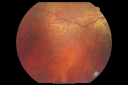
60-year-old woman has cataracts. You saw a significant drusen filled up in the peripheral macula and suggested she come in here for evaluation. She does have some shadows in the side vision she notices periodically since she had a head injury in the 80s. VISUAL ACUITY: OD 20/30, OS 20/20. IOP: OD 12, OS 14. SLIT LAMP EXAM: There is 3+ nuclear sclerosis in both eyes. The left eye has pseudoexfoliation of the lens. EXTENDED OPHTHALMOSCOPY: OD: Vertical C/D is 0.3. There is no posterior vitreous separation. There is 3+ equatorial drusen. OS: Vertical C/D is 0.3. There is no posterior vitreous separation. There is 3+ equatorial drusen. Photos confirm clinical findings. IMPRESSION: 1. EQUATORIAL DRUSEN – BOTH EYES 2. CATARACTS – BOTH EYES 3. PSEUDOEXFOLIATION – LEFT EYE DISCUSSION: I explained to the patient that the equatorial drusen are substantially more than average, but still benign. I asked her to see you back periodically. I told her she does not need to return, unless she should note further problems. She does complain of an unusual side vision change. I would appreciate if you could obtain visual field tests on her if that has not been done already. |
|||||||
|
|
|
|
||||
|
|
|
|
||||
|
|||||||
60-year-old woman has cataracts. You saw a significant drusen filled up in the peripheral macula and suggested she come in here for evaluation. She does have some shadows in the side vision she notices periodically since she had a head injury in the 80s. VISUAL ACUITY: OD 20/30, OS 20/20. IOP: OD 12, OS 14. SLIT LAMP EXAM: There is 3+ nuclear sclerosis in both eyes. The left eye has pseudoexfoliation of the lens. EXTENDED OPHTHALMOSCOPY: OD: Vertical C/D is 0.3. There is no posterior vitreous separation. There is 3+ equatorial drusen. OS: Vertical C/D is 0.3. There is no posterior vitreous separation. There is 3+ equatorial drusen. Photos confirm clinical findings. IMPRESSION: 1. EQUATORIAL DRUSEN – BOTH EYES 2. CATARACTS – BOTH EYES 3. PSEUDOEXFOLIATION – LEFT EYE DISCUSSION: I explained to the patient that the equatorial drusen are substantially more than average, but still benign. I asked her to see you back periodically. I told her she does not need to return, unless she should note further problems. She does complain of an unusual side vision change. I would appreciate if you could obtain visual field tests on her if that has not been done already.