Retina Gallery ~ Full Sized Retina Images

Library of Free, Non-Copyrighted Retina Images and Videos

Choisissez votre langue:


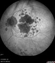
87 year old female. Severe macular bleed 15 years ago OD. Now with disciform scar. Left eye has progressing geographic atrophy. VA OD: HM, OS: 5/200. IOP normal OU

discOD-GAOS_cjlor_081622_02.jpg
Optic Disc Melanocytoma - Atypicalvu 359 fois45 year old female with normal vision and optic nerve lesion in the left eye. VA 20/16 OU. Extensive testing was normal including brain and orbit MRI. The lesion has been stable for 5 years.00000
(0 votes)
discOD-GAOS_cjlor_081622_04.jpg
Optic Disc Melanocytoma - Atypicalvu 323 fois45 year old female with normal vision and optic nerve lesion in the left eye. VA 20/16 OU. Extensive testing was normal including brain and orbit MRI. The lesion has been stable for 5 years.00000
(0 votes)
discOD-GAOS_cjlor_081622_01.jpg
Optic Disc Melanocytoma - Atypicalvu 310 fois45 year old female with normal vision and optic nerve lesion in the left eye. VA 20/16 OU. Extensive testing was normal including brain and orbit MRI. The lesion has been stable for 5 years.00000
(0 votes)
discOD-GAOS_cjlor_081622_05.jpg
Optic Disc Melanocytoma - Atypicalvu 286 fois45 year old female with normal vision and optic nerve lesion in the left eye. VA 20/16 OU. Extensive testing was normal including brain and orbit MRI. The lesion has been stable for 5 years.00000
(0 votes)
discOD-GAOS_cjlor_081622_06.jpg
Optic Disc Melanocytoma - Atypicalvu 307 fois45 year old female with normal vision and optic nerve lesion in the left eye. VA 20/16 OU. Extensive testing was normal including brain and orbit MRI. The lesion has been stable for 5 years.00000
(0 votes)
discOD-GAOS_cjlor_081622_07.jpg
Optic Disc Melanocytoma - Atypicalvu 295 fois45 year old female with normal vision and optic nerve lesion in the left eye. VA 20/16 OU. Extensive testing was normal including brain and orbit MRI. The lesion has been stable for 5 years.00000
(0 votes)
discOD-GAOS_cjlor_081622_08.jpg
Optic Disc Melanocytoma - Atypicalvu 324 fois45 year old female with normal vision and optic nerve lesion in the left eye. VA 20/16 OU. Extensive testing was normal including brain and orbit MRI. The lesion has been stable for 5 years.00000
(0 votes)
discOD-GAOS_cjlor_081622_10.jpg
Optic Disc Melanocytoma - Atypicalvu 301 fois45 year old female with normal vision and optic nerve lesion in the left eye. VA 20/16 OU. Extensive testing was normal including brain and orbit MRI. The lesion has been stable for 5 years.00000
(0 votes)
discOD-GAOS_cjlor_081622_09.jpg
Optic Disc Melanocytoma - Atypicalvu 291 fois45 year old female with normal vision and optic nerve lesion in the left eye. VA 20/16 OU. Extensive testing was normal including brain and orbit MRI. The lesion has been stable for 5 years.00000
(0 votes)
discOD-GAOS_cjlor_081622_11.jpg
Optic Disc Melanocytoma - Atypicalvu 334 fois45 year old female with normal vision and optic nerve lesion in the left eye. VA 20/16 OU. Extensive testing was normal including brain and orbit MRI. The lesion has been stable for 5 years.00000
(0 votes)
discOD-GAOS_cjlor_081622_03.jpg
Optic Disc Melanocytoma - Atypicalvu 325 fois45 year old female with normal vision and optic nerve lesion in the left eye. VA 20/16 OU. Extensive testing was normal including brain and orbit MRI. The lesion has been stable for 5 years.00000
(0 votes)
 
11 photos sur 1 page(s)

87 year old female. Severe macular bleed 15 years ago OD. Now with disciform scar. Left eye has progressing geographic atrophy. VA OD: HM, OS: 5/200. IOP normal OU