Retina Gallery ~ Full Sized Retina Images

Library of Free, Non-Copyrighted Retina Images and Videos

Choose your language:


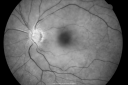
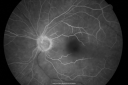
69-year-old man has a central retinal artery occlusion in his left eye with severe vision loss December 14th. I did an anterior chamber tap December 15th. His vision in that eye is still poor. I had him return for re-evaluation mostly because of the tap and also to make sure he did not start to develop rubeosis. His vision in the left eye is still poor. VISUAL ACUITY: Vision OS is light perception. IOP: 9. There is no rubeosis. There is a posterior chamber intraocular lens in good position. EXTENDED OPHTHALMOSCOPY: OS: Vertical C/D ratio is 0.8. The nerve is pale. The whitening of the retina looks to have subsided. IMPRESSION: 1. CENTRAL RETINAL ARTERY OCCLUSION – LEFT EYE

cao_phwa01.png
760 views00000
(0 votes)
cao_phwa02.png
Central Retinal Artery Occlusion less than 24 hours - 69 year old man VA light perception1011 views00000
(0 votes)
cao_phwa03.png
Central Retinal Artery Occlusion less than 24 hours - 69 year old man VA light perception806 views55555
(1 votes)
cao_phwa04.png
Central Retinal Artery Occlusion less than 24 hours - 69 year old man VA light perception584 views00000
(0 votes)
cao_phwa05.png
Central Retinal Artery Occlusion less than 24 hours - 69 year old man VA light perception627 views00000
(0 votes)
cao_phwa06.png
Central Retinal Artery Occlusion less than 24 hours - 69 year old man VA light perception478 views00000
(0 votes)
cao_phwa07.png
Central Retinal Artery Occlusion less than 24 hours - 69 year old man VA light perception527 views00000
(0 votes)
cao_phwa08.png
Central Retinal Artery Occlusion less than 24 hours - 69 year old man VA light perception482 views00000
(0 votes)
cao_phwa09.png
Central Retinal Artery Occlusion less than 24 hours - 69 year old man VA light perception468 views00000
(0 votes)
cao_phwa10.png
441 views00000
(0 votes)
cao_phwa11.png
476 views00000
(0 votes)
cao_phwa12.png
Central Retinal Artery Occlusion less than 24 hours - 69 year old man VA light perception487 views00000
(0 votes)
cao_phwa13.png
Central Retinal Artery Occlusion less than 24 hours - 69 year old man VA light perception589 views55555
(1 votes)
cao_phwa14.png
Central Retinal Artery Occlusion less than 24 hours - 69 year old man VA light perception433 views00000
(0 votes)
cao_phwa15.png
Central Retinal Artery Occlusion less than 24 hours - 69 year old man VA light perception469 views00000
(0 votes)
cao_phwa16.png
Central Retinal Artery Occlusion less than 24 hours - 69 year old man VA light perception490 views00000
(0 votes)
cao_phwa17.png
Central Retinal Artery Occlusion less than 24 hours - 69 year old man VA light perception490 views00000
(0 votes)
cao_phwa18.png
Central Retinal Artery Occlusion less than 24 hours - 69 year old man VA light perception477 views00000
(0 votes)
cao_phwa19.png
Central Retinal Artery Occlusion less than 24 hours - 69 year old man VA light perception442 views00000
(0 votes)
cao_phwa20.png
Central Retinal Artery Occlusion less than 24 hours - 69 year old man VA light perception500 views00000
(0 votes)
cao_phwa21.png
Central Retinal Artery Occlusion less than 24 hours - 69 year old man VA light perception503 views00000
(0 votes)
cao_phwa22.png
Central Retinal Artery Occlusion less than 24 hours - 69 year old man VA light perception455 views00000
(0 votes)
cao_phwa23.png
Central Retinal Artery Occlusion less than 24 hours - 69 year old man VA light perception408 views00000
(0 votes)
cao_phwa24.png
Central Retinal Artery Occlusion less than 24 hours - 69 year old man VA light perception522 views00000
(0 votes)
cao_phwa25.png
Central Retinal Artery Occlusion less than 24 hours - 69 year old man VA light perception684 views55555
(2 votes)
cao_phwa26.png
Central Retinal Artery Occlusion less than 24 hours - 69 year old man VA light perception542 views00000
(0 votes)
   
26 files on 1 page(s)

69-year-old man has a central retinal artery occlusion in his left eye with severe vision loss December 14th. I did an anterior chamber tap December 15th. His vision in that eye is still poor. I had him return for re-evaluation mostly because of the tap and also to make sure he did not start to develop rubeosis. His vision in the left eye is still poor. VISUAL ACUITY: Vision OS is light perception. IOP: 9. There is no rubeosis. There is a posterior chamber intraocular lens in good position. EXTENDED OPHTHALMOSCOPY: OS: Vertical C/D ratio is 0.8. The nerve is pale. The whitening of the retina looks to have subsided. IMPRESSION: 1. CENTRAL RETINAL ARTERY OCCLUSION – LEFT EYE