Pistol to Temple - Shockwave Retinal Damage
|
|
66-year-old woman was seen in the office on 11/24/2010. She had a gunshot wound to the head on August 12, 2010, with the entry wound just on the right side, just about below her temporal, toward the lower part of the temporal and the exit wound on the left side just above her cheek, a little behind the eye. Since then she recovered and did not need any surgery. She did have a post discharge seizure, which was brief and she had low blood pressure at the time. She had bruising of both lids. When that all cleared she realized her vision was poor in both eyes. She has seen a neuro-ophthalmologist who thought she had traumatic optic neuropathy. Because of the macular irregularities, she suggested she come here for an evaluation as well.
VISUAL ACUITY: OD light perception, OS 20/400. IOP: OD 11, OS 12.
SLIT EXAMINATION: There is 1+ nuclear sclerosis in both eyes.
EXTENDED OPHTHALMOSCOPY:
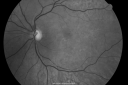
OD: Vertical C/D ratio is 0.6. There is 1+ pallor to the optic nerve. There is posterior vitreous separation. There are pigment epithelial irregularities in the macula.
OS: Vertical C/D ratio is 0.6. There is posterior vitreous separation. There are pigment irregularities in the macula.
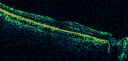
OCT SCAN: The OCT scan shows retinal atrophy in both eyes, predominately of the inner retina with full thickness retinal atrophy centrally. The average central thickness in the right eye is 122 microns, the left eye 80 microns.
NERVE FIBER LAYER SCANS: Also show nerve fiber layer atrophy in each eye with the thickness in the right eye being 46 microns and the left eye 77 microns.
Photos confirm clinical findings.
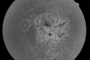
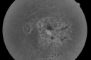
FLUORESCEIN ANGIOGRAM: Shows the irregular macula hyperfluorescence in each eye from the nongeographic central pigment epithelial atrophy. There is no evidence of any leakage or fluid.
IMPRESSION:
1. ISCHEMIC RETINOPATHY – BOTH EYES
2. CENTRAL RETINAL ARTERY OCCLUSION – BOTH EYES
3. PROBABLE OPHTHALMIC ARTERY OCCLUSION – RIGHT EYE
4. POSSIBLE TRAUMATIC OPTIC NEUROPATHY
DISCUSSION: I explained to the patient that the predominate findings on the testing today is extremely thin retinas, both in the macula and the periphery as well as nerve fiber layer atrophy. This is consistent with either traumatic injury causing traumatic optic neuropathy, but I think probably given the rapidity of the vision loss, this may very well have been from an ischemic event. Either way at this point unfortunately I do not have anything to do for her eyes. I explained to her there is no treatment. We have to either replace or resurrect damaged retina. I suggested however, she return for a check in six months. I asked her to see you back regularly. I gave her recommendations for low vision evaluations as well as counseling that might be helpful to her adapting to her current vision state.
|
|
|

vista 612 veces66-year-old woman had a gunshot wound to the head on August 12, 2010, with the entry wound just on the right side, just about below her temple, toward the lower part of the temple and the exit wound on the left side just above her cheek, a little behind the eye. Since then she recovered and did not need any surgery. When that all cleared she realized her vision was poor in both eyes. She has seen a neuro-ophthalmologist who thought she had traumatic optic neuropathy. OD light perception, OS 20/400. 
(1 votos)
|
|

vista 518 veces66-year-old woman had a gunshot wound to the head on August 12, 2010, with the entry wound just on the right side, just about below her temple, toward the lower part of the temple and the exit wound on the left side just above her cheek, a little behind the eye. Since then she recovered and did not need any surgery. When that all cleared she realized her vision was poor in both eyes. She has seen a neuro-ophthalmologist who thought she had traumatic optic neuropathy. OD light perception, OS 20/400. 
(0 votos)
|
|

vista 508 veces66-year-old woman had a gunshot wound to the head on August 12, 2010, with the entry wound just on the right side, just about below her temple, toward the lower part of the temple and the exit wound on the left side just above her cheek, a little behind the eye. Since then she recovered and did not need any surgery. When that all cleared she realized her vision was poor in both eyes. She has seen a neuro-ophthalmologist who thought she had traumatic optic neuropathy. OD light perception, OS 20/400.
(0 votos)
|
|

vista 526 veces66-year-old woman had a gunshot wound to the head on August 12, 2010, with the entry wound just on the right side, just about below her temple, toward the lower part of the temple and the exit wound on the left side just above her cheek, a little behind the eye. Since then she recovered and did not need any surgery. When that all cleared she realized her vision was poor in both eyes. She has seen a neuro-ophthalmologist who thought she had traumatic optic neuropathy. OD light perception, OS 20/400.
(0 votos)
|
|

vista 554 veces66-year-old woman had a gunshot wound to the head on August 12, 2010, with the entry wound just on the right side, just about below her temple, toward the lower part of the temple and the exit wound on the left side just above her cheek, a little behind the eye. Since then she recovered and did not need any surgery. When that all cleared she realized her vision was poor in both eyes. She has seen a neuro-ophthalmologist who thought she had traumatic optic neuropathy. OD light perception, OS 20/400.
(0 votos)
|
|

vista 561 veces66-year-old woman had a gunshot wound to the head on August 12, 2010, with the entry wound just on the right side, just about below her temple, toward the lower part of the temple and the exit wound on the left side just above her cheek, a little behind the eye. Since then she recovered and did not need any surgery. When that all cleared she realized her vision was poor in both eyes. She has seen a neuro-ophthalmologist who thought she had traumatic optic neuropathy. OD light perception, OS 20/400.
(0 votos)
|
|
vista 459 veces66-year-old woman had a gunshot wound to the head on August 12, 2010, with the entry wound just on the right side, just about below her temple, toward the lower part of the temple and the exit wound on the left side just above her cheek, a little behind the eye. Since then she recovered and did not need any surgery. When that all cleared she realized her vision was poor in both eyes. She has seen a neuro-ophthalmologist who thought she had traumatic optic neuropathy. OD light perception, OS 20/400.
(0 votos)
|
|

vista 608 veces66-year-old woman had a gunshot wound to the head on August 12, 2010, with the entry wound just on the right side, just about below her temple, toward the lower part of the temple and the exit wound on the left side just above her cheek, a little behind the eye. Since then she recovered and did not need any surgery. When that all cleared she realized her vision was poor in both eyes. She has seen a neuro-ophthalmologist who thought she had traumatic optic neuropathy. OD light perception, OS 20/400.
(0 votos)
|
|

vista 609 veces66-year-old woman had a gunshot wound to the head on August 12, 2010, with the entry wound just on the right side, just about below her temple, toward the lower part of the temple and the exit wound on the left side just above her cheek, a little behind the eye. Since then she recovered and did not need any surgery. When that all cleared she realized her vision was poor in both eyes. She has seen a neuro-ophthalmologist who thought she had traumatic optic neuropathy. OD light perception, OS 20/400.
(0 votos)
|
|

Pistol to head in attempted suicide 3 Months Ago - Retinal Atrophy - Shockwave Retinopathy VA Right Eye - Light Perception - Left Eye - 20/400vista 502 veces66-year-old woman had a gunshot wound to the head on August 12, 2010, with the entry wound just on the right side, just about below her temple, toward the lower part of the temple and the exit wound on the left side just above her cheek, a little behind the eye. Since then she recovered and did not need any surgery. When that all cleared she realized her vision was poor in both eyes. She has seen a neuro-ophthalmologist who thought she had traumatic optic neuropathy. OD light perception, OS 20/400.
(0 votos)
|
|

Pistol to head in attempted suicide 3 Months Ago - Retinal Atrophy - Shockwave Retinopathy VA Right Eye - Light Perception - Left Eye - 20/400vista 577 veces66-year-old woman had a gunshot wound to the head on August 12, 2010, with the entry wound just on the right side, just about below her temple, toward the lower part of the temple and the exit wound on the left side just above her cheek, a little behind the eye. Since then she recovered and did not need any surgery. When that all cleared she realized her vision was poor in both eyes. She has seen a neuro-ophthalmologist who thought she had traumatic optic neuropathy. OD light perception, OS 20/400.
(1 votos)
|
|

Pistol to head in attempted suicide 3 Months Ago - Retinal Atrophy - Shockwave Retinopathy VA Right Eye - Light Perception - Left Eye - 20/400vista 467 veces66-year-old woman had a gunshot wound to the head on August 12, 2010, with the entry wound just on the right side, just about below her temple, toward the lower part of the temple and the exit wound on the left side just above her cheek, a little behind the eye. Since then she recovered and did not need any surgery. When that all cleared she realized her vision was poor in both eyes. She has seen a neuro-ophthalmologist who thought she had traumatic optic neuropathy. OD light perception, OS 20/400.
(0 votos)
|
|

Pistol to head in attempted suicide 3 Months Ago - Retinal Atrophy - Shockwave Retinopathy VA Right Eye - Light Perception - Left Eye - 20/400vista 430 veces66-year-old woman had a gunshot wound to the head on August 12, 2010, with the entry wound just on the right side, just about below her temple, toward the lower part of the temple and the exit wound on the left side just above her cheek, a little behind the eye. Since then she recovered and did not need any surgery. When that all cleared she realized her vision was poor in both eyes. She has seen a neuro-ophthalmologist who thought she had traumatic optic neuropathy. OD light perception, OS 20/400.
(0 votos)
|
|

Pistol to head in attempted suicide 3 Months Ago - Retinal Atrophy - Shockwave Retinopathy VA Right Eye - Light Perception - Left Eye - 20/400vista 571 veces66-year-old woman had a gunshot wound to the head on August 12, 2010, with the entry wound just on the right side, just about below her temple, toward the lower part of the temple and the exit wound on the left side just above her cheek, a little behind the eye. Since then she recovered and did not need any surgery. When that all cleared she realized her vision was poor in both eyes. She has seen a neuro-ophthalmologist who thought she had traumatic optic neuropathy. OD light perception, OS 20/400.
(1 votos)
|
|

Pistol to head in attempted suicide 3 Months Ago - Retinal Atrophy - Shockwave Retinopathy VA Right Eye - Light Perception - Left Eye - 20/400vista 442 veces66-year-old woman had a gunshot wound to the head on August 12, 2010, with the entry wound just on the right side, just about below her temple, toward the lower part of the temple and the exit wound on the left side just above her cheek, a little behind the eye. Since then she recovered and did not need any surgery. When that all cleared she realized her vision was poor in both eyes. She has seen a neuro-ophthalmologist who thought she had traumatic optic neuropathy. OD light perception, OS 20/400.
(0 votos)
|
|

Pistol to head in attempted suicide 3 Months Ago - Retinal Atrophy - Shockwave Retinopathy VA Right Eye - Light Perception - Left Eye - 20/400vista 533 veces66-year-old woman had a gunshot wound to the head on August 12, 2010, with the entry wound just on the right side, just about below her temple, toward the lower part of the temple and the exit wound on the left side just above her cheek, a little behind the eye. Since then she recovered and did not need any surgery. When that all cleared she realized her vision was poor in both eyes. She has seen a neuro-ophthalmologist who thought she had traumatic optic neuropathy. OD light perception, OS 20/400.
(0 votos)
|
|
Pistol to head in attempted suicide 3 Months Ago - Retinal Atrophy - Shockwave Retinopathy VA Right Eye - Light Perception - Left Eye - 20/400vista 457 veces66-year-old woman had a gunshot wound to the head on August 12, 2010, with the entry wound just on the right side, just about below her temple, toward the lower part of the temple and the exit wound on the left side just above her cheek, a little behind the eye. Since then she recovered and did not need any surgery. When that all cleared she realized her vision was poor in both eyes. She has seen a neuro-ophthalmologist who thought she had traumatic optic neuropathy. OD light perception, OS 20/400.
(0 votos)
|
|

Pistol to head in attempted suicide 3 Months Ago - Retinal Atrophy - Shockwave Retinopathy VA Right Eye - Light Perception - Left Eye - 20/400vista 446 veces66-year-old woman had a gunshot wound to the head on August 12, 2010, with the entry wound just on the right side, just about below her temple, toward the lower part of the temple and the exit wound on the left side just above her cheek, a little behind the eye. Since then she recovered and did not need any surgery. When that all cleared she realized her vision was poor in both eyes. She has seen a neuro-ophthalmologist who thought she had traumatic optic neuropathy. OD light perception, OS 20/400.
(0 votos)
|
|

Pistol to head in attempted suicide 3 Months Ago - Retinal Atrophy - Shockwave Retinopathy VA Right Eye - Light Perception - Left Eye - 20/400vista 445 veces66-year-old woman had a gunshot wound to the head on August 12, 2010, with the entry wound just on the right side, just about below her temple, toward the lower part of the temple and the exit wound on the left side just above her cheek, a little behind the eye. Since then she recovered and did not need any surgery. When that all cleared she realized her vision was poor in both eyes. She has seen a neuro-ophthalmologist who thought she had traumatic optic neuropathy. OD light perception, OS 20/400.
(0 votos)
|
|

Pistol to head in attempted suicide 3 Months Ago - Retinal Atrophy - Shockwave Retinopathy VA Right Eye - Light Perception - Left Eye - 20/400vista 427 veces66-year-old woman had a gunshot wound to the head on August 12, 2010, with the entry wound just on the right side, just about below her temple, toward the lower part of the temple and the exit wound on the left side just above her cheek, a little behind the eye. Since then she recovered and did not need any surgery. When that all cleared she realized her vision was poor in both eyes. She has seen a neuro-ophthalmologist who thought she had traumatic optic neuropathy. OD light perception, OS 20/400.
(0 votos)
|
|

Pistol to head in attempted suicide 3 Months Ago - Retinal Atrophy - Shockwave Retinopathy VA Right Eye - Light Perception - Left Eye - 20/400vista 443 veces66-year-old woman had a gunshot wound to the head on August 12, 2010, with the entry wound just on the right side, just about below her temple, toward the lower part of the temple and the exit wound on the left side just above her cheek, a little behind the eye. Since then she recovered and did not need any surgery. When that all cleared she realized her vision was poor in both eyes. She has seen a neuro-ophthalmologist who thought she had traumatic optic neuropathy. OD light perception, OS 20/400.
(0 votos)
|
|
Pistol to head in attempted suicide 3 Months Ago - Retinal Atrophy - Shockwave Retinopathy VA Right Eye - Light Perception - Left Eye - 20/400vista 483 veces66-year-old woman had a gunshot wound to the head on August 12, 2010, with the entry wound just on the right side, just about below her temple, toward the lower part of the temple and the exit wound on the left side just above her cheek, a little behind the eye. Since then she recovered and did not need any surgery. When that all cleared she realized her vision was poor in both eyes. She has seen a neuro-ophthalmologist who thought she had traumatic optic neuropathy. OD light perception, OS 20/400.
(0 votos)
|
|

Pistol to head in attempted suicide 3 Months Ago - Retinal Atrophy - Shockwave Retinopathy VA Right Eye - Light Perception - Left Eye - 20/400vista 443 veces66-year-old woman had a gunshot wound to the head on August 12, 2010, with the entry wound just on the right side, just about below her temple, toward the lower part of the temple and the exit wound on the left side just above her cheek, a little behind the eye. Since then she recovered and did not need any surgery. When that all cleared she realized her vision was poor in both eyes. She has seen a neuro-ophthalmologist who thought she had traumatic optic neuropathy. OD light perception, OS 20/400.
(0 votos)
|
|

Pistol to head in attempted suicide 3 Months Ago - Retinal Atrophy - Shockwave Retinopathy VA Right Eye - Light Perception - Left Eye - 20/400vista 460 veces66-year-old woman had a gunshot wound to the head on August 12, 2010, with the entry wound just on the right side, just about below her temple, toward the lower part of the temple and the exit wound on the left side just above her cheek, a little behind the eye. Since then she recovered and did not need any surgery. When that all cleared she realized her vision was poor in both eyes. She has seen a neuro-ophthalmologist who thought she had traumatic optic neuropathy. OD light perception, OS 20/400.
(0 votos)
|
|

Pistol to head in attempted suicide 3 Months Ago - Retinal Atrophy - Shockwave Retinopathy VA Right Eye - Light Perception - Left Eye - 20/400vista 596 veces66-year-old woman had a gunshot wound to the head on August 12, 2010, with the entry wound just on the right side, just about below her temple, toward the lower part of the temple and the exit wound on the left side just above her cheek, a little behind the eye. Since then she recovered and did not need any surgery. When that all cleared she realized her vision was poor in both eyes. She has seen a neuro-ophthalmologist who thought she had traumatic optic neuropathy. OD light perception, OS 20/400.
(0 votos)
|
|
Pistol to head in attempted suicide 3 Months Ago - Retinal Atrophy - Shockwave Retinopathy VA Right Eye - Light Perception - Left Eye - 20/400vista 498 veces66-year-old woman had a gunshot wound to the head on August 12, 2010, with the entry wound just on the right side, just about below her temple, toward the lower part of the temple and the exit wound on the left side just above her cheek, a little behind the eye. Since then she recovered and did not need any surgery. When that all cleared she realized her vision was poor in both eyes. She has seen a neuro-ophthalmologist who thought she had traumatic optic neuropathy. OD light perception, OS 20/400.
(0 votos)
|
|

Pistol to head in attempted suicide 3 Months Ago - Retinal Atrophy - Shockwave Retinopathy VA Right Eye - Light Perception - Left Eye - 20/400vista 456 veces66-year-old woman had a gunshot wound to the head on August 12, 2010, with the entry wound just on the right side, just about below her temple, toward the lower part of the temple and the exit wound on the left side just above her cheek, a little behind the eye. Since then she recovered and did not need any surgery. When that all cleared she realized her vision was poor in both eyes. She has seen a neuro-ophthalmologist who thought she had traumatic optic neuropathy. OD light perception, OS 20/400.
(0 votos)
|
|

Pistol to head in attempted suicide 3 Months Ago - Retinal Atrophy - Shockwave Retinopathy VA Right Eye - Light Perception - Left Eye - 20/400vista 525 veces66-year-old woman had a gunshot wound to the head on August 12, 2010, with the entry wound just on the right side, just about below her temple, toward the lower part of the temple and the exit wound on the left side just above her cheek, a little behind the eye. Since then she recovered and did not need any surgery. When that all cleared she realized her vision was poor in both eyes. She has seen a neuro-ophthalmologist who thought she had traumatic optic neuropathy. OD light perception, OS 20/400.
(0 votos)
|
|

Pistol to head in attempted suicide 3 Months Ago - Retinal Atrophy - Shockwave Retinopathy VA Right Eye - Light Perception - Left Eye - 20/400vista 499 veces66-year-old woman had a gunshot wound to the head on August 12, 2010, with the entry wound just on the right side, just about below her temple, toward the lower part of the temple and the exit wound on the left side just above her cheek, a little behind the eye. Since then she recovered and did not need any surgery. When that all cleared she realized her vision was poor in both eyes. She has seen a neuro-ophthalmologist who thought she had traumatic optic neuropathy. OD light perception, OS 20/400.
(0 votos)
|
|

Pistol to head in attempted suicide 3 Months Ago - Retinal Atrophy - Shockwave Retinopathy VA Right Eye - Light Perception - Left Eye - 20/400vista 440 veces66-year-old woman had a gunshot wound to the head on August 12, 2010, with the entry wound just on the right side, just about below her temple, toward the lower part of the temple and the exit wound on the left side just above her cheek, a little behind the eye. Since then she recovered and did not need any surgery. When that all cleared she realized her vision was poor in both eyes. She has seen a neuro-ophthalmologist who thought she had traumatic optic neuropathy. OD light perception, OS 20/400.
(0 votos)
|
|

Pistol to head in attempted suicide 3 Months Ago - Retinal Atrophy - Shockwave Retinopathy VA Right Eye - Light Perception - Left Eye - 20/400vista 469 veces66-year-old woman had a gunshot wound to the head on August 12, 2010, with the entry wound just on the right side, just about below her temple, toward the lower part of the temple and the exit wound on the left side just above her cheek, a little behind the eye. Since then she recovered and did not need any surgery. When that all cleared she realized her vision was poor in both eyes. She has seen a neuro-ophthalmologist who thought she had traumatic optic neuropathy. OD light perception, OS 20/400.
(0 votos)
|
|

Pistol to head in attempted suicide - Retinal Atrophy - Shockwave Retinopathy vista 543 veces66-year-old woman had a gunshot wound to the head on August 12, 2010, with the entry wound just on the right side, just about below her temple, toward the lower part of the temple and the exit wound on the left side just above her cheek, a little behind the eye. Since then she recovered and did not need any surgery. When that all cleared she realized her vision was poor in both eyes. She has seen a neuro-ophthalmologist who thought she had traumatic optic neuropathy. OD light perception, OS 20/400.
(0 votos)
|
|

Pistol to head in attempted suicide - Retinal Atrophy - Shockwave Retinopathy vista 617 veces66-year-old woman had a gunshot wound to the head on August 12, 2010, with the entry wound just on the right side, just about below her temple, toward the lower part of the temple and the exit wound on the left side just above her cheek, a little behind the eye. Since then she recovered and did not need any surgery. When that all cleared she realized her vision was poor in both eyes. She has seen a neuro-ophthalmologist who thought she had traumatic optic neuropathy. OD light perception, OS 20/400.
(0 votos)
|
|

Pistol to head in attempted suicide - Retinal Atrophy - Shockwave Retinopathy vista 682 veces66-year-old woman had a gunshot wound to the head on August 12, 2010, with the entry wound just on the right side, just about below her temple, toward the lower part of the temple and the exit wound on the left side just above her cheek, a little behind the eye. Since then she recovered and did not need any surgery. When that all cleared she realized her vision was poor in both eyes. She has seen a neuro-ophthalmologist who thought she had traumatic optic neuropathy. OD light perception, OS 20/400.
(0 votos)
|
|
|
| 35 archivos en 1 página(s) |
|
66-year-old woman was seen in the office on 11/24/2010. She had a gunshot wound to the head on August 12, 2010, with the entry wound just on the right side, just about below her temporal, toward the lower part of the temporal and the exit wound on the left side just above her cheek, a little behind the eye. Since then she recovered and did not need any surgery. She did have a post discharge seizure, which was brief and she had low blood pressure at the time. She had bruising of both lids. When that all cleared she realized her vision was poor in both eyes. She has seen a neuro-ophthalmologist who thought she had traumatic optic neuropathy. Because of the macular irregularities, she suggested she come here for an evaluation as well.
VISUAL ACUITY: OD light perception, OS 20/400. IOP: OD 11, OS 12.
SLIT EXAMINATION: There is 1+ nuclear sclerosis in both eyes.
EXTENDED OPHTHALMOSCOPY:
OD: Vertical C/D ratio is 0.6. There is 1+ pallor to the optic nerve. There is posterior vitreous separation. There are pigment epithelial irregularities in the macula.
OS: Vertical C/D ratio is 0.6. There is posterior vitreous separation. There are pigment irregularities in the macula.
OCT SCAN: The OCT scan shows retinal atrophy in both eyes, predominately of the inner retina with full thickness retinal atrophy centrally. The average central thickness in the right eye is 122 microns, the left eye 80 microns.
NERVE FIBER LAYER SCANS: Also show nerve fiber layer atrophy in each eye with the thickness in the right eye being 46 microns and the left eye 77 microns.
Photos confirm clinical findings.
FLUORESCEIN ANGIOGRAM: Shows the irregular macula hyperfluorescence in each eye from the nongeographic central pigment epithelial atrophy. There is no evidence of any leakage or fluid.
IMPRESSION:
1. ISCHEMIC RETINOPATHY – BOTH EYES
2. CENTRAL RETINAL ARTERY OCCLUSION – BOTH EYES
3. PROBABLE OPHTHALMIC ARTERY OCCLUSION – RIGHT EYE
4. POSSIBLE TRAUMATIC OPTIC NEUROPATHY
DISCUSSION: I explained to the patient that the predominate findings on the testing today is extremely thin retinas, both in the macula and the periphery as well as nerve fiber layer atrophy. This is consistent with either traumatic injury causing traumatic optic neuropathy, but I think probably given the rapidity of the vision loss, this may very well have been from an ischemic event. Either way at this point unfortunately I do not have anything to do for her eyes. I explained to her there is no treatment. We have to either replace or resurrect damaged retina. I suggested however, she return for a check in six months. I asked her to see you back regularly. I gave her recommendations for low vision evaluations as well as counseling that might be helpful to her adapting to her current vision state.


