CNVM Classic Subfoveal CNVM
|
|
85-year-old woman was seen in the office on 11/8/07. She has noticed decreased vision in the left eye. It is hard to say for how long, maybe a few months. She had her last eye exam about a year ago. She thoughts perhaps it was cataracts, but you saw a problem with the macula and suggested she come here for evaluation.
VISUAL ACUITY: Vision OD is 20/25, OS is 2/200. IOP: 17 OU.
AMSLER GRID: Normal OU.
SLIT LAMP EXAM: Posterior chamber intraocular lens is in good position with open capsule. The left eye has 3+ nuclear sclerosis.
EXTENDED OPHTHALMOSCOPY:
OD: Vertical C/D ratio is 0.3. There is a posterior vitreous separation and rare macular drusen.
OS: Vertical C/D ratio is 0.3. There is a posterior vitreous separation. There is exudative macular detachment with thickening of the pigment epithelium centrally.
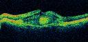
OCT SCAN: The right eye shows a normal foveal contour. The left eye shows macular edema with thickening of the pigment epithelium underlying it.
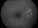
FLUORESCEIN ANGIOGRAPHY: FA shows a classic 4-disc area of subfoveal choroidal neovascular membrane.
IMPRESSION:
1. AGE-RELATED MACULAR DEGENERATION – BOTH EYES
2. CLASSIC SUBFOVEAL CHOROIDAL NEOVASCULAR MEMBRANE – LEFT EYE
3. MACULAR EDEMA – LEFT EYE
DISCUSSION: I explained to the patient that with intravitreal Avastin there is a 90% chance of improving the vision in the left eye. Given that her vision is so poor and that
her other eye is so good, to prevent having to treat her too often, I may also treat her with photodynamic laser. I injected the eye with intravitreal injection of Avastin (1.25 mg/0.05 ml) without any difficulty. She will return for check in one month, sooner should she notice any problem. She will see you regularly.
|

Subfoveal CNVM wet AMD fellow eyevu 796 foisShe has noticed decreased vision in the left eye. It is hard to say for how long, maybe a few months. She had her last eye exam about a year ago. She thoughts perhaps it was cataracts, but you saw a problem with the macula and suggested she come here for evaluation.
VISUAL ACUITY: Vision OD is 20/25, OS is 2/200
(0 votes)
|
|

Subfoveal CNVM wet AMD fellow eye oct line scanvu 681 foisShe has noticed decreased vision in the left eye. It is hard to say for how long, maybe a few months. She had her last eye exam about a year ago. She thoughts perhaps it was cataracts, but you saw a problem with the macula and suggested she come here for evaluation.
VISUAL ACUITY: Vision OD is 20/25, OS is 2/200
(0 votes)
|
|

Subfoveal CNVM wet AMD color photovu 934 foisShe has noticed decreased vision in the left eye. It is hard to say for how long, maybe a few months. She had her last eye exam about a year ago. She thoughts perhaps it was cataracts, but you saw a problem with the macula and suggested she come here for evaluation.
VISUAL ACUITY: Vision OD is 20/25, OS is 2/200
(0 votes)
|
|
Subfoveal CNVM wet AMD oct line scanvu 1012 foisShe has noticed decreased vision in the left eye. It is hard to say for how long, maybe a few months. She had her last eye exam about a year ago. She thoughts perhaps it was cataracts, but you saw a problem with the macula and suggested she come here for evaluation.
VISUAL ACUITY: Vision OD is 20/25, OS is 2/200
(0 votes)
|
|
Subfoveal CNVM wet AMD fellow eyevu 581 foisShe has noticed decreased vision in the left eye. It is hard to say for how long, maybe a few months. She had her last eye exam about a year ago. She thoughts perhaps it was cataracts, but you saw a problem with the macula and suggested she come here for evaluation.
VISUAL ACUITY: Vision OD is 20/25, OS is 2/200
(0 votes)
|
|

Subfoveal CNVM wet AMD early fa full imagevu 799 foisShe has noticed decreased vision in the left eye. It is hard to say for how long, maybe a few months. She had her last eye exam about a year ago. She thoughts perhaps it was cataracts, but you saw a problem with the macula and suggested she come here for evaluation.
VISUAL ACUITY: Vision OD is 20/25, OS is 2/200
(0 votes)
|
|

Subfoveal CNVM wet AMD early favu 594 foisShe has noticed decreased vision in the left eye. It is hard to say for how long, maybe a few months. She had her last eye exam about a year ago. She thoughts perhaps it was cataracts, but you saw a problem with the macula and suggested she come here for evaluation.
VISUAL ACUITY: Vision OD is 20/25, OS is 2/200
(0 votes)
|
|

Subfoveal CNVM wet AMD late fa full imagevu 794 foisShe has noticed decreased vision in the left eye. It is hard to say for how long, maybe a few months. She had her last eye exam about a year ago. She thoughts perhaps it was cataracts, but you saw a problem with the macula and suggested she come here for evaluation.
VISUAL ACUITY: Vision OD is 20/25, OS is 2/200
(0 votes)
|
|

Subfoveal CNVM wet AMD late favu 608 fois85-year-old woman was seen in the office on 11/8/07. She has noticed decreased vision in the left eye. It is hard to say for how long, maybe a few months. She had her last eye exam about a year ago. She thoughts perhaps it was cataracts, but you saw a problem with the macula and suggested she come here for evaluation.
VISUAL ACUITY: Vision OD is 20/25, OS is 2/200
(0 votes)
|
|
|
|
|
|
|
85-year-old woman was seen in the office on 11/8/07. She has noticed decreased vision in the left eye. It is hard to say for how long, maybe a few months. She had her last eye exam about a year ago. She thoughts perhaps it was cataracts, but you saw a problem with the macula and suggested she come here for evaluation.
VISUAL ACUITY: Vision OD is 20/25, OS is 2/200. IOP: 17 OU.
AMSLER GRID: Normal OU.
SLIT LAMP EXAM: Posterior chamber intraocular lens is in good position with open capsule. The left eye has 3+ nuclear sclerosis.
EXTENDED OPHTHALMOSCOPY:
OD: Vertical C/D ratio is 0.3. There is a posterior vitreous separation and rare macular drusen.
OS: Vertical C/D ratio is 0.3. There is a posterior vitreous separation. There is exudative macular detachment with thickening of the pigment epithelium centrally.
OCT SCAN: The right eye shows a normal foveal contour. The left eye shows macular edema with thickening of the pigment epithelium underlying it.
FLUORESCEIN ANGIOGRAPHY: FA shows a classic 4-disc area of subfoveal choroidal neovascular membrane.
IMPRESSION:
1. AGE-RELATED MACULAR DEGENERATION – BOTH EYES
2. CLASSIC SUBFOVEAL CHOROIDAL NEOVASCULAR MEMBRANE – LEFT EYE
3. MACULAR EDEMA – LEFT EYE
DISCUSSION: I explained to the patient that with intravitreal Avastin there is a 90% chance of improving the vision in the left eye. Given that her vision is so poor and that
her other eye is so good, to prevent having to treat her too often, I may also treat her with photodynamic laser. I injected the eye with intravitreal injection of Avastin (1.25 mg/0.05 ml) without any difficulty. She will return for check in one month, sooner should she notice any problem. She will see you regularly.


