Melanoma - Exudative Retinal Detachment
|
|
82-year-old man was seen in the office on November 4, 2010. He had a few falls about two weeks ago. He is still under evaluation because of that. He had three falls within a short period. He did cut his right upper brow and he bled some. He is under ongoing evaluation because of the falls and he still has a pending neurological evaluation. He is on prednisone right now because of swelling of his left hand. He has an appointment with the rheumatologist because of that. He has had aortic valve replacement with a bovine valve as well as double bypass surgery.
After the falls he started checking his vision and noticed there was a veil over the left eye, which he had seen for about two weeks in the superior visual field. You saw a problem with the retina and suggest he come here for an evaluation. His right eye sees fine. He had bladder cancer back in 2007, but it was localized and was treated with radiation and as far as he understands it, there is no problem with any spreading of the bladder cancer.
VISUAL ACUITY: OD 20/25, OS 20/40. IOP: OD 12, OS 11.
SLIT EXAMINATION: The posterior chamber intraocular lens is in good position in both eyes.
EXTENDED OPHTHALMOSCOPY:
OD: Vertical C/D ratio is 0.5. There is posterior vitreous separation. The macula and periphery look healthy.
OS: Vertical C/D ratio is 0.5. There is a posterior vitreous separation. There is an exudate of retinal detachment extending from 4 o’clock around to 9 o’clock. There is an irregular hypo and hyperpigmented tumor, which is fairly flat, but has a broad base on ultrasound. The tumor measure 20x20 millimeters. On the base it stops 2 disc-diameters short of the optic nerve. It is localized just about around 7 o’clock. It does extend anterior to the equator. The A-scan height of it shows to be 2.4 millimeters high and the B-scan height shows it to be about the same and it has low internal reflectivity. Photos confirm clinical findings.
FLUORESCEIN ANGIOGRAPHY: Fluorescein angiography shows that the tumor in the left eye does have its own circulation, with irregular hypo and hyperpigmentation over the surface. There also was a small retinal pigment epithelial tear away from the tumor, just inferotemporal to the macula with hyperfluorescence there.
The right eye was angiographically normal.
IMPRESSION:
1. MELANOMA – LEFT EYE
2. EXUDATIVE RETINAL DETACHMENT – LEFT EYE
3. RECENT FALLS
DISCUSSION: I explained to the patient he does have a choroidal melanoma in the left eye, which is causing an exudate of retinal detachment. It looks like it has been there for sometime and the detachment looks to be chronic. I talked to him about the different therapies, which include enucleation, radioactive plaque therapy or external beam radiation. I told him he could travel and there are several centers that do tumors, but it is difficult for them to travel and he would rather have treatment done locally.
I will contact a radiation oncologist to see how soon we can get a plaque made and also I need to talk to his medical doctor so he can have a systemic evaluation, looking for possible other sources of tumor or possible other places where the tumor might have spread and also to evaluate why he had these falls recently to make sure there is nothing medically that would preclude the plaque placement. The plaque placement is not an emergency, but I would like to do it within the next month or so.
|

Choroidal Melanoma - Exudative Retinal Detachment 82 Year Old Man vista 641 veces82-year-old man who had 3 falls 2 weeks ago. After the falls he started checking his vision and noticed there was a veil over the left eye, which he had seen for about two weeks in the superior visual field. OD 20/25, OS 20/40
(patient was medically never well enough for brachytherapy and died 6 months later from heart disease) 
(0 votos)
|
|

Choroidal Melanoma - Exudative Retinal Detachment 82 Year Old Man vista 689 veces82-year-old man who had 3 falls 2 weeks ago. After the falls he started checking his vision and noticed there was a veil over the left eye, which he had seen for about two weeks in the superior visual field. OD 20/25, OS 20/40
(patient was medically never well enough for brachytherapy and died 6 months later from heart disease)
(0 votos)
|
|
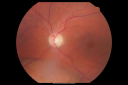
Choroidal Melanoma - Exudative Retinal Detachment 82 Year Old Man vista 726 veces82-year-old man who had 3 falls 2 weeks ago. After the falls he started checking his vision and noticed there was a veil over the left eye, which he had seen for about two weeks in the superior visual field. OD 20/25, OS 20/40
(patient was medically never well enough for brachytherapy and died 6 months later from heart disease)
(0 votos)
|
|
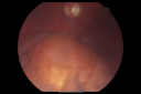
Choroidal Melanoma - Exudative Retinal Detachment 82 Year Old Man vista 665 veces82-year-old man who had 3 falls 2 weeks ago. After the falls he started checking his vision and noticed there was a veil over the left eye, which he had seen for about two weeks in the superior visual field. OD 20/25, OS 20/40
(patient was medically never well enough for brachytherapy and died 6 months later from heart disease)
(0 votos)
|
|

Choroidal Melanoma - Exudative Retinal Detachment 82 Year Old Man vista 573 veces82-year-old man who had 3 falls 2 weeks ago. After the falls he started checking his vision and noticed there was a veil over the left eye, which he had seen for about two weeks in the superior visual field. OD 20/25, OS 20/40
(patient was medically never well enough for brachytherapy and died 6 months later from heart disease)
(0 votos)
|
|

Choroidal Melanoma - Exudative Retinal Detachment 82 Year Old Man vista 564 veces82-year-old man who had 3 falls 2 weeks ago. After the falls he started checking his vision and noticed there was a veil over the left eye, which he had seen for about two weeks in the superior visual field. OD 20/25, OS 20/40
(patient was medically never well enough for brachytherapy and died 6 months later from heart disease)
(0 votos)
|
|

Choroidal Melanoma - Exudative Retinal Detachment 82 Year Old Man vista 486 veces82-year-old man who had 3 falls 2 weeks ago. After the falls he started checking his vision and noticed there was a veil over the left eye, which he had seen for about two weeks in the superior visual field. OD 20/25, OS 20/40
(patient was medically never well enough for brachytherapy and died 6 months later from heart disease)
(0 votos)
|
|

Choroidal Melanoma - Exudative Retinal Detachment 82 Year Old Man vista 513 veces82-year-old man who had 3 falls 2 weeks ago. After the falls he started checking his vision and noticed there was a veil over the left eye, which he had seen for about two weeks in the superior visual field. OD 20/25, OS 20/40
(patient was medically never well enough for brachytherapy and died 6 months later from heart disease)
(0 votos)
|
|

Choroidal Melanoma - Exudative Retinal Detachment 82 Year Old Man vista 487 veces82-year-old man who had 3 falls 2 weeks ago. After the falls he started checking his vision and noticed there was a veil over the left eye, which he had seen for about two weeks in the superior visual field. OD 20/25, OS 20/40
(patient was medically never well enough for brachytherapy and died 6 months later from heart disease)
(0 votos)
|
|

Choroidal Melanoma - Exudative Retinal Detachment 82 Year Old Man vista 502 veces82-year-old man who had 3 falls 2 weeks ago. After the falls he started checking his vision and noticed there was a veil over the left eye, which he had seen for about two weeks in the superior visual field. OD 20/25, OS 20/40
(patient was medically never well enough for brachytherapy and died 6 months later from heart disease)
(0 votos)
|
|

Choroidal Melanoma - Exudative Retinal Detachment 82 Year Old Man vista 488 veces82-year-old man who had 3 falls 2 weeks ago. After the falls he started checking his vision and noticed there was a veil over the left eye, which he had seen for about two weeks in the superior visual field. OD 20/25, OS 20/40
(patient was medically never well enough for brachytherapy and died 6 months later from heart disease)
(0 votos)
|
|
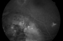
Choroidal Melanoma - Exudative Retinal Detachment 82 Year Old Man vista 454 veces82-year-old man who had 3 falls 2 weeks ago. After the falls he started checking his vision and noticed there was a veil over the left eye, which he had seen for about two weeks in the superior visual field. OD 20/25, OS 20/40
(patient was medically never well enough for brachytherapy and died 6 months later from heart disease)
(0 votos)
|
|

Choroidal Melanoma - Exudative Retinal Detachment 82 Year Old Man vista 473 veces82-year-old man who had 3 falls 2 weeks ago. After the falls he started checking his vision and noticed there was a veil over the left eye, which he had seen for about two weeks in the superior visual field. OD 20/25, OS 20/40
(patient was medically never well enough for brachytherapy and died 6 months later from heart disease)
(0 votos)
|
|

Choroidal Melanoma - Exudative Retinal Detachment 82 Year Old Man vista 457 veces82-year-old man who had 3 falls 2 weeks ago. After the falls he started checking his vision and noticed there was a veil over the left eye, which he had seen for about two weeks in the superior visual field. OD 20/25, OS 20/40
(patient was medically never well enough for brachytherapy and died 6 months later from heart disease)
(0 votos)
|
|
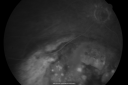
Choroidal Melanoma - Exudative Retinal Detachment 82 Year Old Man vista 441 veces82-year-old man who had 3 falls 2 weeks ago. After the falls he started checking his vision and noticed there was a veil over the left eye, which he had seen for about two weeks in the superior visual field. OD 20/25, OS 20/40
(patient was medically never well enough for brachytherapy and died 6 months later from heart disease)
(0 votos)
|
|
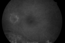
Choroidal Melanoma - Exudative Retinal Detachment 82 Year Old Man vista 440 veces82-year-old man who had 3 falls 2 weeks ago. After the falls he started checking his vision and noticed there was a veil over the left eye, which he had seen for about two weeks in the superior visual field. OD 20/25, OS 20/40
(patient was medically never well enough for brachytherapy and died 6 months later from heart disease)
(0 votos)
|
|

Choroidal Melanoma - Exudative Retinal Detachment 82 Year Old Man vista 477 veces82-year-old man who had 3 falls 2 weeks ago. After the falls he started checking his vision and noticed there was a veil over the left eye, which he had seen for about two weeks in the superior visual field. OD 20/25, OS 20/40
(patient was medically never well enough for brachytherapy and died 6 months later from heart disease)
(0 votos)
|
|

Choroidal Melanoma - Exudative Retinal Detachment 82 Year Old Man vista 493 veces82-year-old man who had 3 falls 2 weeks ago. After the falls he started checking his vision and noticed there was a veil over the left eye, which he had seen for about two weeks in the superior visual field. OD 20/25, OS 20/40
(patient was medically never well enough for brachytherapy and died 6 months later from heart disease)
(0 votos)
|
|

Choroidal Melanoma - Exudative Retinal Detachment 82 Year Old Man vista 528 veces82-year-old man who had 3 falls 2 weeks ago. After the falls he started checking his vision and noticed there was a veil over the left eye, which he had seen for about two weeks in the superior visual field. OD 20/25, OS 20/40
(patient was medically never well enough for brachytherapy and died 6 months later from heart disease)
(0 votos)
|
|

Choroidal Melanoma - Exudative Retinal Detachment 82 Year Old Man vista 549 veces82-year-old man who had 3 falls 2 weeks ago. After the falls he started checking his vision and noticed there was a veil over the left eye, which he had seen for about two weeks in the superior visual field. OD 20/25, OS 20/40
(patient was medically never well enough for brachytherapy and died 6 months later from heart disease)
(0 votos)
|
|

Choroidal Melanoma - Exudative Retinal Detachment 82 Year Old Man B Scan Ultrasound Longitudinalvista 833 veces82-year-old man who had 3 falls 2 weeks ago. After the falls he started checking his vision and noticed there was a veil over the left eye, which he had seen for about two weeks in the superior visual field. OD 20/25, OS 20/40
(patient was medically never well enough for brachytherapy and died 6 months later from heart disease)
(0 votos)
|
|
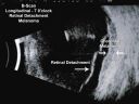
Choroidal Melanoma - Exudative Retinal Detachment 82 Year Old Man B Scan Ultrasound Longitudinalvista 1170 veces82-year-old man who had 3 falls 2 weeks ago. After the falls he started checking his vision and noticed there was a veil over the left eye, which he had seen for about two weeks in the superior visual field. OD 20/25, OS 20/40
(patient was medically never well enough for brachytherapy and died 6 months later from heart disease)
(0 votos)
|
|

Choroidal Melanoma - Exudative Retinal Detachment 82 Year Old Man B Scan Ultrasound Transvercevista 896 veces82-year-old man who had 3 falls 2 weeks ago. After the falls he started checking his vision and noticed there was a veil over the left eye, which he had seen for about two weeks in the superior visual field. OD 20/25, OS 20/40
(patient was medically never well enough for brachytherapy and died 6 months later from heart disease)
(0 votos)
|
|

Choroidal Melanoma - Exudative Retinal Detachment 82 Year Old Man B Scan - Transversevista 1308 veces82-year-old man who had 3 falls 2 weeks ago. After the falls he started checking his vision and noticed there was a veil over the left eye, which he had seen for about two weeks in the superior visual field. OD 20/25, OS 20/40
(patient was medically never well enough for brachytherapy and died 6 months later from heart disease)
(0 votos)
|
|
| 24 archivos en 1 página(s) |
|
82-year-old man was seen in the office on November 4, 2010. He had a few falls about two weeks ago. He is still under evaluation because of that. He had three falls within a short period. He did cut his right upper brow and he bled some. He is under ongoing evaluation because of the falls and he still has a pending neurological evaluation. He is on prednisone right now because of swelling of his left hand. He has an appointment with the rheumatologist because of that. He has had aortic valve replacement with a bovine valve as well as double bypass surgery.
After the falls he started checking his vision and noticed there was a veil over the left eye, which he had seen for about two weeks in the superior visual field. You saw a problem with the retina and suggest he come here for an evaluation. His right eye sees fine. He had bladder cancer back in 2007, but it was localized and was treated with radiation and as far as he understands it, there is no problem with any spreading of the bladder cancer.
VISUAL ACUITY: OD 20/25, OS 20/40. IOP: OD 12, OS 11.
SLIT EXAMINATION: The posterior chamber intraocular lens is in good position in both eyes.
EXTENDED OPHTHALMOSCOPY:
OD: Vertical C/D ratio is 0.5. There is posterior vitreous separation. The macula and periphery look healthy.
OS: Vertical C/D ratio is 0.5. There is a posterior vitreous separation. There is an exudate of retinal detachment extending from 4 o’clock around to 9 o’clock. There is an irregular hypo and hyperpigmented tumor, which is fairly flat, but has a broad base on ultrasound. The tumor measure 20x20 millimeters. On the base it stops 2 disc-diameters short of the optic nerve. It is localized just about around 7 o’clock. It does extend anterior to the equator. The A-scan height of it shows to be 2.4 millimeters high and the B-scan height shows it to be about the same and it has low internal reflectivity. Photos confirm clinical findings.
FLUORESCEIN ANGIOGRAPHY: Fluorescein angiography shows that the tumor in the left eye does have its own circulation, with irregular hypo and hyperpigmentation over the surface. There also was a small retinal pigment epithelial tear away from the tumor, just inferotemporal to the macula with hyperfluorescence there.
The right eye was angiographically normal.
IMPRESSION:
1. MELANOMA – LEFT EYE
2. EXUDATIVE RETINAL DETACHMENT – LEFT EYE
3. RECENT FALLS
DISCUSSION: I explained to the patient he does have a choroidal melanoma in the left eye, which is causing an exudate of retinal detachment. It looks like it has been there for sometime and the detachment looks to be chronic. I talked to him about the different therapies, which include enucleation, radioactive plaque therapy or external beam radiation. I told him he could travel and there are several centers that do tumors, but it is difficult for them to travel and he would rather have treatment done locally.
I will contact a radiation oncologist to see how soon we can get a plaque made and also I need to talk to his medical doctor so he can have a systemic evaluation, looking for possible other sources of tumor or possible other places where the tumor might have spread and also to evaluate why he had these falls recently to make sure there is nothing medically that would preclude the plaque placement. The plaque placement is not an emergency, but I would like to do it within the next month or so.


