Ischemic Optic Neuropathy - AION - Choroidal Ischemia - Non-Arteritic
|
|
68-year-old woman was seen in the office on 5/27/2011. She had a Bell’s palsy in the right eye in 2000. The right eye has always had some trouble because of that but then she noticed central vision loss in the right eye about a week ago. It is getting a little bit better. The left eye sees fine. She had an ear infection also at the same time. She has no headaches, no fevers, no jaw claudication, and no weakness or recent constitutional symptoms or weight change suggestive of a giant cell arteritis.
VISUAL ACUITY: OD 20/160, OS 20/25. IOP: 15 OU. The Amsler grid is normal in both eyes.
SLIT LAMP EXAMINATION: There is 2+ nuclear sclerosis in both eyes.
EXTENDED OPHTHALMOSCOPY:
OD: Vertical C/D ratio is 0.1. There is posterior vitreous separation. There is a swelling in the inferior pole of the optic nerve. The nerve is reasonably pink though.
OS: Vertical C/D ratio is 0.1. There is posterior vitreous separation. The macula and periphery look healthy.
Photos confirm clinical findings.
FLUORESCEIN ANGIOGRAM: Does show some choroidal hyper perfusion around the optic nerve in the very early frames and then everything fills normally. In the late frames, there is leakage around the optic nerve typical for ischemic optic neuropathy.
IMPRESSION:
1. NON ARTERITIC ANTERIOR ISCHEMIC OPTIC NEUROPATHY – RIGHT EYE
2. CATARACTS – BOTH EYES
DISCUSSION: I explained to the patient she does have ischemic optic neuropathy in the right eye. She is leaving for Maryland Monday. Today is Friday. I gave her the name of a neuroophthalmologist to see near her home in the meantime. I told her I would be happy to see her back when she is back in town. There is a possibility this may get better. She has some difficulty tolerating aspirin. I asked her to try taking an enteric-coated baby aspirin every other day just for now to protect her other eye.
|

Non-Arteritic Anterior Ischemic Optic Neuropathy - Peripapillary Choroidal Ischemiavu 775 fois68-year-old woman noticed central vision loss in the right eye about a week ago. It is getting a little bit better. OD 20/160, OS 20/25.
(0 votes)
|
|

Non-Arteritic Anterior Ischemic Optic Neuropathy - Peripapillary Choroidal Ischemiavu 715 fois68-year-old woman noticed central vision loss in the right eye about a week ago. It is getting a little bit better. OD 20/160, OS 20/25.
(0 votes)
|
|

Non-Arteritic Anterior Ischemic Optic Neuropathy - Peripapillary Choroidal Ischemiavu 668 fois68-year-old woman noticed central vision loss in the right eye about a week ago. It is getting a little bit better. OD 20/160, OS 20/25.
(0 votes)
|
|

Non-Arteritic Anterior Ischemic Optic Neuropathy - Peripapillary Choroidal Ischemiavu 738 fois68-year-old woman noticed central vision loss in the right eye about a week ago. It is getting a little bit better. OD 20/160, OS 20/25.
(0 votes)
|
|

Non-Arteritic Anterior Ischemic Optic Neuropathy - Peripapillary Choroidal Ischemiavu 788 fois68-year-old woman noticed central vision loss in the right eye about a week ago. It is getting a little bit better. OD 20/160, OS 20/25.
(0 votes)
|
|

Non-Arteritic Anterior Ischemic Optic Neuropathy - Peripapillary Choroidal Ischemiavu 739 fois68-year-old woman noticed central vision loss in the right eye about a week ago. It is getting a little bit better. OD 20/160, OS 20/25.
(0 votes)
|
|

Non-Arteritic Anterior Ischemic Optic Neuropathy - Peripapillary Choroidal Ischemiavu 788 fois68-year-old woman noticed central vision loss in the right eye about a week ago. It is getting a little bit better. OD 20/160, OS 20/25.
(0 votes)
|
|
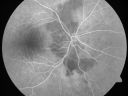
Non-Arteritic Anterior Ischemic Optic Neuropathy - Peripapillary Choroidal Ischemiavu 653 fois68-year-old woman noticed central vision loss in the right eye about a week ago. It is getting a little bit better. OD 20/160, OS 20/25.
(0 votes)
|
|

Non-Arteritic Anterior Ischemic Optic Neuropathy - Peripapillary Choroidal Ischemiavu 918 fois68-year-old woman noticed central vision loss in the right eye about a week ago. It is getting a little bit better. OD 20/160, OS 20/25.
(0 votes)
|
|

Non-Arteritic Anterior Ischemic Optic Neuropathy - Peripapillary Choroidal Ischemiavu 725 fois68-year-old woman noticed central vision loss in the right eye about a week ago. It is getting a little bit better. OD 20/160, OS 20/25.
(0 votes)
|
|
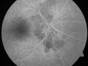
Non-Arteritic Anterior Ischemic Optic Neuropathy - Peripapillary Choroidal Ischemiavu 744 fois68-year-old woman noticed central vision loss in the right eye about a week ago. It is getting a little bit better. OD 20/160, OS 20/25.
(0 votes)
|
|

Non-Arteritic Anterior Ischemic Optic Neuropathy - Peripapillary Choroidal Ischemiavu 688 fois68-year-old woman noticed central vision loss in the right eye about a week ago. It is getting a little bit better. OD 20/160, OS 20/25.
(0 votes)
|
|

Non-Arteritic Anterior Ischemic Optic Neuropathy - Peripapillary Choroidal Ischemiavu 679 fois68-year-old woman noticed central vision loss in the right eye about a week ago. It is getting a little bit better. OD 20/160, OS 20/25.
(0 votes)
|
|

Non-Arteritic Anterior Ischemic Optic Neuropathy - Peripapillary Choroidal Ischemiavu 566 fois68-year-old woman noticed central vision loss in the right eye about a week ago. It is getting a little bit better. OD 20/160, OS 20/25.
(0 votes)
|
|

Non-Arteritic Anterior Ischemic Optic Neuropathy - Peripapillary Choroidal Ischemiavu 604 fois68-year-old woman noticed central vision loss in the right eye about a week ago. It is getting a little bit better. OD 20/160, OS 20/25.
(0 votes)
|
|
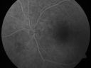
Non-Arteritic Anterior Ischemic Optic Neuropathy - Peripapillary Choroidal Ischemiavu 726 fois68-year-old woman noticed central vision loss in the right eye about a week ago. It is getting a little bit better. OD 20/160, OS 20/25.
(0 votes)
|
|
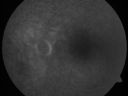
Non-Arteritic Anterior Ischemic Optic Neuropathy - Peripapillary Choroidal Ischemiavu 593 fois68-year-old woman noticed central vision loss in the right eye about a week ago. It is getting a little bit better. OD 20/160, OS 20/25.
(0 votes)
|
|

Non-Arteritic Anterior Ischemic Optic Neuropathy - Peripapillary Choroidal Ischemiavu 871 fois68-year-old woman noticed central vision loss in the right eye about a week ago. It is getting a little bit better. OD 20/160, OS 20/25.
(1 votes)
|
|
|
|
|
|
68-year-old woman was seen in the office on 5/27/2011. She had a Bell’s palsy in the right eye in 2000. The right eye has always had some trouble because of that but then she noticed central vision loss in the right eye about a week ago. It is getting a little bit better. The left eye sees fine. She had an ear infection also at the same time. She has no headaches, no fevers, no jaw claudication, and no weakness or recent constitutional symptoms or weight change suggestive of a giant cell arteritis.
VISUAL ACUITY: OD 20/160, OS 20/25. IOP: 15 OU. The Amsler grid is normal in both eyes.
SLIT LAMP EXAMINATION: There is 2+ nuclear sclerosis in both eyes.
EXTENDED OPHTHALMOSCOPY:
OD: Vertical C/D ratio is 0.1. There is posterior vitreous separation. There is a swelling in the inferior pole of the optic nerve. The nerve is reasonably pink though.
OS: Vertical C/D ratio is 0.1. There is posterior vitreous separation. The macula and periphery look healthy.
Photos confirm clinical findings.
FLUORESCEIN ANGIOGRAM: Does show some choroidal hyper perfusion around the optic nerve in the very early frames and then everything fills normally. In the late frames, there is leakage around the optic nerve typical for ischemic optic neuropathy.
IMPRESSION:
1. NON ARTERITIC ANTERIOR ISCHEMIC OPTIC NEUROPATHY – RIGHT EYE
2. CATARACTS – BOTH EYES
DISCUSSION: I explained to the patient she does have ischemic optic neuropathy in the right eye. She is leaving for Maryland Monday. Today is Friday. I gave her the name of a neuroophthalmologist to see near her home in the meantime. I told her I would be happy to see her back when she is back in town. There is a possibility this may get better. She has some difficulty tolerating aspirin. I asked her to try taking an enteric-coated baby aspirin every other day just for now to protect her other eye.


