Fundus Flavimaculatus - Stargardt Macular Dystrophy
|
|
61-year-old woman was seen in the office on October 10, 2011. She has had decreasing vision for about the last five years. She doesn’t smoke. She takes eye vitamins and fish oil.
VISUAL ACUITY: OD 20/50, OS 20/200. IOP: 13 OU.
SLIT EXAMINATION: There is 1+ nuclear sclerosis in both eyes.
EXTENDED OPHTHALMOSCOPY:
OD: Vertical C/D ratio is 0.5. There are pisciform lesions on the macula with some atrophy centrally.
OS: Vertical C/D ratio is 0.5. There are pisciform lesions on the macula with some atrophy centrally.
FUNDUS PHOTOGRAPHY: The infrared images do show areas of hypo reflectivity corresponding to pigment epithelial atrophy.
FUNDUS AUTO FLUORESCENCE: The fundus auto fluorescence show areas of hyper auto fluorescence inside the pisiform lesions and the fovea unfortunately is dark centrally.
SPECTRALIS-SD-OCT SCAN: The OCT scan shows retinal atrophy in the center in each eye.
IMPRESSION:
1. FUNDUS FLAVIMACULATUS (STARGARDT'S DISEASE) – BOTH EYES
DISCUSSION: I explained to the patient unfortunately she does have a macular dystrophy causing vision loss and retinal atrophy in each eye. At the moment there is no treatment, but there are trials started, actually right now in California for cell transplantation and maybe in a few years we will have something that can help her.
|
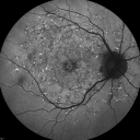
Fundus Flavimaculatus - Stargardt Disease - 20/50 OD 20/200 OS 61 Year old Fundus Autofluorescence odvu 2254 fois61-year-old decreasing vision for about the last five years. OD 20/50, OS 20/200.
Pisciform Lesions and Macular Atrophy
(0 votes)
|
|

Fundus Flavimaculatus - Stargardt Disease - 20/50 OD 20/200 OS 61 Year old Fundus Autofluorescence odvu 1293 fois61-year-old decreasing vision for about the last five years. OD 20/50, OS 20/200.
Pisciform Lesions and Macular Atrophy
(0 votes)
|
|

Fundus Flavimaculatus - Stargardt Disease - 20/50 OD 20/200 OS 61 Year old Fundus Autofluorescence osvu 1701 fois61-year-old decreasing vision for about the last five years. OD 20/50, OS 20/200.
Pisciform Lesions and Macular Atrophy
(0 votes)
|
|

Fundus Flavimaculatus - Stargardt Disease - 20/50 OD 20/200 OS 61 Year old Fundus Autofluorescence osvu 1380 fois61-year-old decreasing vision for about the last five years. OD 20/50, OS 20/200.
Pisciform Lesions and Macular Atrophy
(0 votes)
|
|

Fundus Flavimaculatus - Stargardt Disease - 20/50 OD 20/200 OS 61 Year old Color Photovu 1786 fois61-year-old decreasing vision for about the last five years. OD 20/50, OS 20/200.
Pisciform Lesions and Macular Atrophy
(1 votes)
|
|

Fundus Flavimaculatus - Stargardt Disease - 20/50 OD 20/200 OS 61 Year old Color Photovu 1217 fois61-year-old decreasing vision for about the last five years. OD 20/50, OS 20/200.
Pisciform Lesions and Macular Atrophy
(0 votes)
|
|

Fundus Flavimaculatus - Stargardt Disease - 20/50 OD 20/200 OS 61 Year old Color Photovu 962 fois61-year-old decreasing vision for about the last five years. OD 20/50, OS 20/200.
Pisciform Lesions and Macular Atrophy
(0 votes)
|
|

Fundus Flavimaculatus - Stargardt Disease - 20/50 OD 20/200 OS 61 Year old Color Photovu 1093 fois61-year-old decreasing vision for about the last five years. OD 20/50, OS 20/200.
Pisciform Lesions and Macular Atrophy
(0 votes)
|
|
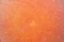
Fundus Flavimaculatus - Stargardt Disease - 20/50 OD 20/200 OS 61 Year old Color Photovu 1000 fois61-year-old decreasing vision for about the last five years. OD 20/50, OS 20/200.
Pisciform Lesions and Macular Atrophy
(0 votes)
|
|

Fundus Flavimaculatus - Stargardt Disease - 20/50 OD 20/200 OS 61 Year old Color Photovu 1432 fois61-year-old decreasing vision for about the last five years. OD 20/50, OS 20/200.
Pisciform Lesions and Macular Atrophy
(0 votes)
|
|

Fundus Flavimaculatus - Stargardt Disease - 20/50 OD 20/200 OS 61 Year old Color Photovu 1067 fois61-year-old decreasing vision for about the last five years. OD 20/50, OS 20/200.
Pisciform Lesions and Macular Atrophy
(0 votes)
|
|

Fundus Flavimaculatus - Stargardt Disease - 20/50 OD 20/200 OS 61 Year old Infrared Image ODvu 1248 fois61-year-old decreasing vision for about the last five years. OD 20/50, OS 20/200.
Pisciform Lesions and Macular Atrophy
(0 votes)
|
|
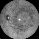
Fundus Flavimaculatus - Stargardt Disease - 20/50 OD 20/200 OS 61 Year old Infrared Image OSvu 1102 fois61-year-old decreasing vision for about the last five years. OD 20/50, OS 20/200.
Pisciform Lesions and Macular Atrophy
(0 votes)
|
|
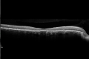
Fundus Flavimaculatus - Stargardt Disease - 20/50 OD 20/200 OS 61 Year old - SD OCT ODvu 1337 fois61-year-old decreasing vision for about the last five years. OD 20/50, OS 20/200.
Pisciform Lesions and Macular Atrophy
(0 votes)
|
|

Fundus Flavimaculatus - Stargardt Disease - 20/50 OD 20/200 OS 61 Year old - SD - OCT OSvu 2116 fois61-year-old decreasing vision for about the last five years. OD 20/50, OS 20/200.
Pisciform Lesions and Macular Atrophy
(0 votes)
|
|
|
|
|
61-year-old woman was seen in the office on October 10, 2011. She has had decreasing vision for about the last five years. She doesn’t smoke. She takes eye vitamins and fish oil.
VISUAL ACUITY: OD 20/50, OS 20/200. IOP: 13 OU.
SLIT EXAMINATION: There is 1+ nuclear sclerosis in both eyes.
EXTENDED OPHTHALMOSCOPY:
OD: Vertical C/D ratio is 0.5. There are pisciform lesions on the macula with some atrophy centrally.
OS: Vertical C/D ratio is 0.5. There are pisciform lesions on the macula with some atrophy centrally.
FUNDUS PHOTOGRAPHY: The infrared images do show areas of hypo reflectivity corresponding to pigment epithelial atrophy.
FUNDUS AUTO FLUORESCENCE: The fundus auto fluorescence show areas of hyper auto fluorescence inside the pisiform lesions and the fovea unfortunately is dark centrally.
SPECTRALIS-SD-OCT SCAN: The OCT scan shows retinal atrophy in the center in each eye.
IMPRESSION:
1. FUNDUS FLAVIMACULATUS (STARGARDT'S DISEASE) – BOTH EYES
DISCUSSION: I explained to the patient unfortunately she does have a macular dystrophy causing vision loss and retinal atrophy in each eye. At the moment there is no treatment, but there are trials started, actually right now in California for cell transplantation and maybe in a few years we will have something that can help her.


