Juxtafovoveal Telangiectasis - Group 2a - MacTel - Previous SRNVM Left Eye - Macular Hole Right Eye
|
|
76-year-old woman has juxtafoveal retinal telangiectasis. She had Stage five neovascularization in the left eye I treated with injections between September of 2007 and October of 2008 with Avastin (3 years ago). Since then she has not had a recurrence. Her vision remains good. The left eye is her better eye. She does notice the distance vision declining slightly.
VISUAL ACUITY: OD 20/400, OS 20/40. IOP: 10 OU. The posterior chamber intraocular lens is in good position in both eyes.
EXTENDED OPHTHALMOSCOPY:
OD: Vertical C/D ratio is 0.1. There is hyperpigmentation temporal to the fovea.
OS: Vertical C/D ratio is 0.1. There is hyperpigmentation temporal to the fovea. Photos confirm clinical findings.
FLUORESCEIN ANGIOGRAPHY: Fluorescein angiography shows diffuse hyperfluorescence in an annular fashion around the macula in each eye with hypofluorescence corresponding to the pigment spots.
FUNDUS AUTO FLUORESCENCE: Shows hypofluorescence corresponding to the pigment spots.
SPECTRALIS-SD-OCT SCAN: The OCT scan shows typical intraretinal cysts in each eye, but there is no evidence of any increased retinal thickness in either eye and there is no evidence of any recurrent neovascularization in the right eye. The right eye does have a true full thickness macular hole.
IMPRESSION:
1. MACULAR HOLE – RIGHT EYE
2. JUXTAFOVEAL RETINAL TELANGIECTASIS – BOTH EYES
3. INVOLUTED SUBRETINAL CHOROIDAL NEOVASCULAR MEMBRANE – LEFT EYE
DISCUSSION: I explained to the patient the maculae both look about stable. I asked her to return for a check in six months or sooner if she should notice a problem and see you periodically.
|
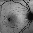
Group 2a Juxtafoveal Telangiectasis - MacTel - Stage 5 left eye - Macular Hole Right eye728 views76-year-old woman has juxtafoveal retinal telangiectasis. She had Stage five neovascularization in the left eye I treated with injections between September of 2007 and October of 2008 with Avastin (3 years ago). OCT scan today shows macular hole right eye. VISUAL ACUITY: OD 20/400, OS 20/40. 
(0 votes)
|
|

Group 2a Juxtafoveal Telangiectasis - MacTel - Stage 5 left eye - Macular Hole Right eye473 views76-year-old woman has juxtafoveal retinal telangiectasis. She had Stage five neovascularization in the left eye I treated with injections between September of 2007 and October of 2008 with Avastin (3 years ago). OCT scan today shows macular hole right eye. VISUAL ACUITY: OD 20/400, OS 20/40.
(0 votes)
|
|

Group 2a Juxtafoveal Telangiectasis - MacTel - Stage 5 left eye - Macular Hole Right eye590 views76-year-old woman has juxtafoveal retinal telangiectasis. She had Stage five neovascularization in the left eye I treated with injections between September of 2007 and October of 2008 with Avastin (3 years ago). OCT scan today shows macular hole right eye. VISUAL ACUITY: OD 20/400, OS 20/40.
(0 votes)
|
|
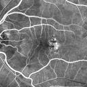
Group 2a Juxtafoveal Telangiectasis - MacTel - Stage 5 left eye - Macular Hole Right eye534 views76-year-old woman has juxtafoveal retinal telangiectasis. She had Stage five neovascularization in the left eye I treated with injections between September of 2007 and October of 2008 with Avastin (3 years ago). OCT scan today shows macular hole right eye. VISUAL ACUITY: OD 20/400, OS 20/40.
(0 votes)
|
|
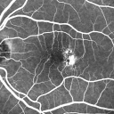
Group 2a Juxtafoveal Telangiectasis - MacTel - Stage 5 left eye - Macular Hole Right eye522 views76-year-old woman has juxtafoveal retinal telangiectasis. She had Stage five neovascularization in the left eye I treated with injections between September of 2007 and October of 2008 with Avastin (3 years ago). OCT scan today shows macular hole right eye. VISUAL ACUITY: OD 20/400, OS 20/40.
(0 votes)
|
|
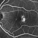
Group 2a Juxtafoveal Telangiectasis - MacTel - Stage 5 left eye - Macular Hole Right eye547 views76-year-old woman has juxtafoveal retinal telangiectasis. She had Stage five neovascularization in the left eye I treated with injections between September of 2007 and October of 2008 with Avastin (3 years ago). OCT scan today shows macular hole right eye. VISUAL ACUITY: OD 20/400, OS 20/40.
(0 votes)
|
|
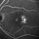
Group 2a Juxtafoveal Telangiectasis - MacTel - Stage 5 left eye - Macular Hole Right eye500 views76-year-old woman has juxtafoveal retinal telangiectasis. She had Stage five neovascularization in the left eye I treated with injections between September of 2007 and October of 2008 with Avastin (3 years ago). OCT scan today shows macular hole right eye. VISUAL ACUITY: OD 20/400, OS 20/40.
(0 votes)
|
|
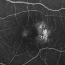
Group 2a Juxtafoveal Telangiectasis - MacTel - Stage 5 left eye - Macular Hole Right eye555 views76-year-old woman has juxtafoveal retinal telangiectasis. She had Stage five neovascularization in the left eye I treated with injections between September of 2007 and October of 2008 with Avastin (3 years ago). OCT scan today shows macular hole right eye. VISUAL ACUITY: OD 20/400, OS 20/40.
(0 votes)
|
|
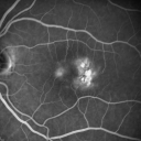
Group 2a Juxtafoveal Telangiectasis - MacTel - Stage 5 left eye - Macular Hole Right eye500 views76-year-old woman has juxtafoveal retinal telangiectasis. She had Stage five neovascularization in the left eye I treated with injections between September of 2007 and October of 2008 with Avastin (3 years ago). OCT scan today shows macular hole right eye. VISUAL ACUITY: OD 20/400, OS 20/40.
(0 votes)
|
|
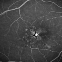
Group 2a Juxtafoveal Telangiectasis - MacTel - Stage 5 left eye - Macular Hole Right eye476 views76-year-old woman has juxtafoveal retinal telangiectasis. She had Stage five neovascularization in the left eye I treated with injections between September of 2007 and October of 2008 with Avastin (3 years ago). OCT scan today shows macular hole right eye. VISUAL ACUITY: OD 20/400, OS 20/40.
(0 votes)
|
|
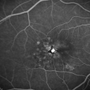
Group 2a Juxtafoveal Telangiectasis - MacTel - Stage 5 left eye - Macular Hole Right eye486 views76-year-old woman has juxtafoveal retinal telangiectasis. She had Stage five neovascularization in the left eye I treated with injections between September of 2007 and October of 2008 with Avastin (3 years ago). OCT scan today shows macular hole right eye. VISUAL ACUITY: OD 20/400, OS 20/40.
(0 votes)
|
|
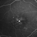
Group 2a Juxtafoveal Telangiectasis - MacTel - Stage 5 left eye - Macular Hole Right eye460 views76-year-old woman has juxtafoveal retinal telangiectasis. She had Stage five neovascularization in the left eye I treated with injections between September of 2007 and October of 2008 with Avastin (3 years ago). OCT scan today shows macular hole right eye. VISUAL ACUITY: OD 20/400, OS 20/40.
(0 votes)
|
|
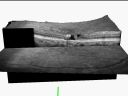
Group 2a Juxtafoveal Telangiectasis - MacTel - Stage 5 left eye - Macular Hole Right eye537 views76-year-old woman has juxtafoveal retinal telangiectasis. She had Stage five neovascularization in the left eye I treated with injections between September of 2007 and October of 2008 with Avastin (3 years ago). OCT scan today shows macular hole right eye. VISUAL ACUITY: OD 20/400, OS 20/40.
(0 votes)
|
|
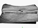
Group 2a Juxtafoveal Telangiectasis - MacTel - Stage 5 left eye - Macular Hole Right eye544 views76-year-old woman has juxtafoveal retinal telangiectasis. She had Stage five neovascularization in the left eye I treated with injections between September of 2007 and October of 2008 with Avastin (3 years ago). OCT scan today shows macular hole right eye. VISUAL ACUITY: OD 20/400, OS 20/40.
(0 votes)
|
|
|
|
|
|
76-year-old woman has juxtafoveal retinal telangiectasis. She had Stage five neovascularization in the left eye I treated with injections between September of 2007 and October of 2008 with Avastin (3 years ago). Since then she has not had a recurrence. Her vision remains good. The left eye is her better eye. She does notice the distance vision declining slightly.
VISUAL ACUITY: OD 20/400, OS 20/40. IOP: 10 OU. The posterior chamber intraocular lens is in good position in both eyes.
EXTENDED OPHTHALMOSCOPY:
OD: Vertical C/D ratio is 0.1. There is hyperpigmentation temporal to the fovea.
OS: Vertical C/D ratio is 0.1. There is hyperpigmentation temporal to the fovea. Photos confirm clinical findings.
FLUORESCEIN ANGIOGRAPHY: Fluorescein angiography shows diffuse hyperfluorescence in an annular fashion around the macula in each eye with hypofluorescence corresponding to the pigment spots.
FUNDUS AUTO FLUORESCENCE: Shows hypofluorescence corresponding to the pigment spots.
SPECTRALIS-SD-OCT SCAN: The OCT scan shows typical intraretinal cysts in each eye, but there is no evidence of any increased retinal thickness in either eye and there is no evidence of any recurrent neovascularization in the right eye. The right eye does have a true full thickness macular hole.
IMPRESSION:
1. MACULAR HOLE – RIGHT EYE
2. JUXTAFOVEAL RETINAL TELANGIECTASIS – BOTH EYES
3. INVOLUTED SUBRETINAL CHOROIDAL NEOVASCULAR MEMBRANE – LEFT EYE
DISCUSSION: I explained to the patient the maculae both look about stable. I asked her to return for a check in six months or sooner if she should notice a problem and see you periodically.


