Melanoma - Medium Choroidal
|
|
33-year-old man with a history of decreased in the right eye.
He had blurring in the eye about a year ago. He reports that it improved spontaneously until it was “nearly resolvedâ€. He said he had some persistent distortion, but that over the past couple of weeks he has noticed this dark spot recurring in the right eye, the left eye is unaffected. He does not have any flashes, floaters or change in his peripheral visual acuity. He denies any ophthalmic pain.
Past ophthalmic history is negative for retinal laser surgery or trauma. He has not had an eye examination in about fifteen years. Past medical history is negative for any chronic conditions. Family history is negative for retinal detachment or blindness of unknown cause. He is a non smoker and takes no ophthalmic medications. Complete history and review of systems are recorded in the chart.
OPHTHALMIC EXAMINATION: Visual acuity without correction 20/80 OD, 20/16 OS. No improvement with pinhole. IOP: 12 OD, 11 OS.
SLIT LAMP EXAMINATION: Biomicroscopy reveals quiet anterior segments with a 4+ deep quiet chambers and a clear lens OU.
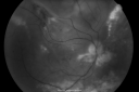
POSTERIOR SEGMENT EXAMINATION: Shows a clear view with no PVD in either eye. C/D ratio is 0.2 OU. Large retinal vessels are unremarkable. The macula of the right eye has a brown choroidal lesion with pigment overlying the surface and orange spots along its lateral surface. The left macula is normal.
EXTENDED OPHTHALMOSCOPY: The retinal periphery of the right eye shows a 12x8x5 mm high choroidal mass. This mass is associated with inferior serous retinal detachment elevating the neurosensory retina from the equator anteriorly on the right side. There appears to be guttering from the mass. The left eye has a flat and intact periphery with no retinal tears, breaks or detachments.
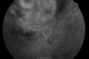
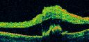
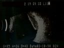
B-SCAN: B-scan ecography of the ultrasound of the right eye shows a mass about 5 mm high with a moderate to low internal reflectivity on A-scan echogram. There is serous fluid inferiorly on the echogram. Fundus photography documents presence of choroidal mass.
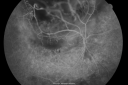
FLUORESCEIN ANGIOGRAM: Red free photographs correspond to clinical findings. Early phase angiography shows independent choroidal circulation within the mass of the right side. Retinal vasculature feels normal. As the study progresses there is broad hyperfluorescence of the mass as its internal circulation fills.
IMPRESSION:
1. CHOROIDAL MASS ON THE RIGHT SIDE WITH INDEPENDENT INTERNAL CIRCULATION ON ANGIOGRAPHY, MOST LIKELY MALIGNANT MELANOMA
2. SEROUS RETINAL DETACHMENT RIGHT SIDE ASSOCIATED WITH CHOROIDAL MASS INDICATIVE OF ACTIVITY AND GROWTH
DISCUSSION: I explained the difference with diagnosis and the problem is indeed choroidal malignant melanoma.
He needs a systemic work-up for metastic lesions, including an examination of his central nervous system, liver, and pulmonary system as well as any other tests deemed necessary by his primary care doctor or referring oncologist.
The patient is going to consider brachytherapy with radioactive iodine, either with our practice or with academic center. He is going to come back in about two weeks so we can document the growth of the lesion and confirm the diagnosis and we will consider scheduling him for surgery at that time.
|
|
|
|
|
|
|
|
|
|
|
|
|
|
|
|
|
|
|
|
|
|
|
|
|

Choroidal Melanoma Mediumvu 759 fois33-year-old man with a history of decreased in the right eye. He had blurring in the eye about a year ago. He reports that it improved spontaneously until it was “nearly resolvedâ€. He said he had some persistent distortion, but that over the past couple of weeks he has noticed this dark spot recurring in the right eye, the left eye is unaffected. He does not have any flashes, floaters or change in his peripheral visual acuity. 20/80 OD, 20/16 OS. No improvement with pinhole. IOP: 12 OD, 11 OS.
POSTERIOR SEGMENT EXAMINATION: Shows a clear view with no PVD in either eye. C/D ratio is 0.2 OU. Large retinal vessels are unremarkable. The macula of the right eye has a brown choroidal lesion with pigment overlying the surface and orange spots along its lateral surface. The left macula is normal.
B-SCAN: B-scan ecography of the ultrasound of the right eye shows a mass about 5 mm high with a moderate to low internal reflectivity on A-scan echogram. There is serous fluid inferiorly on the echogram
(1 votes)
|
|

Choroidal Melanoma Mediumvu 565 fois33-year-old man with a history of decreased in the right eye. He had blurring in the eye about a year ago. He reports that it improved spontaneously until it was “nearly resolvedâ€. He said he had some persistent distortion, but that over the past couple of weeks he has noticed this dark spot recurring in the right eye, the left eye is unaffected. He does not have any flashes, floaters or change in his peripheral visual acuity. 20/80 OD, 20/16 OS. No improvement with pinhole. IOP: 12 OD, 11 OS.
POSTERIOR SEGMENT EXAMINATION: Shows a clear view with no PVD in either eye. C/D ratio is 0.2 OU. Large retinal vessels are unremarkable. The macula of the right eye has a brown choroidal lesion with pigment overlying the surface and orange spots along its lateral surface. The left macula is normal.
B-SCAN: B-scan ecography of the ultrasound of the right eye shows a mass about 5 mm high with a moderate to low internal reflectivity on A-scan echogram. There is serous fluid inferiorly on the echogram
(0 votes)
|
|

Choroidal Melanoma Mediumvu 524 fois33-year-old man with a history of decreased in the right eye. He had blurring in the eye about a year ago. He reports that it improved spontaneously until it was “nearly resolvedâ€. He said he had some persistent distortion, but that over the past couple of weeks he has noticed this dark spot recurring in the right eye, the left eye is unaffected. He does not have any flashes, floaters or change in his peripheral visual acuity. 20/80 OD, 20/16 OS. No improvement with pinhole. IOP: 12 OD, 11 OS.
POSTERIOR SEGMENT EXAMINATION: Shows a clear view with no PVD in either eye. C/D ratio is 0.2 OU. Large retinal vessels are unremarkable. The macula of the right eye has a brown choroidal lesion with pigment overlying the surface and orange spots along its lateral surface. The left macula is normal.
B-SCAN: B-scan ecography of the ultrasound of the right eye shows a mass about 5 mm high with a moderate to low internal reflectivity on A-scan echogram. There is serous fluid inferiorly on the echogram
(0 votes)
|
|

Choroidal Melanoma Mediumvu 522 fois33-year-old man with a history of decreased in the right eye. He had blurring in the eye about a year ago. He reports that it improved spontaneously until it was “nearly resolvedâ€. He said he had some persistent distortion, but that over the past couple of weeks he has noticed this dark spot recurring in the right eye, the left eye is unaffected. He does not have any flashes, floaters or change in his peripheral visual acuity. 20/80 OD, 20/16 OS. No improvement with pinhole. IOP: 12 OD, 11 OS.
POSTERIOR SEGMENT EXAMINATION: Shows a clear view with no PVD in either eye. C/D ratio is 0.2 OU. Large retinal vessels are unremarkable. The macula of the right eye has a brown choroidal lesion with pigment overlying the surface and orange spots along its lateral surface. The left macula is normal.
B-SCAN: B-scan ecography of the ultrasound of the right eye shows a mass about 5 mm high with a moderate to low internal reflectivity on A-scan echogram. There is serous fluid inferiorly on the echogram
(0 votes)
|
|

vu 582 fois33-year-old man with a history of decreased in the right eye. He had blurring in the eye about a year ago. He reports that it improved spontaneously until it was “nearly resolvedâ€. He said he had some persistent distortion, but that over the past couple of weeks he has noticed this dark spot recurring in the right eye, the left eye is unaffected. He does not have any flashes, floaters or change in his peripheral visual acuity. 20/80 OD, 20/16 OS. No improvement with pinhole. IOP: 12 OD, 11 OS.
POSTERIOR SEGMENT EXAMINATION: Shows a clear view with no PVD in either eye. C/D ratio is 0.2 OU. Large retinal vessels are unremarkable. The macula of the right eye has a brown choroidal lesion with pigment overlying the surface and orange spots along its lateral surface. The left macula is normal.
B-SCAN: B-scan ecography of the ultrasound of the right eye shows a mass about 5 mm high with a moderate to low internal reflectivity on A-scan echogram. There is serous fluid inferiorly on the echogram
(0 votes)
|
|

Choroidal Melanoma Mediumvu 570 fois33-year-old man with a history of decreased in the right eye. He had blurring in the eye about a year ago. He reports that it improved spontaneously until it was “nearly resolvedâ€. He said he had some persistent distortion, but that over the past couple of weeks he has noticed this dark spot recurring in the right eye, the left eye is unaffected. He does not have any flashes, floaters or change in his peripheral visual acuity. 20/80 OD, 20/16 OS. No improvement with pinhole. IOP: 12 OD, 11 OS.
POSTERIOR SEGMENT EXAMINATION: Shows a clear view with no PVD in either eye. C/D ratio is 0.2 OU. Large retinal vessels are unremarkable. The macula of the right eye has a brown choroidal lesion with pigment overlying the surface and orange spots along its lateral surface. The left macula is normal.
B-SCAN: B-scan ecography of the ultrasound of the right eye shows a mass about 5 mm high with a moderate to low internal reflectivity on A-scan echogram. There is serous fluid inferiorly on the echogram
(0 votes)
|
|

Choroidal Melanoma Mediumvu 615 fois33-year-old man with a history of decreased in the right eye. He had blurring in the eye about a year ago. He reports that it improved spontaneously until it was “nearly resolvedâ€. He said he had some persistent distortion, but that over the past couple of weeks he has noticed this dark spot recurring in the right eye, the left eye is unaffected. He does not have any flashes, floaters or change in his peripheral visual acuity. 20/80 OD, 20/16 OS. No improvement with pinhole. IOP: 12 OD, 11 OS.
POSTERIOR SEGMENT EXAMINATION: Shows a clear view with no PVD in either eye. C/D ratio is 0.2 OU. Large retinal vessels are unremarkable. The macula of the right eye has a brown choroidal lesion with pigment overlying the surface and orange spots along its lateral surface. The left macula is normal.
B-SCAN: B-scan ecography of the ultrasound of the right eye shows a mass about 5 mm high with a moderate to low internal reflectivity on A-scan echogram. There is serous fluid inferiorly on the echogram
(0 votes)
|
|

Choroidal Melanoma Mediumvu 527 fois33-year-old man with a history of decreased in the right eye. He had blurring in the eye about a year ago. He reports that it improved spontaneously until it was “nearly resolvedâ€. He said he had some persistent distortion, but that over the past couple of weeks he has noticed this dark spot recurring in the right eye, the left eye is unaffected. He does not have any flashes, floaters or change in his peripheral visual acuity. 20/80 OD, 20/16 OS. No improvement with pinhole. IOP: 12 OD, 11 OS.
POSTERIOR SEGMENT EXAMINATION: Shows a clear view with no PVD in either eye. C/D ratio is 0.2 OU. Large retinal vessels are unremarkable. The macula of the right eye has a brown choroidal lesion with pigment overlying the surface and orange spots along its lateral surface. The left macula is normal.
B-SCAN: B-scan ecography of the ultrasound of the right eye shows a mass about 5 mm high with a moderate to low internal reflectivity on A-scan echogram. There is serous fluid inferiorly on the echogram
(0 votes)
|
|

Choroidal Melanoma Mediumvu 518 fois33-year-old man with a history of decreased in the right eye. He had blurring in the eye about a year ago. He reports that it improved spontaneously until it was “nearly resolvedâ€. He said he had some persistent distortion, but that over the past couple of weeks he has noticed this dark spot recurring in the right eye, the left eye is unaffected. He does not have any flashes, floaters or change in his peripheral visual acuity. 20/80 OD, 20/16 OS. No improvement with pinhole. IOP: 12 OD, 11 OS.
POSTERIOR SEGMENT EXAMINATION: Shows a clear view with no PVD in either eye. C/D ratio is 0.2 OU. Large retinal vessels are unremarkable. The macula of the right eye has a brown choroidal lesion with pigment overlying the surface and orange spots along its lateral surface. The left macula is normal.
B-SCAN: B-scan ecography of the ultrasound of the right eye shows a mass about 5 mm high with a moderate to low internal reflectivity on A-scan echogram. There is serous fluid inferiorly on the echogram
(0 votes)
|
|

Choroidal Melanoma Mediumvu 519 fois33-year-old man with a history of decreased in the right eye. He had blurring in the eye about a year ago. He reports that it improved spontaneously until it was “nearly resolvedâ€. He said he had some persistent distortion, but that over the past couple of weeks he has noticed this dark spot recurring in the right eye, the left eye is unaffected. He does not have any flashes, floaters or change in his peripheral visual acuity. 20/80 OD, 20/16 OS. No improvement with pinhole. IOP: 12 OD, 11 OS.
POSTERIOR SEGMENT EXAMINATION: Shows a clear view with no PVD in either eye. C/D ratio is 0.2 OU. Large retinal vessels are unremarkable. The macula of the right eye has a brown choroidal lesion with pigment overlying the surface and orange spots along its lateral surface. The left macula is normal.
B-SCAN: B-scan ecography of the ultrasound of the right eye shows a mass about 5 mm high with a moderate to low internal reflectivity on A-scan echogram. There is serous fluid inferiorly on the echogram
(0 votes)
|
|

Choroidal Melanoma Mediumvu 508 fois33-year-old man with a history of decreased in the right eye. He had blurring in the eye about a year ago. He reports that it improved spontaneously until it was “nearly resolvedâ€. He said he had some persistent distortion, but that over the past couple of weeks he has noticed this dark spot recurring in the right eye, the left eye is unaffected. He does not have any flashes, floaters or change in his peripheral visual acuity. 20/80 OD, 20/16 OS. No improvement with pinhole. IOP: 12 OD, 11 OS.
POSTERIOR SEGMENT EXAMINATION: Shows a clear view with no PVD in either eye. C/D ratio is 0.2 OU. Large retinal vessels are unremarkable. The macula of the right eye has a brown choroidal lesion with pigment overlying the surface and orange spots along its lateral surface. The left macula is normal.
B-SCAN: B-scan ecography of the ultrasound of the right eye shows a mass about 5 mm high with a moderate to low internal reflectivity on A-scan echogram. There is serous fluid inferiorly on the echogram
(0 votes)
|
|

Choroidal Melanoma Mediumvu 513 fois33-year-old man with a history of decreased in the right eye. He had blurring in the eye about a year ago. He reports that it improved spontaneously until it was “nearly resolvedâ€. He said he had some persistent distortion, but that over the past couple of weeks he has noticed this dark spot recurring in the right eye, the left eye is unaffected. He does not have any flashes, floaters or change in his peripheral visual acuity. 20/80 OD, 20/16 OS. No improvement with pinhole. IOP: 12 OD, 11 OS.
POSTERIOR SEGMENT EXAMINATION: Shows a clear view with no PVD in either eye. C/D ratio is 0.2 OU. Large retinal vessels are unremarkable. The macula of the right eye has a brown choroidal lesion with pigment overlying the surface and orange spots along its lateral surface. The left macula is normal.
B-SCAN: B-scan ecography of the ultrasound of the right eye shows a mass about 5 mm high with a moderate to low internal reflectivity on A-scan echogram. There is serous fluid inferiorly on the echogram
(0 votes)
|
|

Choroidal Melanoma Mediumvu 492 fois33-year-old man with a history of decreased in the right eye. He had blurring in the eye about a year ago. He reports that it improved spontaneously until it was “nearly resolvedâ€. He said he had some persistent distortion, but that over the past couple of weeks he has noticed this dark spot recurring in the right eye, the left eye is unaffected. He does not have any flashes, floaters or change in his peripheral visual acuity. 20/80 OD, 20/16 OS. No improvement with pinhole. IOP: 12 OD, 11 OS.
POSTERIOR SEGMENT EXAMINATION: Shows a clear view with no PVD in either eye. C/D ratio is 0.2 OU. Large retinal vessels are unremarkable. The macula of the right eye has a brown choroidal lesion with pigment overlying the surface and orange spots along its lateral surface. The left macula is normal.
B-SCAN: B-scan ecography of the ultrasound of the right eye shows a mass about 5 mm high with a moderate to low internal reflectivity on A-scan echogram. There is serous fluid inferiorly on the echogram
(0 votes)
|
|

Choroidal Melanoma Mediumvu 500 fois33-year-old man with a history of decreased in the right eye. He had blurring in the eye about a year ago. He reports that it improved spontaneously until it was “nearly resolvedâ€. He said he had some persistent distortion, but that over the past couple of weeks he has noticed this dark spot recurring in the right eye, the left eye is unaffected. He does not have any flashes, floaters or change in his peripheral visual acuity. 20/80 OD, 20/16 OS. No improvement with pinhole. IOP: 12 OD, 11 OS.
POSTERIOR SEGMENT EXAMINATION: Shows a clear view with no PVD in either eye. C/D ratio is 0.2 OU. Large retinal vessels are unremarkable. The macula of the right eye has a brown choroidal lesion with pigment overlying the surface and orange spots along its lateral surface. The left macula is normal.
B-SCAN: B-scan ecography of the ultrasound of the right eye shows a mass about 5 mm high with a moderate to low internal reflectivity on A-scan echogram. There is serous fluid inferiorly on the echogram
(0 votes)
|
|

Choroidal Melanoma Mediumvu 526 fois33-year-old man with a history of decreased in the right eye. He had blurring in the eye about a year ago. He reports that it improved spontaneously until it was “nearly resolvedâ€. He said he had some persistent distortion, but that over the past couple of weeks he has noticed this dark spot recurring in the right eye, the left eye is unaffected. He does not have any flashes, floaters or change in his peripheral visual acuity. 20/80 OD, 20/16 OS. No improvement with pinhole. IOP: 12 OD, 11 OS.
POSTERIOR SEGMENT EXAMINATION: Shows a clear view with no PVD in either eye. C/D ratio is 0.2 OU. Large retinal vessels are unremarkable. The macula of the right eye has a brown choroidal lesion with pigment overlying the surface and orange spots along its lateral surface. The left macula is normal.
B-SCAN: B-scan ecography of the ultrasound of the right eye shows a mass about 5 mm high with a moderate to low internal reflectivity on A-scan echogram. There is serous fluid inferiorly on the echogram
(0 votes)
|
|

Choroidal Melanoma Mediumvu 539 fois33-year-old man with a history of decreased in the right eye. He had blurring in the eye about a year ago. He reports that it improved spontaneously until it was “nearly resolvedâ€. He said he had some persistent distortion, but that over the past couple of weeks he has noticed this dark spot recurring in the right eye, the left eye is unaffected. He does not have any flashes, floaters or change in his peripheral visual acuity. 20/80 OD, 20/16 OS. No improvement with pinhole. IOP: 12 OD, 11 OS.
POSTERIOR SEGMENT EXAMINATION: Shows a clear view with no PVD in either eye. C/D ratio is 0.2 OU. Large retinal vessels are unremarkable. The macula of the right eye has a brown choroidal lesion with pigment overlying the surface and orange spots along its lateral surface. The left macula is normal.
B-SCAN: B-scan ecography of the ultrasound of the right eye shows a mass about 5 mm high with a moderate to low internal reflectivity on A-scan echogram. There is serous fluid inferiorly on the echogram
(0 votes)
|
|
Choroidal Melanoma Mediumvu 611 fois33-year-old man with a history of decreased in the right eye. He had blurring in the eye about a year ago. He reports that it improved spontaneously until it was “nearly resolvedâ€. He said he had some persistent distortion, but that over the past couple of weeks he has noticed this dark spot recurring in the right eye, the left eye is unaffected. He does not have any flashes, floaters or change in his peripheral visual acuity. 20/80 OD, 20/16 OS. No improvement with pinhole. IOP: 12 OD, 11 OS.
POSTERIOR SEGMENT EXAMINATION: Shows a clear view with no PVD in either eye. C/D ratio is 0.2 OU. Large retinal vessels are unremarkable. The macula of the right eye has a brown choroidal lesion with pigment overlying the surface and orange spots along its lateral surface. The left macula is normal.
B-SCAN: B-scan ecography of the ultrasound of the right eye shows a mass about 5 mm high with a moderate to low internal reflectivity on A-scan echogram. There is serous fluid inferiorly on the echogram
(0 votes)
|
|

Choroidal Melanoma Mediumvu 525 fois33-year-old man with a history of decreased in the right eye. He had blurring in the eye about a year ago. He reports that it improved spontaneously until it was “nearly resolvedâ€. He said he had some persistent distortion, but that over the past couple of weeks he has noticed this dark spot recurring in the right eye, the left eye is unaffected. He does not have any flashes, floaters or change in his peripheral visual acuity. 20/80 OD, 20/16 OS. No improvement with pinhole. IOP: 12 OD, 11 OS.
POSTERIOR SEGMENT EXAMINATION: Shows a clear view with no PVD in either eye. C/D ratio is 0.2 OU. Large retinal vessels are unremarkable. The macula of the right eye has a brown choroidal lesion with pigment overlying the surface and orange spots along its lateral surface. The left macula is normal.
B-SCAN: B-scan ecography of the ultrasound of the right eye shows a mass about 5 mm high with a moderate to low internal reflectivity on A-scan echogram. There is serous fluid inferiorly on the echogram
(0 votes)
|
|

Choroidal Melanoma Mediumvu 595 fois33-year-old man with a history of decreased in the right eye. He had blurring in the eye about a year ago. He reports that it improved spontaneously until it was “nearly resolvedâ€. He said he had some persistent distortion, but that over the past couple of weeks he has noticed this dark spot recurring in the right eye, the left eye is unaffected. He does not have any flashes, floaters or change in his peripheral visual acuity. 20/80 OD, 20/16 OS. No improvement with pinhole. IOP: 12 OD, 11 OS.
POSTERIOR SEGMENT EXAMINATION: Shows a clear view with no PVD in either eye. C/D ratio is 0.2 OU. Large retinal vessels are unremarkable. The macula of the right eye has a brown choroidal lesion with pigment overlying the surface and orange spots along its lateral surface. The left macula is normal.
B-SCAN: B-scan ecography of the ultrasound of the right eye shows a mass about 5 mm high with a moderate to low internal reflectivity on A-scan echogram. There is serous fluid inferiorly on the echogram
(0 votes)
|
|

Choroidal Melanoma Mediumvu 689 fois33-year-old man with a history of decreased in the right eye. He had blurring in the eye about a year ago. He reports that it improved spontaneously until it was “nearly resolvedâ€. He said he had some persistent distortion, but that over the past couple of weeks he has noticed this dark spot recurring in the right eye, the left eye is unaffected. He does not have any flashes, floaters or change in his peripheral visual acuity. 20/80 OD, 20/16 OS. No improvement with pinhole. IOP: 12 OD, 11 OS.
POSTERIOR SEGMENT EXAMINATION: Shows a clear view with no PVD in either eye. C/D ratio is 0.2 OU. Large retinal vessels are unremarkable. The macula of the right eye has a brown choroidal lesion with pigment overlying the surface and orange spots along its lateral surface. The left macula is normal.
B-SCAN: B-scan ecography of the ultrasound of the right eye shows a mass about 5 mm high with a moderate to low internal reflectivity on A-scan echogram. There is serous fluid inferiorly on the echogram
(0 votes)
|
|
Choroidal Melanoma Mediumvu 697 fois33-year-old man with a history of decreased in the right eye. He had blurring in the eye about a year ago. He reports that it improved spontaneously until it was “nearly resolvedâ€. He said he had some persistent distortion, but that over the past couple of weeks he has noticed this dark spot recurring in the right eye, the left eye is unaffected. He does not have any flashes, floaters or change in his peripheral visual acuity. 20/80 OD, 20/16 OS. No improvement with pinhole. IOP: 12 OD, 11 OS.
POSTERIOR SEGMENT EXAMINATION: Shows a clear view with no PVD in either eye. C/D ratio is 0.2 OU. Large retinal vessels are unremarkable. The macula of the right eye has a brown choroidal lesion with pigment overlying the surface and orange spots along its lateral surface. The left macula is normal.
B-SCAN: B-scan ecography of the ultrasound of the right eye shows a mass about 5 mm high with a moderate to low internal reflectivity on A-scan echogram. There is serous fluid inferiorly on the echogram
(0 votes)
|
|

Choroidal Melanoma Mediumvu 786 fois33-year-old man with a history of decreased in the right eye. He had blurring in the eye about a year ago. He reports that it improved spontaneously until it was “nearly resolvedâ€. He said he had some persistent distortion, but that over the past couple of weeks he has noticed this dark spot recurring in the right eye, the left eye is unaffected. He does not have any flashes, floaters or change in his peripheral visual acuity. 20/80 OD, 20/16 OS. No improvement with pinhole. IOP: 12 OD, 11 OS.
POSTERIOR SEGMENT EXAMINATION: Shows a clear view with no PVD in either eye. C/D ratio is 0.2 OU. Large retinal vessels are unremarkable. The macula of the right eye has a brown choroidal lesion with pigment overlying the surface and orange spots along its lateral surface. The left macula is normal.
B-SCAN: B-scan ecography of the ultrasound of the right eye shows a mass about 5 mm high with a moderate to low internal reflectivity on A-scan echogram. There is serous fluid inferiorly on the echogram
(0 votes)
|
|
|
|
|
|
|
33-year-old man with a history of decreased in the right eye.
He had blurring in the eye about a year ago. He reports that it improved spontaneously until it was “nearly resolvedâ€. He said he had some persistent distortion, but that over the past couple of weeks he has noticed this dark spot recurring in the right eye, the left eye is unaffected. He does not have any flashes, floaters or change in his peripheral visual acuity. He denies any ophthalmic pain.
Past ophthalmic history is negative for retinal laser surgery or trauma. He has not had an eye examination in about fifteen years. Past medical history is negative for any chronic conditions. Family history is negative for retinal detachment or blindness of unknown cause. He is a non smoker and takes no ophthalmic medications. Complete history and review of systems are recorded in the chart.
OPHTHALMIC EXAMINATION: Visual acuity without correction 20/80 OD, 20/16 OS. No improvement with pinhole. IOP: 12 OD, 11 OS.
SLIT LAMP EXAMINATION: Biomicroscopy reveals quiet anterior segments with a 4+ deep quiet chambers and a clear lens OU.
POSTERIOR SEGMENT EXAMINATION: Shows a clear view with no PVD in either eye. C/D ratio is 0.2 OU. Large retinal vessels are unremarkable. The macula of the right eye has a brown choroidal lesion with pigment overlying the surface and orange spots along its lateral surface. The left macula is normal.
EXTENDED OPHTHALMOSCOPY: The retinal periphery of the right eye shows a 12x8x5 mm high choroidal mass. This mass is associated with inferior serous retinal detachment elevating the neurosensory retina from the equator anteriorly on the right side. There appears to be guttering from the mass. The left eye has a flat and intact periphery with no retinal tears, breaks or detachments.
B-SCAN: B-scan ecography of the ultrasound of the right eye shows a mass about 5 mm high with a moderate to low internal reflectivity on A-scan echogram. There is serous fluid inferiorly on the echogram. Fundus photography documents presence of choroidal mass.
FLUORESCEIN ANGIOGRAM: Red free photographs correspond to clinical findings. Early phase angiography shows independent choroidal circulation within the mass of the right side. Retinal vasculature feels normal. As the study progresses there is broad hyperfluorescence of the mass as its internal circulation fills.
IMPRESSION:
1. CHOROIDAL MASS ON THE RIGHT SIDE WITH INDEPENDENT INTERNAL CIRCULATION ON ANGIOGRAPHY, MOST LIKELY MALIGNANT MELANOMA
2. SEROUS RETINAL DETACHMENT RIGHT SIDE ASSOCIATED WITH CHOROIDAL MASS INDICATIVE OF ACTIVITY AND GROWTH
DISCUSSION: I explained the difference with diagnosis and the problem is indeed choroidal malignant melanoma.
He needs a systemic work-up for metastic lesions, including an examination of his central nervous system, liver, and pulmonary system as well as any other tests deemed necessary by his primary care doctor or referring oncologist.
The patient is going to consider brachytherapy with radioactive iodine, either with our practice or with academic center. He is going to come back in about two weeks so we can document the growth of the lesion and confirm the diagnosis and we will consider scheduling him for surgery at that time.