Unknown - White Spots Periphery - Recurrent CME - Maybe Retinitis Pigmentosa without Pigment
|
|
46-year-old woman was seen in the office on January 23, 2012. She has a family history of retinitis pigmentosa. There is five children all together and to her knowledge three of her siblings have been diagnosed with retinitis pigmentosa. Her mother and father were seen and as far as everyone can tell, neither of them has it, although there was some thought they might be carriers. They were evaluated twenty five years ago and since then as far as she knows, there has not been a lot of testing done.
She has noticed a substantial decline in vision in both eyes over the last month or two. Previous to that the vision was good and she notices a dark spot in the central vision over the last few weeks, more in the left eye than the right eye. She does notice some problem with glare. She was skiing the other day and had some difficulty functioning and her night vision has been not quite right for sometime. She has not had a peripheral vision test for a while and you noticed on a retinal scan recently her maculae are abnormal. Also she had a peripheral iridotomy laser back in 1993 for shallow angles.
VISUAL ACUITY: OD 20/40, OS 20/50. IOP: OD 11, OS 13. The anterior chamber is shallow in both eyes and she has peripheral iridotomies. She has 2+ nuclear sclerotic cataracts.
EXTENDED OPHTHALMOSCOPY:
OD: Vertical C/D ratio is 0.3. There is no posterior vitreous separation. The fovea looks irregular. There are unusual looking white spots in the mid periphery, which radiate out and look to be superficial, in fact some of them are in front of the retinal vessels.
OS: Vertical C/D ratio is 0.3. There is no posterior vitreous separation. The macula looks irregular. Again, there is some superficial white spots radiating out in the periphery, but again they look to be in front of the retinal vessels. Her nerves are not pale and her retinal arteries are only minimally attenuated. Photos confirm clinical findings.
SPECTRALIS-SD-OCT SCAN: The OCT scan shows macular edema in both eyes with a serous macular detachment in the left eye. There are intraretinal cysts and retinal atrophy in the far periphery. The OCT scan does show atrophy of the outer retina.
FLUORESCEIN ANGIOGRAPHY: Fluorescein angiography shows hyperfluorescence in a pattern consistent with retinitis pigmentosa variance, where there is sparing of the central macula with increased hyperfluorescence in the periphery.
FUNDUS AUTO FLUORESCENCE: The fundus auto fluorescence images show patches of hypo auto fluorescence, predominately in the superior periphery with some hyper auto fluorescence around the macula in the mid periphery.
IMPRESSION:
1. RETINAL DYSTROPHY – BOTH EYES WITH WHITE FLECKS
2. POSSIBLE RETINITIS PUNCTATA ALBESCENS
3. FAMILY HISTORY OF RETINITIS PIGMENTOSA
DISCUSSION: I explained to the patient with her strong family history of retinitis pigmentosa, even though she has no pigment spots in the eye, she does have areas of retinal atrophy and geographic characteristics consistent with a retinal dystrophy and I think it would be reasonable for her to consider at least blood testing, if not a electroretinogram or both. I have given her information on the Carver Lab out in Iowa, in addition she may go to Bascom Palmer for further evaluation. In the meantime to treat her macular edema, I started her on Diamox 500 mg twice a day, which does work in retinitis dystrophic macular edema. Also topical angiotensin converting enzyme inhibitor will help it if she has trouble tolerating the systemic ones and I asked her to return for a check in one months or sooner if she should notice a problem.
As of 2/3/13, Patient initially responded to oral and topical ACE inhibitors, but then broke through therapy and required posterior subtenon's kenalog injections to clear edema in the right eye: 6/7/12, 12/17/12, and 1/23/13; left eye 7/5/12
Genetic testing from Carver Lab was negative for 16 genes previously associated with AR RP (ARRP,ABCA4, CERKL, CNGA1, CRB1, CHDDS, EYS, LRAT, MAK, NR2E3, PDE6B, RDH12, RLBP1, RPE65, SAG, TULP1, and USH2A.
|

Retinitis Pigmentosa Sine Pigmento or Unknown Dystrophy - Peripheral Superficial White Spots and Recurrent CME874 views46-year-old woman three of her 5 siblings have been diagnosed with retinitis pigmentosa. Her mother and father were seen and as far as everyone can tell, neither of them has it, although there was some thought they might be carriers. They were evaluated twenty five years ago and since then as far as she knows, there has not been a lot of testing done.
She has noticed a substantial decline in vision in both eyes over the last month or two. OD 20/40, OS 20/50. IOP: Both eyes have CME
(0 votes)
|
|

Retinitis Pigmentosa Sine Pigmento or Unknown Dystrophy - Peripheral Superficial White Spots and Recurrent CME835 views46-year-old woman three of her 5 siblings have been diagnosed with retinitis pigmentosa. Her mother and father were seen and as far as everyone can tell, neither of them has it, although there was some thought they might be carriers. They were evaluated twenty five years ago and since then as far as she knows, there has not been a lot of testing done.
She has noticed a substantial decline in vision in both eyes over the last month or two. OD 20/40, OS 20/50. IOP: Both eyes have CME
(0 votes)
|
|

Retinitis Pigmentosa Sine Pigmento or Unknown Dystrophy - Peripheral Superficial White Spots and Recurrent CME756 views46-year-old woman three of her 5 siblings have been diagnosed with retinitis pigmentosa. Her mother and father were seen and as far as everyone can tell, neither of them has it, although there was some thought they might be carriers. They were evaluated twenty five years ago and since then as far as she knows, there has not been a lot of testing done.
She has noticed a substantial decline in vision in both eyes over the last month or two. OD 20/40, OS 20/50. IOP: Both eyes have CME
(0 votes)
|
|

Retinitis Pigmentosa Sine Pigmento or Unknown Dystrophy - Peripheral Superficial White Spots and Recurrent CME904 views46-year-old woman three of her 5 siblings have been diagnosed with retinitis pigmentosa. Her mother and father were seen and as far as everyone can tell, neither of them has it, although there was some thought they might be carriers. They were evaluated twenty five years ago and since then as far as she knows, there has not been a lot of testing done.
She has noticed a substantial decline in vision in both eyes over the last month or two. OD 20/40, OS 20/50. IOP: Both eyes have CME
(0 votes)
|
|

Retinitis Pigmentosa Sine Pigmento or Unknown Dystrophy - Peripheral Superficial White Spots and Recurrent CME710 views46-year-old woman three of her 5 siblings have been diagnosed with retinitis pigmentosa. Her mother and father were seen and as far as everyone can tell, neither of them has it, although there was some thought they might be carriers. They were evaluated twenty five years ago and since then as far as she knows, there has not been a lot of testing done.
She has noticed a substantial decline in vision in both eyes over the last month or two. OD 20/40, OS 20/50. IOP: Both eyes have CME
(0 votes)
|
|

Retinitis Pigmentosa Sine Pigmento or Unknown Dystrophy - Peripheral Superficial White Spots and Recurrent CME772 views46-year-old woman three of her 5 siblings have been diagnosed with retinitis pigmentosa. Her mother and father were seen and as far as everyone can tell, neither of them has it, although there was some thought they might be carriers. They were evaluated twenty five years ago and since then as far as she knows, there has not been a lot of testing done.
She has noticed a substantial decline in vision in both eyes over the last month or two. OD 20/40, OS 20/50. IOP: Both eyes have CME
(0 votes)
|
|

Retinitis Pigmentosa Sine Pigmento or Unknown Dystrophy - Peripheral Superficial White Spots and Recurrent CME826 views46-year-old woman three of her 5 siblings have been diagnosed with retinitis pigmentosa. Her mother and father were seen and as far as everyone can tell, neither of them has it, although there was some thought they might be carriers. They were evaluated twenty five years ago and since then as far as she knows, there has not been a lot of testing done.
She has noticed a substantial decline in vision in both eyes over the last month or two. OD 20/40, OS 20/50. IOP: Both eyes have CME
(0 votes)
|
|

Retinitis Pigmentosa Sine Pigmento or Unknown Dystrophy - Peripheral Superficial White Spots and Recurrent CME682 views46-year-old woman three of her 5 siblings have been diagnosed with retinitis pigmentosa. Her mother and father were seen and as far as everyone can tell, neither of them has it, although there was some thought they might be carriers. They were evaluated twenty five years ago and since then as far as she knows, there has not been a lot of testing done.
She has noticed a substantial decline in vision in both eyes over the last month or two. OD 20/40, OS 20/50. IOP: Both eyes have CME
(0 votes)
|
|

Retinitis Pigmentosa Sine Pigmento or Unknown Dystrophy - Peripheral Superficial White Spots and Recurrent CME730 views46-year-old woman three of her 5 siblings have been diagnosed with retinitis pigmentosa. Her mother and father were seen and as far as everyone can tell, neither of them has it, although there was some thought they might be carriers. They were evaluated twenty five years ago and since then as far as she knows, there has not been a lot of testing done.
She has noticed a substantial decline in vision in both eyes over the last month or two. OD 20/40, OS 20/50. IOP: Both eyes have CME
(0 votes)
|
|

Retinitis Pigmentosa Sine Pigmento or Unknown Dystrophy - Peripheral Superficial White Spots and Recurrent CME754 views46-year-old woman three of her 5 siblings have been diagnosed with retinitis pigmentosa. Her mother and father were seen and as far as everyone can tell, neither of them has it, although there was some thought they might be carriers. They were evaluated twenty five years ago and since then as far as she knows, there has not been a lot of testing done.
She has noticed a substantial decline in vision in both eyes over the last month or two. OD 20/40, OS 20/50. IOP: Both eyes have CME
(0 votes)
|
|

Retinitis Pigmentosa Sine Pigmento or Unknown Dystrophy - Peripheral Superficial White Spots and Recurrent CME - FAF700 views46-year-old woman three of her 5 siblings have been diagnosed with retinitis pigmentosa. Her mother and father were seen and as far as everyone can tell, neither of them has it, although there was some thought they might be carriers. They were evaluated twenty five years ago and since then as far as she knows, there has not been a lot of testing done.
She has noticed a substantial decline in vision in both eyes over the last month or two. OD 20/40, OS 20/50. IOP: Both eyes have CME
(0 votes)
|
|
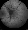
Retinitis Pigmentosa Sine Pigmento or Unknown Dystrophy - Peripheral Superficial White Spots and Recurrent CME - FAF804 views46-year-old woman three of her 5 siblings have been diagnosed with retinitis pigmentosa. Her mother and father were seen and as far as everyone can tell, neither of them has it, although there was some thought they might be carriers. They were evaluated twenty five years ago and since then as far as she knows, there has not been a lot of testing done.
She has noticed a substantial decline in vision in both eyes over the last month or two. OD 20/40, OS 20/50. IOP: Both eyes have CME
(0 votes)
|
|

Retinitis Pigmentosa Sine Pigmento or Unknown Dystrophy - Peripheral Superficial White Spots and Recurrent CME - FAF672 views46-year-old woman three of her 5 siblings have been diagnosed with retinitis pigmentosa. Her mother and father were seen and as far as everyone can tell, neither of them has it, although there was some thought they might be carriers. They were evaluated twenty five years ago and since then as far as she knows, there has not been a lot of testing done.
She has noticed a substantial decline in vision in both eyes over the last month or two. OD 20/40, OS 20/50. IOP: Both eyes have CME
(0 votes)
|
|

Retinitis Pigmentosa Sine Pigmento or Unknown Dystrophy - Peripheral Superficial White Spots and Recurrent CME - FAF714 views46-year-old woman three of her 5 siblings have been diagnosed with retinitis pigmentosa. Her mother and father were seen and as far as everyone can tell, neither of them has it, although there was some thought they might be carriers. They were evaluated twenty five years ago and since then as far as she knows, there has not been a lot of testing done.
She has noticed a substantial decline in vision in both eyes over the last month or two. OD 20/40, OS 20/50. IOP: Both eyes have CME
(0 votes)
|
|

Retinitis Pigmentosa Sine Pigmento or Unknown Dystrophy - Peripheral Superficial White Spots and Recurrent CME597 views46-year-old woman three of her 5 siblings have been diagnosed with retinitis pigmentosa. Her mother and father were seen and as far as everyone can tell, neither of them has it, although there was some thought they might be carriers. They were evaluated twenty five years ago and since then as far as she knows, there has not been a lot of testing done.
She has noticed a substantial decline in vision in both eyes over the last month or two. OD 20/40, OS 20/50. IOP: Both eyes have CME
(0 votes)
|
|

Retinitis Pigmentosa Sine Pigmento or Unknown Dystrophy - Peripheral Superficial White Spots and Recurrent CME609 views46-year-old woman three of her 5 siblings have been diagnosed with retinitis pigmentosa. Her mother and father were seen and as far as everyone can tell, neither of them has it, although there was some thought they might be carriers. They were evaluated twenty five years ago and since then as far as she knows, there has not been a lot of testing done.
She has noticed a substantial decline in vision in both eyes over the last month or two. OD 20/40, OS 20/50. IOP: Both eyes have CME
(0 votes)
|
|

Retinitis Pigmentosa Sine Pigmento or Unknown Dystrophy - Peripheral Superficial White Spots and Recurrent CME641 views46-year-old woman three of her 5 siblings have been diagnosed with retinitis pigmentosa. Her mother and father were seen and as far as everyone can tell, neither of them has it, although there was some thought they might be carriers. They were evaluated twenty five years ago and since then as far as she knows, there has not been a lot of testing done.
She has noticed a substantial decline in vision in both eyes over the last month or two. OD 20/40, OS 20/50. IOP: Both eyes have CME
(0 votes)
|
|

Retinitis Pigmentosa Sine Pigmento or Unknown Dystrophy - Peripheral Superficial White Spots and Recurrent CME634 views46-year-old woman three of her 5 siblings have been diagnosed with retinitis pigmentosa. Her mother and father were seen and as far as everyone can tell, neither of them has it, although there was some thought they might be carriers. They were evaluated twenty five years ago and since then as far as she knows, there has not been a lot of testing done.
She has noticed a substantial decline in vision in both eyes over the last month or two. OD 20/40, OS 20/50. IOP: Both eyes have CME
(0 votes)
|
|

Retinitis Pigmentosa Sine Pigmento or Unknown Dystrophy - Peripheral Superficial White Spots and Recurrent CME - FA Early709 views46-year-old woman three of her 5 siblings have been diagnosed with retinitis pigmentosa. Her mother and father were seen and as far as everyone can tell, neither of them has it, although there was some thought they might be carriers. They were evaluated twenty five years ago and since then as far as she knows, there has not been a lot of testing done.
She has noticed a substantial decline in vision in both eyes over the last month or two. OD 20/40, OS 20/50. IOP: Both eyes have CME
(0 votes)
|
|

Retinitis Pigmentosa Sine Pigmento or Unknown Dystrophy - Peripheral Superficial White Spots and Recurrent CME - FA Early673 views46-year-old woman three of her 5 siblings have been diagnosed with retinitis pigmentosa. Her mother and father were seen and as far as everyone can tell, neither of them has it, although there was some thought they might be carriers. They were evaluated twenty five years ago and since then as far as she knows, there has not been a lot of testing done.
She has noticed a substantial decline in vision in both eyes over the last month or two. OD 20/40, OS 20/50. IOP: Both eyes have CME
(0 votes)
|
|

Retinitis Pigmentosa Sine Pigmento or Unknown Dystrophy - Peripheral Superficial White Spots and Recurrent CME - FA Early654 views46-year-old woman three of her 5 siblings have been diagnosed with retinitis pigmentosa. Her mother and father were seen and as far as everyone can tell, neither of them has it, although there was some thought they might be carriers. They were evaluated twenty five years ago and since then as far as she knows, there has not been a lot of testing done.
She has noticed a substantial decline in vision in both eyes over the last month or two. OD 20/40, OS 20/50. IOP: Both eyes have CME
(0 votes)
|
|

Retinitis Pigmentosa Sine Pigmento or Unknown Dystrophy - Peripheral Superficial White Spots and Recurrent CME - FA Early780 views46-year-old woman three of her 5 siblings have been diagnosed with retinitis pigmentosa. Her mother and father were seen and as far as everyone can tell, neither of them has it, although there was some thought they might be carriers. They were evaluated twenty five years ago and since then as far as she knows, there has not been a lot of testing done.
She has noticed a substantial decline in vision in both eyes over the last month or two. OD 20/40, OS 20/50. IOP: Both eyes have CME
(0 votes)
|
|

Retinitis Pigmentosa Sine Pigmento or Unknown Dystrophy - Peripheral Superficial White Spots and Recurrent CME - FA MID810 views46-year-old woman three of her 5 siblings have been diagnosed with retinitis pigmentosa. Her mother and father were seen and as far as everyone can tell, neither of them has it, although there was some thought they might be carriers. They were evaluated twenty five years ago and since then as far as she knows, there has not been a lot of testing done.
She has noticed a substantial decline in vision in both eyes over the last month or two. OD 20/40, OS 20/50. IOP: Both eyes have CME
(0 votes)
|
|

Retinitis Pigmentosa Sine Pigmento or Unknown Dystrophy - Peripheral Superficial White Spots and Recurrent CME - FA MID622 views46-year-old woman three of her 5 siblings have been diagnosed with retinitis pigmentosa. Her mother and father were seen and as far as everyone can tell, neither of them has it, although there was some thought they might be carriers. They were evaluated twenty five years ago and since then as far as she knows, there has not been a lot of testing done.
She has noticed a substantial decline in vision in both eyes over the last month or two. OD 20/40, OS 20/50. IOP: Both eyes have CME
(0 votes)
|
|

Retinitis Pigmentosa Sine Pigmento or Unknown Dystrophy - Peripheral Superficial White Spots and Recurrent CME - FA MID698 views46-year-old woman three of her 5 siblings have been diagnosed with retinitis pigmentosa. Her mother and father were seen and as far as everyone can tell, neither of them has it, although there was some thought they might be carriers. They were evaluated twenty five years ago and since then as far as she knows, there has not been a lot of testing done.
She has noticed a substantial decline in vision in both eyes over the last month or two. OD 20/40, OS 20/50. IOP: Both eyes have CME
(0 votes)
|
|

Retinitis Pigmentosa Sine Pigmento or Unknown Dystrophy - Peripheral Superficial White Spots and Recurrent CME - FA MID646 views46-year-old woman three of her 5 siblings have been diagnosed with retinitis pigmentosa. Her mother and father were seen and as far as everyone can tell, neither of them has it, although there was some thought they might be carriers. They were evaluated twenty five years ago and since then as far as she knows, there has not been a lot of testing done.
She has noticed a substantial decline in vision in both eyes over the last month or two. OD 20/40, OS 20/50. IOP: Both eyes have CME
(0 votes)
|
|

Retinitis Pigmentosa Sine Pigmento or Unknown Dystrophy - Peripheral Superficial White Spots and Recurrent CME - FA LATE674 views46-year-old woman three of her 5 siblings have been diagnosed with retinitis pigmentosa. Her mother and father were seen and as far as everyone can tell, neither of them has it, although there was some thought they might be carriers. They were evaluated twenty five years ago and since then as far as she knows, there has not been a lot of testing done.
She has noticed a substantial decline in vision in both eyes over the last month or two. OD 20/40, OS 20/50. IOP: Both eyes have CME
(0 votes)
|
|

Retinitis Pigmentosa Sine Pigmento or Unknown Dystrophy - Peripheral Superficial White Spots and Recurrent CME- FA LATE646 views46-year-old woman three of her 5 siblings have been diagnosed with retinitis pigmentosa. Her mother and father were seen and as far as everyone can tell, neither of them has it, although there was some thought they might be carriers. They were evaluated twenty five years ago and since then as far as she knows, there has not been a lot of testing done.
She has noticed a substantial decline in vision in both eyes over the last month or two. OD 20/40, OS 20/50. IOP: Both eyes have CME
(0 votes)
|
|

Retinitis Pigmentosa Sine Pigmento or Unknown Dystrophy - Peripheral Superficial White Spots and Recurrent CME - FA Early599 views46-year-old woman three of her 5 siblings have been diagnosed with retinitis pigmentosa. Her mother and father were seen and as far as everyone can tell, neither of them has it, although there was some thought they might be carriers. They were evaluated twenty five years ago and since then as far as she knows, there has not been a lot of testing done.
She has noticed a substantial decline in vision in both eyes over the last month or two. OD 20/40, OS 20/50. IOP: Both eyes have CME
(0 votes)
|
|

Retinitis Pigmentosa Sine Pigmento or Unknown Dystrophy - Peripheral Superficial White Spots and Recurrent CME - FA Early665 views46-year-old woman three of her 5 siblings have been diagnosed with retinitis pigmentosa. Her mother and father were seen and as far as everyone can tell, neither of them has it, although there was some thought they might be carriers. They were evaluated twenty five years ago and since then as far as she knows, there has not been a lot of testing done.
She has noticed a substantial decline in vision in both eyes over the last month or two. OD 20/40, OS 20/50. IOP: Both eyes have CME
(0 votes)
|
|

Retinitis Pigmentosa Sine Pigmento or Unknown Dystrophy - Peripheral Superficial White Spots and Recurrent CME - FA Early658 views46-year-old woman three of her 5 siblings have been diagnosed with retinitis pigmentosa. Her mother and father were seen and as far as everyone can tell, neither of them has it, although there was some thought they might be carriers. They were evaluated twenty five years ago and since then as far as she knows, there has not been a lot of testing done.
She has noticed a substantial decline in vision in both eyes over the last month or two. OD 20/40, OS 20/50. IOP: Both eyes have CME
(0 votes)
|
|

Retinitis Pigmentosa Sine Pigmento or Unknown Dystrophy - Peripheral Superficial White Spots and Recurrent CME - FA Early693 views46-year-old woman three of her 5 siblings have been diagnosed with retinitis pigmentosa. Her mother and father were seen and as far as everyone can tell, neither of them has it, although there was some thought they might be carriers. They were evaluated twenty five years ago and since then as far as she knows, there has not been a lot of testing done.
She has noticed a substantial decline in vision in both eyes over the last month or two. OD 20/40, OS 20/50. IOP: Both eyes have CME
(0 votes)
|
|

Retinitis Pigmentosa Sine Pigmento or Unknown Dystrophy - Peripheral Superficial White Spots and Recurrent CME - FA Early629 views46-year-old woman three of her 5 siblings have been diagnosed with retinitis pigmentosa. Her mother and father were seen and as far as everyone can tell, neither of them has it, although there was some thought they might be carriers. They were evaluated twenty five years ago and since then as far as she knows, there has not been a lot of testing done.
She has noticed a substantial decline in vision in both eyes over the last month or two. OD 20/40, OS 20/50. IOP: Both eyes have CME
(0 votes)
|
|

Retinitis Pigmentosa Sine Pigmento or Unknown Dystrophy - Peripheral Superficial White Spots and Recurrent CME - FA Early619 views46-year-old woman three of her 5 siblings have been diagnosed with retinitis pigmentosa. Her mother and father were seen and as far as everyone can tell, neither of them has it, although there was some thought they might be carriers. They were evaluated twenty five years ago and since then as far as she knows, there has not been a lot of testing done.
She has noticed a substantial decline in vision in both eyes over the last month or two. OD 20/40, OS 20/50. IOP: Both eyes have CME
(0 votes)
|
|

Retinitis Pigmentosa Sine Pigmento or Unknown Dystrophy - Peripheral Superficial White Spots and Recurrent CME591 views46-year-old woman three of her 5 siblings have been diagnosed with retinitis pigmentosa. Her mother and father were seen and as far as everyone can tell, neither of them has it, although there was some thought they might be carriers. They were evaluated twenty five years ago and since then as far as she knows, there has not been a lot of testing done.
She has noticed a substantial decline in vision in both eyes over the last month or two. OD 20/40, OS 20/50. IOP: Both eyes have CME
(0 votes)
|
|

Retinitis Pigmentosa Sine Pigmento or Unknown Dystrophy - Peripheral Superficial White Spots and Recurrent CME685 views46-year-old woman three of her 5 siblings have been diagnosed with retinitis pigmentosa. Her mother and father were seen and as far as everyone can tell, neither of them has it, although there was some thought they might be carriers. They were evaluated twenty five years ago and since then as far as she knows, there has not been a lot of testing done.
She has noticed a substantial decline in vision in both eyes over the last month or two. OD 20/40, OS 20/50. IOP: Both eyes have CME
(0 votes)
|
|

Retinitis Pigmentosa Sine Pigmento or Unknown Dystrophy - Peripheral Superficial White Spots and Recurrent CME636 views46-year-old woman three of her 5 siblings have been diagnosed with retinitis pigmentosa. Her mother and father were seen and as far as everyone can tell, neither of them has it, although there was some thought they might be carriers. They were evaluated twenty five years ago and since then as far as she knows, there has not been a lot of testing done.
She has noticed a substantial decline in vision in both eyes over the last month or two. OD 20/40, OS 20/50. IOP: Both eyes have CME
(0 votes)
|
|

Retinitis Pigmentosa Sine Pigmento or Unknown Dystrophy - Peripheral Superficial White Spots and Recurrent CME631 views1 month of treatment with diamox 500 mg BID- cme is a little better
(0 votes)
|
|

Retinitis Pigmentosa Sine Pigmento or Unknown Dystrophy - Peripheral Superficial White Spots and Recurrent CME576 views1 month of treatment with diamox 500 mg BID- cme is a little better
(0 votes)
|
|

Retinitis Pigmentosa Sine Pigmento or Unknown Dystrophy - Peripheral Superficial White Spots and Recurrent CME699 views1 month of treatment with diamox 500 mg BID- cme is a little better
(0 votes)
|
|

Retinitis Pigmentosa Sine Pigmento or Unknown Dystrophy - Peripheral Superficial White Spots and Recurrent CME624 views1 month of treatment with diamox 500 mg BID- cme is a little better
(0 votes)
|
|

Retinitis Pigmentosa Sine Pigmento or Unknown Dystrophy - Peripheral Superficial White Spots and Recurrent CME613 views1 month of treatment with diamox 500 mg BID- cme is a little better
(0 votes)
|
|

Retinitis Pigmentosa Sine Pigmento or Unknown Dystrophy - Peripheral Superficial White Spots and Recurrent CME613 views1 month of treatment with diamox 500 mg BID- cme is a little better
(0 votes)
|
|

Retinitis Pigmentosa Sine Pigmento or Unknown Dystrophy - Peripheral Superficial White Spots and Recurrent CME638 views1 month of treatment with diamox 500 mg BID- cme is a little better
(0 votes)
|
|

Retinitis Pigmentosa Sine Pigmento or Unknown Dystrophy - Peripheral Superficial White Spots and Recurrent CME704 views1 month of treatment with diamox 500 mg BID- cme is a little better
(0 votes)
|
|

Retinitis Pigmentosa Sine Pigmento or Unknown Dystrophy - Peripheral Superficial White Spots and Recurrent CME600 views1 month of treatment with diamox 500 mg BID- cme is a little better
(0 votes)
|
|

Retinitis Pigmentosa Sine Pigmento or Unknown Dystrophy - Peripheral Superficial White Spots and Recurrent CME676 views1 month of treatment with diamox 500 mg BID- cme is a little better
(0 votes)
|
|

Retinitis Pigmentosa Sine Pigmento or Unknown Dystrophy - Peripheral Superficial White Spots and Recurrent CME547 views1 month of treatment with diamox 500 mg BID- cme is a little better
(0 votes)
|
|

Retinitis Pigmentosa Sine Pigmento or Unknown Dystrophy - Peripheral Superficial White Spots and Recurrent CME577 views1 month of treatment with diamox 500 mg BID- cme is a little better
(0 votes)
|
|

Retinitis Pigmentosa Sine Pigmento or Unknown Dystrophy - Peripheral Superficial White Spots and Recurrent CME652 viewsCME worse - patient received subtenon's kenalog injection right eye.
(0 votes)
|
|
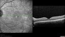
Retinitis Pigmentosa Sine Pigmento or Unknown Dystrophy - Peripheral Superficial White Spots and Recurrent CME779 viewsCME worse - patient received subtenon's kenalog injection right eye.
(0 votes)
|
|

Retinitis Pigmentosa Sine Pigmento or Unknown Dystrophy - Peripheral Superficial White Spots and Recurrent CME779 viewsSubtenon's Kenlalog given right eye today.
(0 votes)
|
|
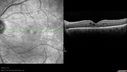
Retinitis Pigmentosa Sine Pigmento or Unknown Dystrophy - Peripheral Superficial White Spots and Recurrent CME845 viewsSubtenon's Kenlalog given right eye today.
(0 votes)
|
|

Retinitis Pigmentosa Sine Pigmento or Unknown Dystrophy - Peripheral Superficial White Spots and Recurrent CME709 viewsComparison of Maps - CME is now cleared
(0 votes)
|
|

Retinitis Pigmentosa Sine Pigmento or Unknown Dystrophy - Peripheral Superficial White Spots and Recurrent CME806 viewsComparison of Maps - CME is now cleared
(0 votes)
|
|
|
|
|
46-year-old woman was seen in the office on January 23, 2012. She has a family history of retinitis pigmentosa. There is five children all together and to her knowledge three of her siblings have been diagnosed with retinitis pigmentosa. Her mother and father were seen and as far as everyone can tell, neither of them has it, although there was some thought they might be carriers. They were evaluated twenty five years ago and since then as far as she knows, there has not been a lot of testing done.
She has noticed a substantial decline in vision in both eyes over the last month or two. Previous to that the vision was good and she notices a dark spot in the central vision over the last few weeks, more in the left eye than the right eye. She does notice some problem with glare. She was skiing the other day and had some difficulty functioning and her night vision has been not quite right for sometime. She has not had a peripheral vision test for a while and you noticed on a retinal scan recently her maculae are abnormal. Also she had a peripheral iridotomy laser back in 1993 for shallow angles.
VISUAL ACUITY: OD 20/40, OS 20/50. IOP: OD 11, OS 13. The anterior chamber is shallow in both eyes and she has peripheral iridotomies. She has 2+ nuclear sclerotic cataracts.
EXTENDED OPHTHALMOSCOPY:
OD: Vertical C/D ratio is 0.3. There is no posterior vitreous separation. The fovea looks irregular. There are unusual looking white spots in the mid periphery, which radiate out and look to be superficial, in fact some of them are in front of the retinal vessels.
OS: Vertical C/D ratio is 0.3. There is no posterior vitreous separation. The macula looks irregular. Again, there is some superficial white spots radiating out in the periphery, but again they look to be in front of the retinal vessels. Her nerves are not pale and her retinal arteries are only minimally attenuated. Photos confirm clinical findings.
SPECTRALIS-SD-OCT SCAN: The OCT scan shows macular edema in both eyes with a serous macular detachment in the left eye. There are intraretinal cysts and retinal atrophy in the far periphery. The OCT scan does show atrophy of the outer retina.
FLUORESCEIN ANGIOGRAPHY: Fluorescein angiography shows hyperfluorescence in a pattern consistent with retinitis pigmentosa variance, where there is sparing of the central macula with increased hyperfluorescence in the periphery.
FUNDUS AUTO FLUORESCENCE: The fundus auto fluorescence images show patches of hypo auto fluorescence, predominately in the superior periphery with some hyper auto fluorescence around the macula in the mid periphery.
IMPRESSION:
1. RETINAL DYSTROPHY – BOTH EYES WITH WHITE FLECKS
2. POSSIBLE RETINITIS PUNCTATA ALBESCENS
3. FAMILY HISTORY OF RETINITIS PIGMENTOSA
DISCUSSION: I explained to the patient with her strong family history of retinitis pigmentosa, even though she has no pigment spots in the eye, she does have areas of retinal atrophy and geographic characteristics consistent with a retinal dystrophy and I think it would be reasonable for her to consider at least blood testing, if not a electroretinogram or both. I have given her information on the Carver Lab out in Iowa, in addition she may go to Bascom Palmer for further evaluation. In the meantime to treat her macular edema, I started her on Diamox 500 mg twice a day, which does work in retinitis dystrophic macular edema. Also topical angiotensin converting enzyme inhibitor will help it if she has trouble tolerating the systemic ones and I asked her to return for a check in one months or sooner if she should notice a problem.
As of 2/3/13, Patient initially responded to oral and topical ACE inhibitors, but then broke through therapy and required posterior subtenon's kenalog injections to clear edema in the right eye: 6/7/12, 12/17/12, and 1/23/13; left eye 7/5/12
Genetic testing from Carver Lab was negative for 16 genes previously associated with AR RP (ARRP,ABCA4, CERKL, CNGA1, CRB1, CHDDS, EYS, LRAT, MAK, NR2E3, PDE6B, RDH12, RLBP1, RPE65, SAG, TULP1, and USH2A.


