Gonioscopy, Scattered Peripheral Anterior Synechiae
|
|
|

Gonioscopy; Scattered Peripheral Anterior Synechiaevu 1288 foisPatient comes in for evaluation for glaucoma. Patient also has a history of Uveitis. Last flare up was back in 1990. Patient's VA was 20/30, Right eye and 20/40-1, Left eye. Slit Lamp Gonioscopy reveals iris bow with scattered PAS around the angles of the anterior chamber. You can also see pigmentation in the trabecular meshwork. Patient will follow up in 3-months. 
(0 votes)
|
|

Gonioscopy; Scattered Peripheral Anterior Synechiaevu 1420 foisPatient comes in for evaluation for glaucoma. Patient also has a history of Uveitis. Last flare up was back in 1990. Patient's VA was 20/30, Right eye and 20/40-1, Left eye. Slit Lamp Gonioscopy reveals iris bow with scattered PAS around the angles of the anterior chamber. You can also see pigmentation in the trabecular meshwork. Patient will follow up in 3-months.
(0 votes)
|
|

Gonioscopy; Scattered Peripheral Anterior Synechiaevu 1150 foisPatient comes in for evaluation for glaucoma. Patient also has a history of Uveitis. Last flare up was back in 1990. Patient's VA was 20/30, Right eye and 20/40-1, Left eye. Slit Lamp Gonioscopy reveals iris bow with scattered PAS around the angles of the anterior chamber. You can also see pigmentation in the trabecular meshwork. Patient will follow up in 3-months.
(0 votes)
|
|
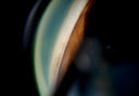
Gonioscopy; Scattered Peripheral Anterior Synechiaevu 1045 foisPatient comes in for evaluation for glaucoma. Patient also has a history of Uveitis. Last flare up was back in 1990. Patient's VA was 20/30, Right eye and 20/40-1, Left eye. Slit Lamp Gonioscopy reveals iris bow with scattered PAS around the angles of the anterior chamber. You can also see pigmentation in the trabecular meshwork. Patient will follow up in 3-months.
(0 votes)
|
|

Gonioscopy; Scattered Peripheral Anterior Synechiaevu 1065 foisPatient comes in for evaluation for glaucoma. Patient also has a history of Uveitis. Last flare up was back in 1990. Patient's VA was 20/30, Right eye and 20/40-1, Left eye. Slit Lamp Gonioscopy reveals iris bow with scattered PAS around the angles of the anterior chamber. You can also see pigmentation in the trabecular meshwork. Patient will follow up in 3-months.
(0 votes)
|
|

Gonioscopy; Scattered Peripheral Anterior Synechiaevu 1130 foisPatient comes in for evaluation for glaucoma. Patient also has a history of Uveitis. Last flare up was back in 1990. Patient's VA was 20/30, Right eye and 20/40-1, Left eye. Slit Lamp Gonioscopy reveals iris bow with scattered PAS around the angles of the anterior chamber. You can also see pigmentation in the trabecular meshwork. Patient will follow up in 3-months.
(0 votes)
|
|

Gonioscopy; Scattered Peripheral Anterior Synechiaevu 1428 foisPatient comes in for evaluation for glaucoma. Patient also has a history of Uveitis. Last flare up was back in 1990. Patient's VA was 20/30, Right eye and 20/40-1, Left eye. Slit Lamp Gonioscopy reveals iris bow with scattered PAS around the angles of the anterior chamber. You can also see pigmentation in the trabecular meshwork. Patient will follow up in 3-months.
(0 votes)
|
|

Gonioscopy; Scattered Peripheral Anterior Synechiaevu 1678 foisPatient comes in for evaluation for glaucoma. Patient also has a history of Uveitis. Last flare up was back in 1990. Patient's VA was 20/30, Right eye and 20/40-1, Left eye. Slit Lamp Gonioscopy reveals iris bow with scattered PAS around the angles of the anterior chamber. You can also see pigmentation in the trabecular meshwork. Patient will follow up in 3-months.
(0 votes)
|
|

Gonioscopy; Scattered Peripheral Anterior Synechiaevu 1415 foisPatient comes in for evaluation for glaucoma. Patient also has a history of Uveitis. Last flare up was back in 1990. Patient's VA was 20/30, Right eye and 20/40-1, Left eye. Slit Lamp Gonioscopy reveals iris bow with scattered PAS around the angles of the anterior chamber. You can also see pigmentation in the trabecular meshwork. Patient will follow up in 3-months.
(0 votes)
|
|

Gonioscopy; Scattered Peripheral Anterior Synechiaevu 1217 foisPatient comes in for evaluation for glaucoma. Patient also has a history of Uveitis. Last flare up was back in 1990. Patient's VA was 20/30, Right eye and 20/40-1, Left eye. Slit Lamp Gonioscopy reveals iris bow with scattered PAS around the angles of the anterior chamber. You can also see pigmentation in the trabecular meshwork. Patient will follow up in 3-months.
(0 votes)
|
|

Gonioscopy; Scattered Peripheral Anterior Synechiaevu 1343 foisPatient comes in for evaluation for glaucoma. Patient also has a history of Uveitis. Last flare up was back in 1990. Patient's VA was 20/30, Right eye and 20/40-1, Left eye. Slit Lamp Gonioscopy reveals iris bow with scattered PAS around the angles of the anterior chamber. You can also see pigmentation in the trabecular meshwork. Patient will follow up in 3-months.
(0 votes)
|
|

Gonioscopy; Scattered Peripheral Anterior Synechiaevu 1778 foisPatient comes in for evaluation for glaucoma. Patient also has a history of Uveitis. Last flare up was back in 1990. Patient's VA was 20/30, Right eye and 20/40-1, Left eye. Slit Lamp Gonioscopy reveals iris bow with scattered PAS around the angles of the anterior chamber. You can also see pigmentation in the trabecular meshwork. Patient will follow up in 3-months.
(0 votes)
|
|

Gonioscopy; Scattered Peripheral Anterior Synechiaevu 1591 foisPatient comes in for evaluation for glaucoma. Patient also has a history of Uveitis. Last flare up was back in 1990. Patient's VA was 20/30, Right eye and 20/40-1, Left eye. Slit Lamp Gonioscopy reveals iris bow with scattered PAS around the angles of the anterior chamber. You can also see pigmentation in the trabecular meshwork. Patient will follow up in 3-months.
(0 votes)
|
|
|
|
|
|
|


