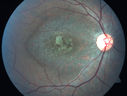
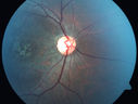
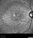
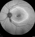
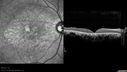
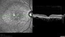
36-year-old man was seen by me in the office on August 23, 2012. I started seeing him years ago. His father in fact is an ophthalmologist. He started to notice vision change sometime in his early twenties. He has had progressing macular atrophy and unfortunately has poor vision in the left eye. The right eye continues to see decently. VISUAL ACUITY: OD 20/40, OS 20/200. IOP: OD 10, OS 12. The lenses are clear. EXTENDED OPHTHALMOSCOPY: OD: Vertical C/D ratio is 0.3. The macula looks atrophic. OS: Vertical C/D ratio is 0.3. The macula looks atrophic. The retinal vessels are fine. FUNDUS PHOTOGRAPHY - INFRA RED: The image shows hypopigmentation centrally. SPECTRALIS-SD-OCT SCAN: The OCT scan shows retinal atrophy in the center of both eyes with loss of the outer retinal layers. The choroid looks unusual in him, almost like the choriocapillaris is thinned out and the large choroidal vessels are more abundant. FUNDUS PHOTOGRAPHY - AUTO FLUORESCENCE: The image shows a ring of hyper auto fluorescence in the posterior pole within the macular arcades, which is a linear bright ring. Inside of that there is some hypo auto fluorescence, but not terribly so and he does seem to have decreased macular pigment in both eyes. IMPRESSION: 1. PROBABLE CONE DYSTROPHY – BOTH EYES AUTOSOMAL RECESSIVE DISCUSSION: I explained to the patient now that genetic testing is available, I think it would be best to characterize his dystrophy toward the goal of having genetic testing done and then ultimately and possibly, if it becomes available, offering him some sort of treatment. |
|||||||
|
|
|
|
||||
|
|
|
|
||||
|
|
|
|
||||
|
|
|
|||||
|
|||||||
36-year-old man was seen by me in the office on August 23, 2012. I started seeing him years ago. His father in fact is an ophthalmologist. He started to notice vision change sometime in his early twenties. He has had progressing macular atrophy and unfortunately has poor vision in the left eye. The right eye continues to see decently. VISUAL ACUITY: OD 20/40, OS 20/200. IOP: OD 10, OS 12. The lenses are clear. EXTENDED OPHTHALMOSCOPY: OD: Vertical C/D ratio is 0.3. The macula looks atrophic. OS: Vertical C/D ratio is 0.3. The macula looks atrophic. The retinal vessels are fine. FUNDUS PHOTOGRAPHY - INFRA RED: The image shows hypopigmentation centrally. SPECTRALIS-SD-OCT SCAN: The OCT scan shows retinal atrophy in the center of both eyes with loss of the outer retinal layers. The choroid looks unusual in him, almost like the choriocapillaris is thinned out and the large choroidal vessels are more abundant. FUNDUS PHOTOGRAPHY - AUTO FLUORESCENCE: The image shows a ring of hyper auto fluorescence in the posterior pole within the macular arcades, which is a linear bright ring. Inside of that there is some hypo auto fluorescence, but not terribly so and he does seem to have decreased macular pigment in both eyes. IMPRESSION: 1. PROBABLE CONE DYSTROPHY – BOTH EYES AUTOSOMAL RECESSIVE DISCUSSION: I explained to the patient now that genetic testing is available, I think it would be best to characterize his dystrophy toward the goal of having genetic testing done and then ultimately and possibly, if it becomes available, offering him some sort of treatment.