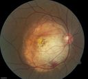
017 - May 2013 Best Image Finalist - Winner - Recurrent PVR Retinal Detachment - Jason Calhoun - Photographer - Mayo Clinic Jacksonville
Reoccurring Retinal Detachmentvista 1106 vecesPatient comes in for second opinion for RD in the right eye. Patient's VA was count fingers @ 2-ft in the right eye and 20/20 in the left eye. Patient is aphakic and has had 5- retinal surgeries in the past in the right eye including a scleral buckle. RD present with the macula off. Will consider surgery with silicone oil. (3 votos)
Ocular Trauma with Retinal Detachmentvista 913 vecesPatient suffered blunt force trauma to the eye resulting total retinal detachment. The retina is visible in the pupillary space. (1 votos)
Melanomavista 791 veces (1 votos)
Choroidal Osteomavista 899 vecesYoung girl with vision loss from osteoma. (0 votos)
Lattice Degenerationvista 1205 vecesAprox 180 degree lattice degeneration in OD of a 17yr old girl. (0 votos)
Macular Hole with Peripheral Laser Scarsvista 1190 vecesDiabetic patient comes in with flashes and floaters in both eyes. Patient's VA is 20/400, right eye and 20/25, left eye. Fundus exam presents with large macular hole with diabetic changes in the retina. Laser scars in the peripheral. (1 votos)
Prominent Posterior Hyaloid with Background Diabetic Retinopathyvista 1290 vecesPatient comes in for follow up on her Diabetic Retinopathy and glaucoma. Patient's VA was 20/30 in the left eye. Fundus exam presents a Posterior Hyaloid with hemorrhage inferiorily. Patient will be seen again in 6-months for follow up. (1 votos)
Choroidal Tumor consistent with Uveal Melanomavista 1172 vecesPatient comes in for second opinion for 2-melanomas in the right eye. Patient's VA is 20/20, both eyes. Fundus exam reveals melanoma superior to the macula which measured at 0.7-mm thick. Second melanoma is nasally which measured at 4.1-mm thick and has low internal reflectivity. Patient will be followed up with MRI and ocular protocol. (1 votos)
Macular Pre-Retinal Hemorrhage vista 1133 vecesPatient complains of loss of vision in his left eye. Patient is diabetic. VA was 20/20, right eye and 20/150, left eye with no improvement pinhole. Fundus exam reveals very large pre retinal hemorrhage in the left eye. Embolism located inferior, nasally just off the optic nerve in the left eye. Patient underwent PRP for treatment of the hemorrhage. (1 votos)