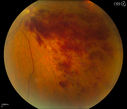
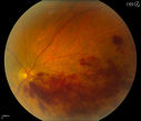
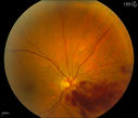
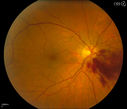
Branch Retinal Vein Occlusion523 x angesehenPatient's VA was 20/40, right eye. Fundus exam shows branch retinal vein occlusion nasally to the optic nerve in the right eye. (0 Bewertungen)
Branch Retinal Vein Occlusion511 x angesehenPatient's VA was 20/40, right eye. Fundus exam shows branch retinal vein occlusion nasally to the optic nerve in the right eye. (0 Bewertungen)
Branch Retinal Vein Occlusion445 x angesehenPatient's VA was 20/40, right eye. Fundus exam shows branch retinal vein occlusion nasally to the optic nerve in the right eye. (0 Bewertungen)
Branch Retinal Vein Occlusion420 x angesehenPatient's VA was 20/40, right eye. Fundus exam shows branch retinal vein occlusion nasally to the optic nerve in the right eye. (0 Bewertungen)
Branch Retinal Vein Occlusion489 x angesehenPatient's VA was 20/40, right eye. Fundus exam shows branch retinal vein occlusion nasally to the optic nerve in the right eye. (0 Bewertungen)
Branch Retinal Vein Occlusion482 x angesehenPatient's VA was 20/40, right eye. Fundus exam shows branch retinal vein occlusion nasally to the optic nerve in the right eye. (0 Bewertungen)
Branch Retinal Vein Occlusion451 x angesehenPatient's VA was 20/40, right eye. Fundus exam shows branch retinal vein occlusion nasally to the optic nerve in the right eye. (0 Bewertungen)
Branch Retinal Vein Occlusion492 x angesehenPatient's VA was 20/40, right eye. Fundus exam shows branch retinal vein occlusion nasally to the optic nerve in the right eye. (0 Bewertungen)