Central Serous Retinopathy - PED and Serous RD
|
|
49-year-old man was doing fine until about two weeks ago when he noticed he was having some difficulty with his vision. He closed his left eye and noticed the right eye was not seeing well. He sees a spot in the central vision with a red halo. He is healthy and his blood pressure is fine. He does not take any steroids.
VISUAL ACUITY: Vision OD is 20/50, OS is 20/20. IOP: OD 17, OS 14.
SLIT LAMP EXAM: Normal with clear lenses.
EXTENDED OPHTHALMOSCOPY:
OD: Vertical C/D ratio is 0.1. There is no posterior vitreous separation. There is a pigment epithelial detachment on the inferotemporal margin of the fovea and there is a serous macular detachment.
OS: Vertical C/D ratio is 0.1. There is no posterior vitreous separation. The macula and periphery look healthy.
OCT SCAN: The right eye shows a pigment epithelial detachment and a serous macular detachment. The left eye had a normal scan.
FLUORESCEIN ANGIOGRAPHY: FA of the right eye shows a leaky subfoveal pigment epithelial detachment with serous macular detachment. The left eye is angiographically normal.
IMPRESSION:
1. CENTRAL SEROUS RETINOPATHY – RIGHT EYE
DISCUSSION: I explained to the patient that he does have central serous retinopathy in the right eye. I asked him to return urgently should he notice any problems, such as substantial decline in vision. I asked him to avoid steroids unless it is absolutely necessary.
Vision returned to 20/20 within 5 months and no recurrences over 2 years.
|

Central Serous Retinopathy - Acutevu 997 fois49-year-old man sees a spot in the central vision with a red halo for 2 weeks OD is 20/50, OS is 20/20
(0 votes)
|
|

Central Serous Retinopathy - Acutevu 875 fois49-year-old man sees a spot in the central vision with a red halo for 2 weeks OD is 20/50, OS is 20/20
(0 votes)
|
|

Central Serous Retinopathy - Acutevu 653 fois49-year-old man sees a spot in the central vision with a red halo for 2 weeks OD is 20/50, OS is 20/20
(0 votes)
|
|
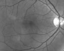
Central Serous Retinopathy - Acutevu 710 fois49-year-old man sees a spot in the central vision with a red halo for 2 weeks OD is 20/50, OS is 20/20
(0 votes)
|
|

Central Serous Retinopathy - Acutevu 785 fois49-year-old man sees a spot in the central vision with a red halo for 2 weeks OD is 20/50, OS is 20/20
(0 votes)
|
|

Central Serous Retinopathy - Acutevu 691 fois49-year-old man sees a spot in the central vision with a red halo for 2 weeks OD is 20/50, OS is 20/20
(0 votes)
|
|

Central Serous Retinopathy - Acutevu 696 fois49-year-old man sees a spot in the central vision with a red halo for 2 weeks OD is 20/50, OS is 20/20
(0 votes)
|
|

Central Serous Retinopathy - Acutevu 605 fois49-year-old man sees a spot in the central vision with a red halo for 2 weeks OD is 20/50, OS is 20/20
(0 votes)
|
|

Central Serous Retinopathy - Acutevu 754 fois49-year-old man sees a spot in the central vision with a red halo for 2 weeks OD is 20/50, OS is 20/20
(0 votes)
|
|

Central Serous Retinopathy - Acutevu 705 fois49-year-old man sees a spot in the central vision with a red halo for 2 weeks OD is 20/50, OS is 20/20
(0 votes)
|
|

Central Serous Retinopathy - Acutevu 864 fois49-year-old man sees a spot in the central vision with a red halo for 2 weeks OD is 20/50, OS is 20/20
(0 votes)
|
|

Central Serous Retinopathy - Acutevu 1364 fois49-year-old man sees a spot in the central vision with a red halo for 2 weeks OD is 20/50, OS is 20/20
(0 votes)
|
|

Central Serous Retinopathy - Acutevu 1046 fois49-year-old man sees a spot in the central vision with a red halo for 2 weeks OD is 20/50, OS is 20/20
(0 votes)
|
|
|
|
|
|
|
49-year-old man was doing fine until about two weeks ago when he noticed he was having some difficulty with his vision. He closed his left eye and noticed the right eye was not seeing well. He sees a spot in the central vision with a red halo. He is healthy and his blood pressure is fine. He does not take any steroids.
VISUAL ACUITY: Vision OD is 20/50, OS is 20/20. IOP: OD 17, OS 14.
SLIT LAMP EXAM: Normal with clear lenses.
EXTENDED OPHTHALMOSCOPY:
OD: Vertical C/D ratio is 0.1. There is no posterior vitreous separation. There is a pigment epithelial detachment on the inferotemporal margin of the fovea and there is a serous macular detachment.
OS: Vertical C/D ratio is 0.1. There is no posterior vitreous separation. The macula and periphery look healthy.
OCT SCAN: The right eye shows a pigment epithelial detachment and a serous macular detachment. The left eye had a normal scan.
FLUORESCEIN ANGIOGRAPHY: FA of the right eye shows a leaky subfoveal pigment epithelial detachment with serous macular detachment. The left eye is angiographically normal.
IMPRESSION:
1. CENTRAL SEROUS RETINOPATHY – RIGHT EYE
DISCUSSION: I explained to the patient that he does have central serous retinopathy in the right eye. I asked him to return urgently should he notice any problems, such as substantial decline in vision. I asked him to avoid steroids unless it is absolutely necessary.
Vision returned to 20/20 within 5 months and no recurrences over 2 years.


