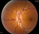
027 - March 2014 - Best Image Finalists - Winner - Bilateral Macular Star - Jason Calhoun - Photographer - Mayo Clinic Jacksonville
telangiectasiavu 904 fois20/20 pt with blurry area in va (0 votes)
AVMvu 1224 foisFP of AVM in OS of a 48 yr old female. (1 votes)
Choroidal Osteomavu 1456 foisPatient with decreased vision within the past 2-years. VA 20/100 in the right eye. Fundus photo shows atrophic scar temporal to the macula in the right eye. (1 votes)
Central Retinal Vein Occlusionvu 1355 foisPatient comes in with mild blurred vision in the right eye. Fundus exam shows CRVO with scattered retinal hemorrhages in the right eye. (1 votes)
Epiretinal Membranevu 1070 foisPatient with a history of pars planitis comes in with decreased vision in the left eye. VA is 20/30 in the left eye. Fundus photos show a rather large epiretinal membrane covering the optic nerve in the left eye. (1 votes)
Bilateral Macular Starvu 1393 foisYoung female patient comes in with blurred vision in both eyes. VA is 20/40 in both eyes. Fundus photos show visible macular star centrally in both eyes. This is a result of Bilateral Neuroretinitis due to cat scratch. (2 votes)
Diabetic Retinopathyvu 1469 foisAnnual diabetic exam. VA 20/30, right eye and 20/50, left eye. Fundus photos shows hemorrhages with cotton wool spots. Exudates are visible around the macula in the right eye. Scattered hemorrhages in the left eye. (1 votes)
Stasis Retinopathy, Central Retinal Vein Occlusionvu 1270 foisYoung male who complains of decreased vision in the left eye. VA is 20/20 in the left eye. Fundus photo shows mild central retinal vein occlusion in the left eye. (1 votes)
Treated Melanomavu 923 foisTreated melanoma in the right eye. VA is light perception in the right eye. (1 votes)