Retina Gallery ~ Full Sized Retina Images

Library of Free, Non-Copyrighted Retina Images and Videos

Choose your language:


67 y.o Female Spot in central vision in left eye started about 1 week ago since then it is not changing. Prior to that the vision was good in the left eye. Vision was the same in the two eyes. Patient had a few little floaters recently. She had ECCE 4-5 years ago and got the crystal lens (expensive lenses). She's dissappointed that the vision is acting up now in one eye. Just moved from Del Ray Beach. She denies headache, jaw claudications, fevers, malaise. No pain behind eyes and no pain on eye movement. Allergies: NKDA. Smoking Status: Never Smoker. Medical Hx: Hypercholesterolemia. Thyroid Disease. Surgical Hx: Hysterectomy. lung sx x2 benign cysts. VA OD: sc20/25 NscJ5 OS: sc20/200 PHNI Nsc20/800 IOP: TP OD: 14 OS: 13

AION_chfr_acuteOS_01.jpg
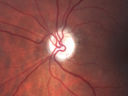
Acute AION OS - Mild disc pallor OD724 viewsSudden vision loss OS x 1 week with optic nerve swelling. Evidence of optic atrophy in right eye. 00000
(0 votes)
AION_chfr_acuteOS_02.jpg
Acute AION OS - Mild disc pallor OD666 viewsSudden vision loss OS x 1 week with optic nerve swelling. Evidence of optic atrophy in right eye. 00000
(0 votes)
AION_chfr_acuteOS_03.jpg
Acute AION OS - Mild disc pallor OD578 viewsSudden vision loss OS x 1 week with optic nerve swelling. Evidence of optic atrophy in right eye. 00000
(0 votes)
AION_chfr_acuteOS_04.jpg
Acute AION OS - Mild disc pallor OD666 viewsSudden vision loss OS x 1 week with optic nerve swelling. Evidence of optic atrophy in right eye. 00000
(0 votes)
AION_chfr_acuteOS_05.JPG
Acute AION OS - Mild disc pallor OD558 viewsSudden vision loss OS x 1 week with optic nerve swelling. Evidence of optic atrophy in right eye. 00000
(0 votes)
AION_chfr_acuteOS_06.jpg
Acute AION OS - Mild disc pallor OD549 viewsSudden vision loss OS x 1 week with optic nerve swelling. Evidence of optic atrophy in right eye. 00000
(0 votes)
AION_chfr_acuteOS_07.jpg
Acute AION OS - Mild disc pallor OD521 viewsSudden vision loss OS x 1 week with optic nerve swelling. Evidence of optic atrophy in right eye. 00000
(0 votes)
AION_chfr_acuteOS_08.jpg
Acute AION OS - Mild disc pallor OD586 viewsSudden vision loss OS x 1 week with optic nerve swelling. Evidence of optic atrophy in right eye. 00000
(0 votes)
AION_chfr_acuteOS_09.jpg
Acute AION OS - Mild disc pallor OD542 viewsSudden vision loss OS x 1 week with optic nerve swelling. Evidence of optic atrophy in right eye. 00000
(0 votes)
AION_chfr_acuteOS_10.jpg
Acute AION OS - Mild disc pallor OD486 viewsSudden vision loss OS x 1 week with optic nerve swelling. Evidence of optic atrophy in right eye. 00000
(0 votes)
AION_chfr_acuteOS_11.jpg
Acute AION OS - Mild disc pallor OD507 viewsSudden vision loss OS x 1 week with optic nerve swelling. Evidence of optic atrophy in right eye. 00000
(0 votes)
AION_chfr_acuteOS_12.jpg
Acute AION OS - Mild disc pallor OD595 viewsSudden vision loss OS x 1 week with optic nerve swelling. Evidence of optic atrophy in right eye. 00000
(0 votes)
AION_chfr_acuteOS_13.jpg
Acute AION OS - Mild disc pallor OD517 viewsSudden vision loss OS x 1 week with optic nerve swelling. Evidence of optic atrophy in right eye. 00000
(0 votes)
AION_chfr_acuteOS_14.jpg
Acute AION OS - Mild disc pallor OD555 viewsSudden vision loss OS x 1 week with optic nerve swelling. Evidence of optic atrophy in right eye. 00000
(0 votes)
AION_chfr_acuteOS_15.jpg
Acute AION OS - Mild disc pallor OD511 viewsSudden vision loss OS x 1 week with optic nerve swelling. Evidence of optic atrophy in right eye. 00000
(0 votes)
AION_chfr_acuteOS_16.jpg
Acute AION OS - Mild disc pallor OD519 viewsSudden vision loss OS x 1 week with optic nerve swelling. Evidence of optic atrophy in right eye. 00000
(0 votes)
AION_chfr_acuteOS_17.jpg
Acute AION OS - Mild disc pallor OD532 viewsSudden vision loss OS x 1 week with optic nerve swelling. Evidence of optic atrophy in right eye. 00000
(0 votes)
AION_chfr_acuteOS_18.jpg
Acute AION OS - Mild disc pallor OD599 viewsSudden vision loss OS x 1 week with optic nerve swelling. Evidence of optic atrophy in right eye. 00000
(0 votes)
AION_chfr_acuteOS_19.JPG
Acute AION OS - Mild disc pallor OD - macular scan shows increasing atrophy in left eye573 views00000
(0 votes)
AION_chfr_acuteOS_20.JPG
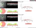
AION - Acute with Follow UP RNFL scan shows edema then atrophy565 views00000
(0 votes)
AION_chfr_acuteOS_21.JPG
Acute AION OS - Follow-up RNFL scan589 views00000
(0 votes)
     
21 files on 1 page(s)

67 y.o Female Spot in central vision in left eye started about 1 week ago since then it is not changing. Prior to that the vision was good in the left eye. Vision was the same in the two eyes. Patient had a few little floaters recently. She had ECCE 4-5 years ago and got the crystal lens (expensive lenses). She's dissappointed that the vision is acting up now in one eye. Just moved from Del Ray Beach. She denies headache, jaw claudications, fevers, malaise. No pain behind eyes and no pain on eye movement. Allergies: NKDA. Smoking Status: Never Smoker. Medical Hx: Hypercholesterolemia. Thyroid Disease. Surgical Hx: Hysterectomy. lung sx x2 benign cysts. VA OD: sc20/25 NscJ5 OS: sc20/200 PHNI Nsc20/800 IOP: TP OD: 14 OS: 13