Retinitis Pigmentosa - Autosomal Recessive
|
|
82 year old man with a sister with RP and no other family members. He has trouble with night vision. VA 20/25 OD, 20/32 OS. Note the symmetry in the FAF images typical for a dystrophy. The photos show bone spicules, arteriol attenuation and optic nerve pallor
|

Retinitis Pigmentosa - Autosomal Recessive - 82 year old man with Good Visionvu 534 foisVA 20/25, 20/32 - Bone spicules in periphery - FAF is very symmetric
(0 votes)
|
|

Retinitis Pigmentosa - Autosomal Recessive - 82 year old man with Good Visionvu 477 foisVA 20/25, 20/32 - Bone spicules in periphery - FAF is very symmetric
(0 votes)
|
|
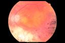
Retinitis Pigmentosa - Autosomal Recessive - 82 year old man with Good Visionvu 539 foisVA 20/25, 20/32 - Bone spicules in periphery - FAF is very symmetric
(0 votes)
|
|
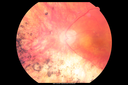
Retinitis Pigmentosa - Autosomal Recessive - 82 year old man with Good Visionvu 530 foisVA 20/25, 20/32 - Bone spicules in periphery - FAF is very symmetric
(0 votes)
|
|

Retinitis Pigmentosa - Autosomal Recessive - 82 year old man with Good Visionvu 551 foisVA 20/25, 20/32 - Bone spicules in periphery - FAF is very symmetric
(0 votes)
|
|

Retinitis Pigmentosa - Autosomal Recessive - 82 year old man with Good Visionvu 525 foisVA 20/25, 20/32 - Bone spicules in periphery - FAF is very symmetric
(0 votes)
|
|

Retinitis Pigmentosa - Autosomal Recessive - 82 year old man with Good Visionvu 470 foisVA 20/25, 20/32 - Bone spicules in periphery - FAF is very symmetric
(0 votes)
|
|

Retinitis Pigmentosa - Autosomal Recessive - 82 year old man with Good Visionvu 461 foisVA 20/25, 20/32 - Bone spicules in periphery - FAF is very symmetric
(0 votes)
|
|

Retinitis Pigmentosa - Autosomal Recessive - 82 year old man with Good Visionvu 475 foisVA 20/25, 20/32 - Bone spicules in periphery - FAF is very symmetric
(0 votes)
|
|
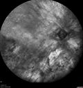
Retinitis Pigmentosa - Autosomal Recessive - 82 year old man with Good Visionvu 519 foisVA 20/25, 20/32 - Bone spicules in periphery - FAF is very symmetric
(0 votes)
|
|

Retinitis Pigmentosa - Autosomal Recessive - 82 year old man with Good Visionvu 456 foisVA 20/25, 20/32 - Bone spicules in periphery - FAF is very symmetric
(0 votes)
|
|

Retinitis Pigmentosa - Autosomal Recessive - 82 year old man with Good Visionvu 482 foisVA 20/25, 20/32 - Bone spicules in periphery - FAF is very symmetric
(0 votes)
|
|

Retinitis Pigmentosa - Autosomal Recessive - 82 year old man with Good Visionvu 452 foisVA 20/25, 20/32 - Bone spicules in periphery - FAF is very symmetric
(0 votes)
|
|

Retinitis Pigmentosa - Autosomal Recessive - 82 year old man with Good Visionvu 485 foisVA 20/25, 20/32 - Bone spicules in periphery - FAF is very symmetric
(0 votes)
|
|

Retinitis Pigmentosa - Autosomal Recessive - 82 year old man with Good Visionvu 480 foisVA 20/25, 20/32 - Bone spicules in periphery - FAF is very symmetric
(0 votes)
|
|
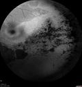
Retinitis Pigmentosa - Autosomal Recessive - 82 year old man with Good Visionvu 467 foisVA 20/25, 20/32 - Bone spicules in periphery - FAF is very symmetric
(0 votes)
|
|

Retinitis Pigmentosa - Autosomal Recessive - 82 year old man with Good Visionvu 537 foisVA 20/25, 20/32 - Bone spicules in periphery - FAF is very symmetric
(0 votes)
|
|
|
|
|
|
|
82 year old man with a sister with RP and no other family members. He has trouble with night vision. VA 20/25 OD, 20/32 OS. Note the symmetry in the FAF images typical for a dystrophy. The photos show bone spicules, arteriol attenuation and optic nerve pallor


