Last additions - Bilateral Branch Retinal Artery Occlusion with Hollenhorst Plaques 67 year old female smoker. Both eyes are developing shadows in the vision. The vision was normal starting recently. May 25th she noticed in the left eye a shadow that developed in the vision that she could not see through that did not move. The shadow is nasal in the bottom half of the vision. Mushroom shaped. This is not getting any better. Then yesterday she got a new shadow in the right eye this time. This is the same type of problem. The shadow in the right eye is more in the bottom half of the vision. The right eye started out worse and has diminshed a little. The right eye shadow is bigger than the shadow in the left eye. Vision 20/25 OD 20/32 OS. Photos show acute bilateral branch retinal artery occlusions.
|

Bilateral Branch Retinal Artery Occlusion - Intraarterial (hollenhorst) plaques visible507 views67 year old with acute visual field loss in each eye. Vision is 20/25 right eye 20/32 left eye. Images show intra-arterial plaques in each eye. The right eye on the optic nerve and the left eye in a branch retinal artery.Feb 06, 2017
|
|

Bilateral Branch Retinal Artery Occlusion - Intraarterial (hollenhorst) plaques visible535 views67 year old with acute visual field loss in each eye. Vision is 20/25 right eye 20/32 left eye. Images show intra-arterial plaques in each eye. The right eye on the optic nerve and the left eye in a branch retinal artery.Feb 06, 2017
|
|

Bilateral Branch Retinal Artery Occlusion - Intraarterial (hollenhorst) plaques visible467 views67 year old with acute visual field loss in each eye. Vision is 20/25 right eye 20/32 left eye. Images show intra-arterial plaques in each eye. The right eye on the optic nerve and the left eye in a branch retinal artery.Feb 06, 2017
|
|

Bilateral Branch Retinal Artery Occlusion - Intraarterial (hollenhorst) plaques visible597 views67 year old with acute visual field loss in each eye. Vision is 20/25 right eye 20/32 left eye. Images show intra-arterial plaques in each eye. The right eye on the optic nerve and the left eye in a branch retinal artery.Feb 06, 2017
|
|

Bilateral Branch Retinal Artery Occlusion - Intraarterial (hollenhorst) plaques visible510 views67 year old with acute visual field loss in each eye. Vision is 20/25 right eye 20/32 left eye. Images show intra-arterial plaques in each eye. The right eye on the optic nerve and the left eye in a branch retinal artery.Feb 06, 2017
|
|

Bilateral Branch Retinal Artery Occlusion - Intraarterial (hollenhorst) plaques visible481 views67 year old with acute visual field loss in each eye. Vision is 20/25 right eye 20/32 left eye. Images show intra-arterial plaques in each eye. The right eye on the optic nerve and the left eye in a branch retinal artery.Feb 06, 2017
|
|

Bilateral Branch Retinal Artery Occlusion - Intraarterial (hollenhorst) plaques visible540 views67 year old with acute visual field loss in each eye. Vision is 20/25 right eye 20/32 left eye. Images show intra-arterial plaques in each eye. The right eye on the optic nerve and the left eye in a branch retinal artery.Feb 06, 2017
|
|

Bilateral Branch Retinal Artery Occlusion - Intraarterial (hollenhorst) plaques visible449 views67 year old with acute visual field loss in each eye. Vision is 20/25 right eye 20/32 left eye. Images show intra-arterial plaques in each eye. The right eye on the optic nerve and the left eye in a branch retinal artery.Feb 06, 2017
|
|

Bilateral Branch Retinal Artery Occlusion - Intraarterial (hollenhorst) plaques visible523 views67 year old with acute visual field loss in each eye. Vision is 20/25 right eye 20/32 left eye. Images show intra-arterial plaques in each eye. The right eye on the optic nerve and the left eye in a branch retinal artery.Feb 06, 2017
|
|

Bilateral Branch Retinal Artery Occlusion - Intraarterial (hollenhorst) plaques visible466 views67 year old with acute visual field loss in each eye. Vision is 20/25 right eye 20/32 left eye. Images show intra-arterial plaques in each eye. The right eye on the optic nerve and the left eye in a branch retinal artery.Feb 06, 2017
|
|

Bilateral Branch Retinal Artery Occlusion - Intraarterial (hollenhorst) plaques visible518 views67 year old with acute visual field loss in each eye. Vision is 20/25 right eye 20/32 left eye. Images show intra-arterial plaques in each eye. The right eye on the optic nerve and the left eye in a branch retinal artery.Feb 06, 2017
|
|

Bilateral Branch Retinal Artery Occlusion - Intraarterial (hollenhorst) plaques visible548 views67 year old with acute visual field loss in each eye. Vision is 20/25 right eye 20/32 left eye. Images show intra-arterial plaques in each eye. The right eye on the optic nerve and the left eye in a branch retinal artery.Feb 06, 2017
|
|

Bilateral Branch Retinal Artery Occlusion - Intraarterial (hollenhorst) plaques visible520 views67 year old with acute visual field loss in each eye. Vision is 20/25 right eye 20/32 left eye. Images show intra-arterial plaques in each eye. The right eye on the optic nerve and the left eye in a branch retinal artery.Feb 06, 2017
|
|

Bilateral Branch Retinal Artery Occlusion - Intraarterial (hollenhorst) plaques visible496 views67 year old with acute visual field loss in each eye. Vision is 20/25 right eye 20/32 left eye. Images show intra-arterial plaques in each eye. The right eye on the optic nerve and the left eye in a branch retinal artery.Feb 06, 2017
|
|

Bilateral Branch Retinal Artery Occlusion - Intraarterial (hollenhorst) plaques visible512 views67 year old with acute visual field loss in each eye. Vision is 20/25 right eye 20/32 left eye. Images show intra-arterial plaques in each eye. The right eye on the optic nerve and the left eye in a branch retinal artery.Feb 06, 2017
|
|

Bilateral Branch Retinal Artery Occlusion - Intraarterial (hollenhorst) plaques visible548 views67 year old with acute visual field loss in each eye. Vision is 20/25 right eye 20/32 left eye. Images show intra-arterial plaques in each eye. The right eye on the optic nerve and the left eye in a branch retinal artery.Feb 06, 2017
|
|

Bilateral Branch Retinal Artery Occlusion - Intraarterial (hollenhorst) plaques visible498 views67 year old with acute visual field loss in each eye. Vision is 20/25 right eye 20/32 left eye. Images show intra-arterial plaques in each eye. The right eye on the optic nerve and the left eye in a branch retinal artery.Feb 06, 2017
|
|
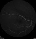
Bilateral Branch Retinal Artery Occlusion - Intraarterial (hollenhorst) plaques visible514 views67 year old with acute visual field loss in each eye. Vision is 20/25 right eye 20/32 left eye. Images show intra-arterial plaques in each eye. The right eye on the optic nerve and the left eye in a branch retinal artery.Feb 06, 2017
|
|

Bilateral Branch Retinal Artery Occlusion - Intraarterial (hollenhorst) plaques visible505 views67 year old with acute visual field loss in each eye. Vision is 20/25 right eye 20/32 left eye. Images show intra-arterial plaques in each eye. The right eye on the optic nerve and the left eye in a branch retinal artery.Feb 06, 2017
|
|

Bilateral Branch Retinal Artery Occlusion - Intraarterial (hollenhorst) plaques visible538 views67 year old with acute visual field loss in each eye. Vision is 20/25 right eye 20/32 left eye. Images show intra-arterial plaques in each eye. The right eye on the optic nerve and the left eye in a branch retinal artery.Feb 06, 2017
|
|

Bilateral Branch Retinal Artery Occlusion - Intraarterial (hollenhorst) plaques visible492 views67 year old with acute visual field loss in each eye. Vision is 20/25 right eye 20/32 left eye. Images show intra-arterial plaques in each eye. The right eye on the optic nerve and the left eye in a branch retinal artery.Feb 06, 2017
|
|

Bilateral Branch Retinal Artery Occlusion - Intraarterial (hollenhorst) plaques visible482 views67 year old with acute visual field loss in each eye. Vision is 20/25 right eye 20/32 left eye. Images show intra-arterial plaques in each eye. The right eye on the optic nerve and the left eye in a branch retinal artery.Feb 06, 2017
|
|

Bilateral Branch Retinal Artery Occlusion - Intraarterial (hollenhorst) plaques visible514 views67 year old with acute visual field loss in each eye. Vision is 20/25 right eye 20/32 left eye. Images show intra-arterial plaques in each eye. The right eye on the optic nerve and the left eye in a branch retinal artery.Feb 06, 2017
|
|

Bilateral Branch Retinal Artery Occlusion - Intraarterial (hollenhorst) plaques visible501 views67 year old with acute visual field loss in each eye. Vision is 20/25 right eye 20/32 left eye. Images show intra-arterial plaques in each eye. The right eye on the optic nerve and the left eye in a branch retinal artery.Feb 06, 2017
|
|

Bilateral Branch Retinal Artery Occlusion - Intraarterial (hollenhorst) plaques visible496 views67 year old with acute visual field loss in each eye. Vision is 20/25 right eye 20/32 left eye. Images show intra-arterial plaques in each eye. The right eye on the optic nerve and the left eye in a branch retinal artery.Feb 06, 2017
|
|

Bilateral Branch Retinal Artery Occlusion - Intraarterial (hollenhorst) plaques visible538 views67 year old with acute visual field loss in each eye. Vision is 20/25 right eye 20/32 left eye. Images show intra-arterial plaques in each eye. The right eye on the optic nerve and the left eye in a branch retinal artery.Feb 06, 2017
|
|

Bilateral Branch Retinal Artery Occlusion - Intraarterial (hollenhorst) plaques visible570 views67 year old with acute visual field loss in each eye. Vision is 20/25 right eye 20/32 left eye. Images show intra-arterial plaques in each eye. The right eye on the optic nerve and the left eye in a branch retinal artery.Feb 06, 2017
|
|

Bilateral Branch Retinal Artery Occlusion - Intraarterial (hollenhorst) plaques visible602 views67 year old with acute visual field loss in each eye. Vision is 20/25 right eye 20/32 left eye. Images show intra-arterial plaques in each eye. The right eye on the optic nerve and the left eye in a branch retinal artery.Feb 06, 2017
|
|

Bilateral Branch Retinal Artery Occlusion - Intraarterial (hollenhorst) plaques visible660 views67 year old with acute visual field loss in each eye. Vision is 20/25 right eye 20/32 left eye. Images show intra-arterial plaques in each eye. The right eye on the optic nerve and the left eye in a branch retinal artery.Feb 06, 2017
|
|
|
|
|
|
|
67 year old female smoker. Both eyes are developing shadows in the vision. The vision was normal starting recently. May 25th she noticed in the left eye a shadow that developed in the vision that she could not see through that did not move. The shadow is nasal in the bottom half of the vision. Mushroom shaped. This is not getting any better. Then yesterday she got a new shadow in the right eye this time. This is the same type of problem. The shadow in the right eye is more in the bottom half of the vision. The right eye started out worse and has diminshed a little. The right eye shadow is bigger than the shadow in the left eye. Vision 20/25 OD 20/32 OS. Photos show acute bilateral branch retinal artery occlusions.


