Last additions - Sturge-Weber Encephalotrigeminal Angiomatosis 61-year-old man was seen in the office on 7/2/2010. He has noticed new increased floaters in the right eye: first about three days ago he had some spots, then two days ago a lot of spots, and then just yesterday an explosion of floaters in the eye. You saw a hemorrhage in the eye and suggested he come here for an evaluation. He does have Sturge-Weber syndrome with a hemangioma on the left side of his face.
VISUAL ACUITY: Vision OD is 20/50, PH 20/30; OS 20/80, PH 20/30. IOP: OD 16, OS 19.
SLIT LAMP EXAM: There is 2+ nuclear sclerosis in both eyes.
EXTENDED OPHTHALMOSCOPY:
OD: Vertical C/D ratio is 0.4. There is 2+ vitreous hemorrhage. There is a posterior vitreous separation. There is a retinal flap tear just anterior to the equator at 9:30.
OS: Vertical C/D ratio is 0.4. There is a posterior vitreous separation. It looks to me like there is a reddish dust to the choroid just on the inferior third of the retina just below the macula and the optic nerve.
IMPRESSION:
1. ACUTE POSTERIOR VITREOUS SEPARATION – RIGHT EYE
2. VITREOUS HEMORRHAGE – RIGHT EYE
3. RETINAL TEAR – RIGHT EYE
4. POSSIBLE CHOROIDAL HEMANGIOMA – LEFT EYE
DISCUSSION: I explained to the patient that there is a vitreous separation in the right eye with vitreous hemorrhage and with a retinal tear. With the retinal tear untreated, there is about a 70-80% chance of having a retinal detachment. With treating, we can reduce the risk of retinal detachment from that tear down to 1%.
I treated him with cryotherapy today. Because he has a vitreous hemorrhage, it is a little difficult to see and also because the defect is in the far periphery.
|

Sturge-Weber with Choroidal Hemangioma Left Eye718 viewsVA 20/25 OU. Left eye treated for glaucoma with drops.Jan 29, 2018
|
|

Hemangioma OS with good vison - 01-29-18563 viewsImages 7 years following initial uploaded imagesJan 29, 2018
|
|

Hemangioma OS with good vison - 01-29-18528 viewsImages 7 years following initial uploaded imagesJan 29, 2018
|
|

Hemangioma OS with good vison - 01-29-18546 viewsImages 7 years following initial uploaded imagesJan 29, 2018
|
|

Hemangioma OS with good vison - 01-29-18576 viewsImages 7 years following initial uploaded imagesJan 29, 2018
|
|
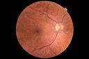
Hemangioma OS with good vison - 01-29-18576 viewsImages 7 years following initial uploaded imagesJan 29, 2018
|
|

Hemangioma OS with good vison - 01-29-18556 viewsImages 7 years following initial uploaded imagesJan 29, 2018
|
|

Hemangioma OS with good vison - 01-29-18506 viewsImages 7 years following initial uploaded imagesJan 29, 2018
|
|
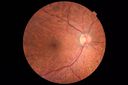
Hemangioma OS with good vison - 01-29-18613 viewsImages 7 years following initial uploaded imagesJan 29, 2018
|
|

Sturge Weber SD OCT EDI showing choroidal thickening687 viewsMay 16, 2016
|
|

Choroidal Hemangioma - Diffuse - Enhanced Depth Imaging Spectral Domain OCT Line Scan - Abnormal Eye931 viewsChoroidal Thickness is 697 Microns from Diffuse Choroidal HemangiomaFeb 12, 2014
|
|

Choroidal Hemangioma - Diffuse - Enhanced Depth Imaging Spectral Domain OCT Line Scan - Abnormal Eye673 viewsChoroidal Thickness is 697 Microns from Diffuse Choroidal HemangiomaFeb 12, 2014
|
|

Choroidal Hemangioma - Diffuse - Enhanced Depth Imaging Spectral Domain OCT Line Scan - Normal Eye817 viewsNormal Choroidal Thickness in Right EyeFeb 12, 2014
|
|

Choroidal Hemangioma - Diffuse - Enhanced Depth Imaging Spectral Domain OCT Line Scan - Normal Eye674 viewsNormal Choroidal Thickness in Right EyeFeb 12, 2014
|
|
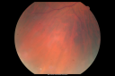
Sturge-Weber Encephalotigeminal Angiomatosis - Facial Hemangioma and Asymptomatic Ipsilateral Diffuse Choroidal Hemangioma852 views61-year-old man with Sturge-Weber syndrome with a hemangioma on the left side of his face.
VISUAL ACUITY: Vision OD is 20/50, PH 20/30; OS 20/80, PH 20/30. IOP: OD 16, OS 19.
Jun 28, 2011
|
|
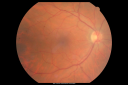
Sturge-Weber Encephalotigeminal Angiomatosis - Facial Hemangioma and Asymptomatic Ipsilateral Diffuse Choroidal Hemangioma935 views61-year-old man with Sturge-Weber syndrome with a hemangioma on the left side of his face.
VISUAL ACUITY: Vision OD is 20/50, PH 20/30; OS 20/80, PH 20/30. IOP: OD 16, OS 19.
Jun 28, 2011
|
|

Sturge-Weber Encephalotigeminal Angiomatosis - Facial Hemangioma and Asymptomatic Ipsilateral Diffuse Choroidal Hemangioma943 views61-year-old man with Sturge-Weber syndrome with a hemangioma on the left side of his face.
VISUAL ACUITY: Vision OD is 20/50, PH 20/30; OS 20/80, PH 20/30. IOP: OD 16, OS 19.
Jun 28, 2011
|
|

Sturge-Weber Encephalotigeminal Angiomatosis - Facial Hemangioma and Asymptomatic Ipsilateral Diffuse Choroidal Hemangioma857 views61-year-old man with Sturge-Weber syndrome with a hemangioma on the left side of his face.
VISUAL ACUITY: Vision OD is 20/50, PH 20/30; OS 20/80, PH 20/30. IOP: OD 16, OS 19.
Jun 28, 2011
|
|
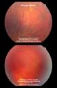
Sturge-Weber Encephalotigeminal Angiomatosis - Facial Hemangioma and Asymptomatic Ipsilateral Diffuse Choroidal Hemangioma1526 views61-year-old man with Sturge-Weber syndrome with a hemangioma on the left side of his face.
VISUAL ACUITY: Vision OD is 20/50, PH 20/30; OS 20/80, PH 20/30. IOP: OD 16, OS 19.
Jun 28, 2011
|
|

Sturge-Weber Encephalotigeminal Angiomatosis - Facial Hemangioma and Asymptomatic Ipsilateral Diffuse Choroidal Hemangioma1293 views61-year-old man with Sturge-Weber syndrome with a hemangioma on the left side of his face.
VISUAL ACUITY: Vision OD is 20/50, PH 20/30; OS 20/80, PH 20/30. IOP: OD 16, OS 19.
Jun 28, 2011
|
|

Sturge-Weber Encephalotigeminal Angiomatosis - Facial Hemangioma and Asymptomatic Ipsilateral Diffuse Choroidal Hemangioma2017 views61-year-old man with Sturge-Weber syndrome with a hemangioma on the left side of his face.
VISUAL ACUITY: Vision OD is 20/50, PH 20/30; OS 20/80, PH 20/30. IOP: OD 16, OS 19.
Jun 28, 2011
|
|

Sturge-Weber Encephalotigeminal Angiomatosis - Facial Hemangioma and Asymptomatic Ipsilateral Diffuse Choroidal Hemangioma1227 views61-year-old man with Sturge-Weber syndrome with a hemangioma on the left side of his face.
VISUAL ACUITY: Vision OD is 20/50, PH 20/30; OS 20/80, PH 20/30. IOP: OD 16, OS 19.
Jun 28, 2011
|
|

Sturge-Weber Encephalotigeminal Angiomatosis - Facial Hemangioma and Asymptomatic Ipsilateral Diffuse Choroidal Hemangioma994 views61-year-old man with Sturge-Weber syndrome with a hemangioma on the left side of his face.
VISUAL ACUITY: Vision OD is 20/50, PH 20/30; OS 20/80, PH 20/30. IOP: OD 16, OS 19.
Jun 28, 2011
|
|

Sturge-Weber Encephalotigeminal Angiomatosis - Facial Hemangioma and Asymptomatic Ipsilateral Diffuse Choroidal Hemangioma979 views61-year-old man with Sturge-Weber syndrome with a hemangioma on the left side of his face.
VISUAL ACUITY: Vision OD is 20/50, PH 20/30; OS 20/80, PH 20/30. IOP: OD 16, OS 19.
Jun 28, 2011
|
|

Sturge-Weber Encephalotigeminal Angiomatosis - Facial Hemangioma and Ipsilateral Diffuse Choroidal Hemangioma1020 views61-year-old man with Sturge-Weber syndrome with a hemangioma on the left side of his face.
VISUAL ACUITY: Vision OD is 20/50, PH 20/30; OS 20/80, PH 20/30. IOP: OD 16, OS 19.
Jun 25, 2011
|
|

Sturge-Weber Encephalotigeminal Angiomatosis - Facial Hemangioma and Ipsilateral Diffuse Choroidal Hemangioma1404 views61-year-old man with Sturge-Weber syndrome with a hemangioma on the left side of his face.
VISUAL ACUITY: Vision OD is 20/50, PH 20/30; OS 20/80, PH 20/30. IOP: OD 16, OS 19.
Jun 25, 2011
|
|
|
|
|
|
61-year-old man was seen in the office on 7/2/2010. He has noticed new increased floaters in the right eye: first about three days ago he had some spots, then two days ago a lot of spots, and then just yesterday an explosion of floaters in the eye. You saw a hemorrhage in the eye and suggested he come here for an evaluation. He does have Sturge-Weber syndrome with a hemangioma on the left side of his face.
VISUAL ACUITY: Vision OD is 20/50, PH 20/30; OS 20/80, PH 20/30. IOP: OD 16, OS 19.
SLIT LAMP EXAM: There is 2+ nuclear sclerosis in both eyes.
EXTENDED OPHTHALMOSCOPY:
OD: Vertical C/D ratio is 0.4. There is 2+ vitreous hemorrhage. There is a posterior vitreous separation. There is a retinal flap tear just anterior to the equator at 9:30.
OS: Vertical C/D ratio is 0.4. There is a posterior vitreous separation. It looks to me like there is a reddish dust to the choroid just on the inferior third of the retina just below the macula and the optic nerve.
IMPRESSION:
1. ACUTE POSTERIOR VITREOUS SEPARATION – RIGHT EYE
2. VITREOUS HEMORRHAGE – RIGHT EYE
3. RETINAL TEAR – RIGHT EYE
4. POSSIBLE CHOROIDAL HEMANGIOMA – LEFT EYE
DISCUSSION: I explained to the patient that there is a vitreous separation in the right eye with vitreous hemorrhage and with a retinal tear. With the retinal tear untreated, there is about a 70-80% chance of having a retinal detachment. With treating, we can reduce the risk of retinal detachment from that tear down to 1%.
I treated him with cryotherapy today. Because he has a vitreous hemorrhage, it is a little difficult to see and also because the defect is in the far periphery.


