Last additions - Post Lasik Epithelial Ingrowth - Visual Axis 58-year-old man had a vitrectomy for a macular pucker in the right eye almost two years ago. His vision was good after you removed the cells from his flap but then recently the cells have regrown and his vision has declined again.
VISUAL ACUITY: Vision OD is 20/100. IOP: OD is 18. There are cells within the visual axis in the cornea. The posterior chamber intraocular lens is in good position with an open capsule.
EXTENDED OPHTHALMOSCOPY:
OD: Vertical C/D ratio is 0.5. The macula looks smooth.
OCT SCAN: The OCT scan shows an average central foveal thickness of 321 microns.
IMPRESSION:
1. STATUS POST VITRECTOMY FOR MACULAR PUCKER – RIGHT EYE
2. Post Lasik Epithelial Ingrowth
DISCUSSION: I explained to the patient that the retina looks lovely but I think his vision is probably limited by his cornea.
|
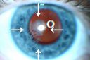
Post Lasik Epithelial Ingrowth773 views58-year-old man had a vitrectomy for a macular pucker in the right eye almost two years ago. His vision was good after his cornea surgeon removed the cells from his flap but then recently the cells have regrown and his vision has declined again.
OD is 20/100.Dec 13, 2011
|
|
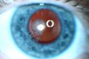
Post Lasik Epithelial Ingrowth745 views58-year-old man had a vitrectomy for a macular pucker in the right eye almost two years ago. His vision was good after his cornea surgeon removed the cells from his flap but then recently the cells have regrown and his vision has declined again.
OD is 20/100.Dec 13, 2011
|
|
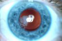
Post Lasik Epithelial Ingrowth952 views58-year-old man had a vitrectomy for a macular pucker in the right eye almost two years ago. His vision was good after his cornea surgeon removed the cells from his flap but then recently the cells have regrown and his vision has declined again.
OD is 20/100.Dec 13, 2011
|
|
|
|
|
58-year-old man had a vitrectomy for a macular pucker in the right eye almost two years ago. His vision was good after you removed the cells from his flap but then recently the cells have regrown and his vision has declined again.
VISUAL ACUITY: Vision OD is 20/100. IOP: OD is 18. There are cells within the visual axis in the cornea. The posterior chamber intraocular lens is in good position with an open capsule.
EXTENDED OPHTHALMOSCOPY:
OD: Vertical C/D ratio is 0.5. The macula looks smooth.
OCT SCAN: The OCT scan shows an average central foveal thickness of 321 microns.
IMPRESSION:
1. STATUS POST VITRECTOMY FOR MACULAR PUCKER – RIGHT EYE
2. Post Lasik Epithelial Ingrowth
DISCUSSION: I explained to the patient that the retina looks lovely but I think his vision is probably limited by his cornea.


