Last additions - Retinal Detachment
|
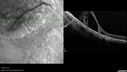
Retinoschisis - Superotemporally402 viewsSome line scans show RD and some showed retinoschisis. Might have underlying RD that re-attached itself.Oct 02, 2017
|
|
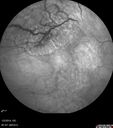
Retinoschisis - Superotemporally392 viewsSome line scans show RD and some showed retinoschisis. Might have underlying RD that re-attached itself.Oct 02, 2017
|
|
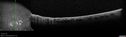
Retinoschisis - Superotemporally383 viewsSome line scans show RD and some showed retinoschisis. Might have underlying RD that re-attached itself.Oct 02, 2017
|
|

Retinoschisis - Superotemporally386 viewsSome line scans show RD and some showed retinoschisis. Might have underlying RD that re-attached itself.Oct 02, 2017
|
|
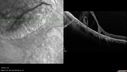
Retinoschisis - Superotemporally467 viewsSome line scans show RD and some showed retinoschisis. Might have underlying RD that re-attached itself.Oct 02, 2017
|
|
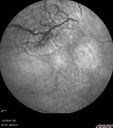
Retinoschisis - Superotemporally443 viewsSome line scans show RD and some showed retinoschisis. Might have underlying RD that re-attached itself.Oct 02, 2017
|
|

Operculated tear with fresh blood554 viewsAug 15, 2017
|
|
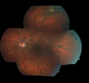
Wagner Syndrome 704 views33 year old man with family history of Wagner syndrome - the left eye had RD repair. The right eye has posterior peripheral pigmented lesions captured here superiorlyJun 30, 2017
|
|

Wagner Syndrome 579 views33 year old man with family history of Wagner syndrome - the left eye had RD repair. The right eye has posterior peripheral pigmented lesions captured here superiorlyJun 30, 2017
|
|
414 viewsMay 02, 2016
|
|

410 viewsMay 02, 2016
|
|

395 viewsMay 02, 2016
|
|

437 viewsMay 02, 2016
|
|

485 viewsMay 02, 2016
|
|

448 viewsMay 02, 2016
|
|

388 viewsMay 02, 2016
|
|

399 viewsMay 02, 2016
|
|

528 viewsMay 02, 2016
|
|

Giant Retinal Tear Retinal Detachment509 views13 year old african american girl high myope. These intra-operative photos show parts of the procedure for this giant tear detachment. The video is on youtube. You can see the center of the macula with no overlying retina.May 02, 2016
|
|

Giant Retinal Tear Retinal Detachment515 views13 year old african american girl high myope. These intra-operative photos show parts of the procedure for this giant tear detachment. The video is on youtube. You can see the center of the macula with no overlying retina.May 02, 2016
|
|

Giant Retinal Tear Retinal Detachment514 views13 year old african american girl high myope. These intra-operative photos show parts of the procedure for this giant tear detachment. The video is on youtube. You can see the center of the macula with no overlying retina.May 02, 2016
|
|

Giant Retinal Tear Retinal Detachment529 views13 year old african american girl high myope. These intra-operative photos show parts of the procedure for this giant tear detachment. The video is on youtube. You can see the center of the macula with no overlying retina.May 02, 2016
|
|

Giant Retinal Tear Retinal Detachment478 views13 year old african american girl high myope. These intra-operative photos show parts of the procedure for this giant tear detachment. The video is on youtube. You can see the center of the macula with no overlying retina.May 02, 2016
|
|

Giant Retinal Tear Retinal Detachment476 views13 year old african american girl high myope. These intra-operative photos show parts of the procedure for this giant tear detachment. The video is on youtube. You can see the center of the macula with no overlying retina.May 02, 2016
|
|

Giant Retinal Tear Retinal Detachment564 views13 year old african american girl high myope. These intra-operative photos show parts of the procedure for this giant tear detachment. The video is on youtube. You can see the center of the macula with no overlying retina.May 02, 2016
|
|

Giant Retinal Tear Retinal Detachment504 views13 year old african american girl high myope. These intra-operative photos show parts of the procedure for this giant tear detachment. The video is on youtube. You can see the center of the macula with no overlying retina.May 02, 2016
|
|

Retinal Tear with Laser Treatment464 viewsPatient comes in with blurry vision and mild floaters in the left eye. Fundus photo shows retinal tear with fluid present in the superior nasal quadrant of the left eye. Laser Retinopexy was performed to fix the retinal tear. Sep 08, 2014
|
|

Retinal Tear with Laser Treatment371 viewsPatient comes in with blurry vision and mild floaters in the left eye. Fundus photo shows retinal tear with fluid present in the superior nasal quadrant of the left eye. Laser Retinopexy was performed to fix the retinal tear. Sep 08, 2014
|
|

Retinal Tear with Laser Treatment1255 viewsPatient comes in with blurry vision and mild floaters in the left eye. Fundus photo shows retinal tear with fluid present in the superior nasal quadrant of the left eye. Laser Retinopexy was performed to fix the retinal tear. Sep 08, 2014
|
|

Old Retinal Detachment Repair with Silicone Oil1032 viewsPatient comes in for eval on glaucoma. Patients VA in the right eye was CF in the right eye and 20/50. left eye. Fundus photography shows old retinal detachment with scattered laser scars in the periphery. Silicone oil visible in the macula as well. Jul 24, 2014
|
|

360-Degree Scleral Buckle1747 viewsPatient comes in with double vision from recent retinal detachment surgery. Optos shows 360 degree scleral buckle with retina attached. No holes or breaks visible. Patient had fresnel prism added on to lens to correct the double vision.Jul 18, 2014
|
|

Bullous Retinal Detachment459 viewsPatient comes in with a curtain down in his vision in the right eye. Patient was hand motion. Fundus photograph shows retinal detachment with macula detached temporal superiorly in the right eye. There were 3-retinal tears between 10-12-o'clock. Schedule scleral buckle to fix RD.Jul 14, 2014
|
|

Bullous Retinal Detachment1047 viewsPatient comes in with a curtain down in his vision in the right eye. Patient was hand motion. Fundus photograph shows retinal detachment with macula detached temporal superiorly in the right eye. There were 3-retinal tears between 10-12-o'clock. Schedule scleral buckle to fix RD.Jul 14, 2014
|
|

Bullous Retinal Detachment445 viewsPatient comes in with a curtain down in his vision in the right eye. Patient was hand motion. Fundus photograph shows retinal detachment with macula detached temporal superiorly in the right eye. There were 3-retinal tears between 10-12-o'clock. Schedule scleral buckle to fix RD.Jul 14, 2014
|
|

Large Retinal Detachment with PVR717 viewsPatient with poor vision in the left eye. Fundus photography shows rhegamatogenous retinal detachment with proliferative vitreoretinopathy in the left eye. Silicone oil is also present in the left eye. Apr 16, 2014
|
|

Large Retinal Detachment with PVR1317 viewsPatient with poor vision in the left eye. Fundus photography shows rhegamatogenous retinal detachment with proliferative vitreoretinopathy in the left eye. Silicone oil is also present in the left eye. Apr 16, 2014
|
|

Large Retinal Detachment with PVR564 viewsPatient with poor vision in the left eye. Fundus photography shows rhegamatogenous retinal detachment with proliferative vitreoretinopathy in the left eye. Silicone oil is also present in the left eye. Apr 16, 2014
|
|

Retinal Detachment with Macula Detached1011 viewsPatient had sudden loss of vision in the left eye. VA is 20/60 in the left eye. Fundus exam reveals retinal detachment inferiorly from 3-9 o'clock with tear at 3:30. Patient underwent scleral buckle in the left eye.Feb 17, 2014
|
|
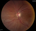
Retinal Detachment with Macula Detached478 viewsPatient had sudden loss of vision in the left eye. VA is 20/60 in the left eye. Fundus exam reveals retinal detachment inferiorly from 3-9 o'clock with tear at 3:30. Patient underwent scleral buckle in the left eye.Feb 17, 2014
|
|

Retinal Detachment - Treated Horseshoe Tear on segmental buckle1113 viewssegmental buckleOct 31, 2013
|
|

Rhegmatogenous Retinal Detachment437 viewsPatient comes in for second opinion on RD, right eye. VA is NLP in the right eye. Fundus photography shows inferior retinal detachment with holes and subretinal fibrosis. No further surgery is suggested at this time. Oct 11, 2013
|
|
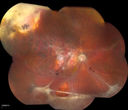
Rhegmatogenous Retinal Detachment845 viewsPatient comes in for second opinion on RD, right eye. VA is NLP in the right eye. Fundus photography shows inferior retinal detachment with holes and subretinal fibrosis. No further surgery is suggested at this time. Oct 11, 2013
|
|

Retinal Detachment with Retinal Tear1017 viewsPatient comes in with complaints of floaters in the right eye. VA was 20/40 with no improvement. Fundus exam shows retinal detachment from 9-12 o'clock with hole at 10:30 posteriorly. Pneumatic Retinopexy was performed with C3F8 Gas bubble and laser around the retinal tear in the right eye. Sep 30, 2013
|
|

Retinal Detachment with Retinal Tear436 viewsPatient comes in with complaints of floaters in the right eye. VA was 20/40 with no improvement. Fundus exam shows retinal detachment from 9-12 o'clock with hole at 10:30 posteriorly. Pneumatic Retinopexy was performed with C3F8 Gas bubble and laser around the retinal tear in the right eye. Sep 30, 2013
|
|

Retinal Detachment with Macula Detached.980 viewsPatient went to the ER over the weekend with loss of vision in the right eye. VA was count fingers in the right eye. Noticed a curtain in the right eye. Horseshoe tear was seen at 10-o'clock temporally, right eye. Proceeded with surgery the next day.Jul 15, 2013
|
|

Lattice Degeneration1185 viewsAprox 180 degree lattice degeneration in OD of a 17yr old girl.May 13, 2013
|
|

Buckle removed453 viewsSilicon implant had been extruded for 2 yearsMay 04, 2013
|
|

Deep skin tunel remains after silicon buckle removed417 viewssuperior lid skin tunnel made by 2 years of erosion of the buckleMay 04, 2013
|
|

Implant removed from the skin tunel528 viewsthe extruded piece of silicon is seen after removal from the skin tunnel. It is still attached to the sclera, through a conjunctival holeMay 04, 2013
|
|
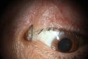
Silicon implant is seen inserted in to the superior lid skin472 viewsanother view of the implant intruded in the lid skinMay 04, 2013
|
|

Silicon implant is seen inserted in to the superior lid skin474 viewsPatient with several months of discomfort, discharge, FB sensation after scleral buckle surgery.May 04, 2013
|
|

Grade B Proliferative Vitreo-retinopathy - Pigment in Anterior Vitreous - Chronic Retinal Detachment1047 views72 year old woman - just noticed vision loss.Apr 22, 2013
|
|
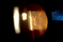
Grade B Proliferative Vitreo-retinopathy - Pigment in Anterior Vitreous - Chronic Retinal Detachment668 views72 year old woman - just noticed vision loss.Apr 22, 2013
|
|

Grade B Proliferative Vitreo-retinopathy - Pigment in Anterior Vitreous - Chronic Retinal Detachment753 views72 year old woman - just noticed vision loss.Apr 22, 2013
|
|

Reoccurring Retinal Detachment484 viewsPatient comes in for second opinion for RD in the right eye. Patient's VA was count fingers @ 2-ft in the right eye and 20/20 in the left eye. Patient is aphakic and has had 5- retinal surgeries in the past in the right eye including a scleral buckle. RD present with the macula off. Will consider surgery with silicone oil. Apr 09, 2013
|
|
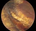
Reoccurring Retinal Detachment421 viewsPatient comes in for second opinion for RD in the right eye. Patient's VA was count fingers @ 2-ft in the right eye and 20/20 in the left eye. Patient is aphakic and has had 5- retinal surgeries in the past in the right eye including a scleral buckle. RD present with the macula off. Will consider surgery with silicone oil. Apr 09, 2013
|
|
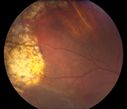
Reoccurring Retinal Detachment440 viewsPatient comes in for second opinion for RD in the right eye. Patient's VA was count fingers @ 2-ft in the right eye and 20/20 in the left eye. Patient is aphakic and has had 5- retinal surgeries in the past in the right eye including a scleral buckle. RD present with the macula off. Will consider surgery with silicone oil. Apr 09, 2013
|
|

Reoccurring Retinal Detachment395 viewsPatient comes in for second opinion for RD in the right eye. Patient's VA was count fingers @ 2-ft in the right eye and 20/20 in the left eye. Patient is aphakic and has had 5- retinal surgeries in the past in the right eye including a scleral buckle. RD present with the macula off. Will consider surgery with silicone oil. Apr 09, 2013
|
|

Reoccurring Retinal Detachment414 viewsPatient comes in for second opinion for RD in the right eye. Patient's VA was count fingers @ 2-ft in the right eye and 20/20 in the left eye. Patient is aphakic and has had 5- retinal surgeries in the past in the right eye including a scleral buckle. RD present with the macula off. Will consider surgery with silicone oil. Apr 09, 2013
|
|

Reoccurring Retinal Detachment411 viewsPatient comes in for second opinion for RD in the right eye. Patient's VA was count fingers @ 2-ft in the right eye and 20/20 in the left eye. Patient is aphakic and has had 5- retinal surgeries in the past in the right eye including a scleral buckle. RD present with the macula off. Will consider surgery with silicone oil. Apr 09, 2013
|
|

Reoccurring Retinal Detachment455 viewsPatient comes in for second opinion for RD in the right eye. Patient's VA was count fingers @ 2-ft in the right eye and 20/20 in the left eye. Patient is aphakic and has had 5- retinal surgeries in the past in the right eye including a scleral buckle. RD present with the macula off. Will consider surgery with silicone oil. Apr 09, 2013
|
|

Reoccurring Retinal Detachment374 viewsPatient comes in for second opinion for RD in the right eye. Patient's VA was count fingers @ 2-ft in the right eye and 20/20 in the left eye. Patient is aphakic and has had 5- retinal surgeries in the past in the right eye including a scleral buckle. RD present with the macula off. Will consider surgery with silicone oil. Apr 09, 2013
|
|

Reoccurring Retinal Detachment467 viewsPatient comes in for second opinion for RD in the right eye. Patient's VA was count fingers @ 2-ft in the right eye and 20/20 in the left eye. Patient is aphakic and has had 5- retinal surgeries in the past in the right eye including a scleral buckle. RD present with the macula off. Will consider surgery with silicone oil. Apr 09, 2013
|
|

Reoccurring Retinal Detachment383 viewsPatient comes in for second opinion for RD in the right eye. Patient's VA was count fingers @ 2-ft in the right eye and 20/20 in the left eye. Patient is aphakic and has had 5- retinal surgeries in the past in the right eye including a scleral buckle. RD present with the macula off. Will consider surgery with silicone oil. Apr 09, 2013
|
|

Reoccurring Retinal Detachment404 viewsPatient comes in for second opinion for RD in the right eye. Patient's VA was count fingers @ 2-ft in the right eye and 20/20 in the left eye. Patient is aphakic and has had 5- retinal surgeries in the past in the right eye including a scleral buckle. RD present with the macula off. Will consider surgery with silicone oil. Apr 09, 2013
|
|
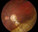
Reoccurring Retinal Detachment438 viewsPatient comes in for second opinion for RD in the right eye. Patient's VA was count fingers @ 2-ft in the right eye and 20/20 in the left eye. Patient is aphakic and has had 5- retinal surgeries in the past in the right eye including a scleral buckle. RD present with the macula off. Will consider surgery with silicone oil. Apr 09, 2013
|
|
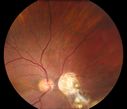
Reoccurring Retinal Detachment408 viewsPatient comes in for second opinion for RD in the right eye. Patient's VA was count fingers @ 2-ft in the right eye and 20/20 in the left eye. Patient is aphakic and has had 5- retinal surgeries in the past in the right eye including a scleral buckle. RD present with the macula off. Will consider surgery with silicone oil. Apr 09, 2013
|
|

Reoccurring Retinal Detachment375 viewsPatient comes in for second opinion for RD in the right eye. Patient's VA was count fingers @ 2-ft in the right eye and 20/20 in the left eye. Patient is aphakic and has had 5- retinal surgeries in the past in the right eye including a scleral buckle. RD present with the macula off. Will consider surgery with silicone oil. Apr 09, 2013
|
|

Reoccurring Retinal Detachment408 viewsPatient comes in for second opinion for RD in the right eye. Patient's VA was count fingers @ 2-ft in the right eye and 20/20 in the left eye. Patient is aphakic and has had 5- retinal surgeries in the past in the right eye including a scleral buckle. RD present with the macula off. Will consider surgery with silicone oil. Apr 09, 2013
|
|

Reoccurring Retinal Detachment1088 viewsPatient comes in for second opinion for RD in the right eye. Patient's VA was count fingers @ 2-ft in the right eye and 20/20 in the left eye. Patient is aphakic and has had 5- retinal surgeries in the past in the right eye including a scleral buckle. RD present with the macula off. Will consider surgery with silicone oil. Apr 09, 2013
|
|

Reoccurring Retinal Detachment530 viewsPatient comes in for second opinion for RD in the right eye. Patient's VA was count fingers @ 2-ft in the right eye and 20/20 in the left eye. Patient is aphakic and has had 5- retinal surgeries in the past in the right eye including a scleral buckle. RD present with the macula off. Will consider surgery with silicone oil. Apr 09, 2013
|
|

Proleferative Vitreoretinopathy - Panretinal Retinoschisis 499 viewsApr 03, 2013
|
|

Proleferative Vitreoretinopathy - Panretinal Retinoschisis 493 viewsApr 03, 2013
|
|

Proleferative Vitreoretinopathy - Panretinal Retinoschisis 503 viewsApr 03, 2013
|
|

Proleferative Vitreoretinopathy - Panretinal Retinoschisis 523 viewsApr 03, 2013
|
|

Proleferative Vitreoretinopathy - Panretinal Retinoschisis 481 viewsApr 03, 2013
|
|

Proleferative Vitreoretinopathy - Panretinal Retinoschisis 485 viewsApr 03, 2013
|
|

Proleferative Vitreoretinopathy - Panretinal Retinoschisis 491 viewsApr 03, 2013
|
|

Proleferative Vitreoretinopathy - Panretinal Retinoschisis 536 viewsApr 03, 2013
|
|

Proleferative Vitreoretinopathy - Panretinal Retinoschisis 468 viewsFluorescein Angiogram shows Diffuse Leakage from retinal vessels and CME.Apr 03, 2013
|
|

Proleferative Vitreoretinopathy - Panretinal Retinoschisis 458 viewsFluorescein Angiogram shows Diffuse Leakage from retinal vessels and CME.Apr 03, 2013
|
|
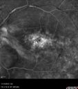
Proleferative Vitreoretinopathy - Panretinal Retinoschisis 489 viewsFluorescein Angiogram shows Diffuse Leakage from retinal vessels and CME.Apr 03, 2013
|
|
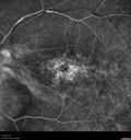
Proleferative Vitreoretinopathy - Panretinal Retinoschisis 499 viewsFluorescein Angiogram shows Diffuse Leakage from retinal vessels and CME.Apr 03, 2013
|
|

Proleferative Vitreoretinopathy - Panretinal Retinoschisis 495 viewsFluorescein Angiogram shows Diffuse Leakage from retinal vessels and CME.Apr 03, 2013
|
|

Proleferative Vitreoretinopathy - Panretinal Retinoschisis 513 viewsFluorescein Angiogram shows Diffuse Leakage from retinal vessels and CME.Apr 03, 2013
|
|

Proleferative Vitreoretinopathy - Panretinal Retinoschisis - VIDEO TAKES TIME TO LOAD472 viewsApr 03, 2013
|
|

Proleferative Vitreoretinopathy - Panretinal Retinoschisis 528 viewsApr 03, 2013
|
|

Proleferative Vitreoretinopathy - Panretinal Retinoschisis 574 viewsApr 03, 2013
|
|

Proleferative Vitreoretinopathy - Panretinal Retinoschisis 633 viewsApr 03, 2013
|
|

Proleferative Vitreoretinopathy - Panretinal Retinoschisis 584 viewsApr 03, 2013
|
|

Proleferative Vitreoretinopathy - Panretinal Retinoschisis 558 viewsApr 03, 2013
|
|

Proleferative Vitreoretinopathy - Panretinal Retinoschisis 553 viewsApr 03, 2013
|
|

Silicone Oil Droplets on Silicone Intraocular Lens477 views64 Year Old Woman 1 week after Oil removal 20/80 vision.Feb 28, 2013
|
|

Rubeosis after Silicone Oil Removal Complex RD Repair with Peripheral 180 Degree Retinotomy (Inferiorly)584 viewsFeb 28, 2013
|
|

Asymptomatic Laser Demarcated Retinal Detachment has Spontaneously Re-attached 6 Months After Laser994 viewsSpontaneously Re-attached Detached RetinaFeb 21, 2013
|
|

RD466 viewsRDFeb 11, 2013
|
|
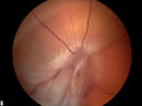
RD with PVR1453 viewsRD with Proliferative Vitreoretinopathy Feb 11, 2013
|
|

RD811 viewsInferior RDFeb 11, 2013
|
|

RD - OLD468 viewsRDFeb 11, 2013
|
|

RD992 viewsRDFeb 11, 2013
|
|
| 491 files on 5 page(s) |
 |
3 | |
|


