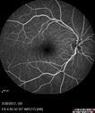
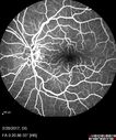
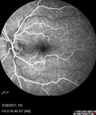
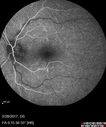
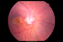
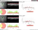
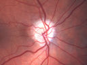
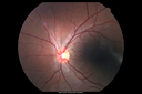
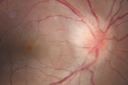
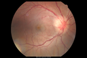
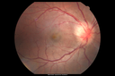
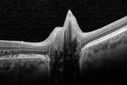
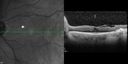
Toxic Optic Neuropathy - Ethambutol521 x angesehen15 months of Ethambutol with rapid vision loss OU. VA 20/100 OD (post cataract sugery ) 20/200 OS with dense cataract. There is temporal RNFL loss in both eyes19. Januar 2018
Toxic Optic Neuropathy - Ethambutol479 x angesehen15 months of Ethambutol with rapid vision loss OU. VA 20/100 OD (post cataract sugery ) 20/200 OS with dense cataract. There is temporal RNFL loss in both eyes19. Januar 2018
Toxic Optic Neuropathy - Ethambutol515 x angesehen15 months of Ethambutol with rapid vision loss OU. VA 20/100 OD (post cataract sugery ) 20/200 OS with dense cataract. There is temporal RNFL loss in both eyes19. Januar 2018
Syphilitic Optic Neuritis - possible Gumma514 x angesehen82 year old woman with vision loss in the left eye 20/32 and positive FTA-ABS (negative RPR). - this may be a gumma07. April 2017
Syphilitic Optic Neuritis - possible Gumma450 x angesehen82 year old woman with vision loss in the left eye 20/32 and positive FTA-ABS (negative RPR). - this may be a gumma07. April 2017
Syphilitic Optic Neuritis - possible Gumma516 x angesehen82 year old woman with vision loss in the left eye 20/32 and positive FTA-ABS (negative RPR). - this may be a gumma07. April 2017
Syphilitic Optic Neuritis - possible Gumma477 x angesehen82 year old woman with vision loss in the left eye 20/32 and positive FTA-ABS (negative RPR). - this may be a gumma07. April 2017
Syphilitic Optic Neuritis - possible Gumma467 x angesehen82 year old woman with vision loss in the left eye 20/32 and positive FTA-ABS (negative RPR). - this may be a gumma07. April 2017
Syphilitic Optic Neuritis - possible Gumma488 x angesehen82 year old woman with vision loss in the left eye 20/32 and positive FTA-ABS (negative RPR). - this may be a gumma07. April 2017
Syphilitic Optic Neuritis - possible Gumma488 x angesehen82 year old woman with vision loss in the left eye 20/32 and positive FTA-ABS (negative RPR). - this may be a gumma07. April 2017
Syphilitic Optic Neuritis - possible Gumma457 x angesehen82 year old woman with vision loss in the left eye 20/32 and positive FTA-ABS (negative RPR). - this may be a gumma07. April 2017
Syphilitic Optic Neuritis - possible Gumma489 x angesehen82 year old woman with vision loss in the left eye 20/32 and positive FTA-ABS (negative RPR). - this may be a gumma07. April 2017
Syphilitic Optic Neuritis - possible Gumma475 x angesehen82 year old woman with vision loss in the left eye 20/32 and positive FTA-ABS (negative RPR). - this may be a gumma07. April 2017
Syphilitic Optic Neuritis - possible Gumma433 x angesehen82 year old woman with vision loss in the left eye 20/32 and positive FTA-ABS (negative RPR). - this may be a gumma07. April 2017
Syphilitic Optic Neuritis - possible Gumma408 x angesehen82 year old woman with vision loss in the left eye 20/32 and positive FTA-ABS (negative RPR). - this may be a gumma07. April 2017
Syphilitic Optic Neuritis - possible Gumma429 x angesehen82 year old woman with vision loss in the left eye 20/32 and positive FTA-ABS (negative RPR). - this may be a gumma07. April 2017
Syphilitic Optic Neuritis - possible Gumma411 x angesehen82 year old woman with vision loss in the left eye 20/32 and positive FTA-ABS (negative RPR). - this may be a gumma07. April 2017
Syphilitic Optic Neuritis - possible Gumma419 x angesehen82 year old woman with vision loss in the left eye 20/32 and positive FTA-ABS (negative RPR). - this may be a gumma07. April 2017
Syphilitic Optic Neuritis - possible Gumma486 x angesehen82 year old woman with vision loss in the left eye 20/32 and positive FTA-ABS (negative RPR). - this may be a gumma07. April 2017
Syphilitic Optic Neuritis - possible Gumma610 x angesehen82 year old woman with vision loss in the left eye 20/32 and positive FTA-ABS (negative RPR). - this may be a gumma07. April 2017
Syphilitic Optic Neuritis - possible Gumma545 x angesehen82 year old woman with vision loss in the left eye 20/32 and positive FTA-ABS (negative RPR). - this may be a gumma07. April 2017
Syphilitic Optic Neuritis - possible Gumma620 x angesehen82 year old woman with vision loss in the left eye 20/32 and positive FTA-ABS (negative RPR). - this may be a gumma07. April 2017
Syphilitic Optic Neuritis - possible Gumma547 x angesehen82 year old woman with vision loss in the left eye 20/32 and positive FTA-ABS (negative RPR). - this may be a gumma07. April 2017
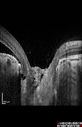
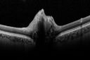
Optic Nerve Pit Retinal Detachment - SD OCT of Nerve EDI shows Pit583 x angesehen9 Year old with morning glory disc who developed nerve pit RD. Raster scans of the optic nerve on EDI setting show the communication between the subretinal space and the fluid around the cerebrospinal fluid surrounding the optic nerve sheath.06. März 2017
Optic Nerve Pit Retinal Detachment - SD OCT of Nerve EDI shows Pit526 x angesehen9 Year old with morning glory disc who developed nerve pit RD. Raster scans of the optic nerve on EDI setting show the communication between the subretinal space and the fluid around the cerebrospinal fluid surrounding the optic nerve sheath.06. März 2017
Optic Nerve Pit Retinal Detachment - SD OCT of Nerve EDI shows Pit475 x angesehen9 Year old with morning glory disc who developed nerve pit RD. Raster scans of the optic nerve on EDI setting show the communication between the subretinal space and the fluid around the cerebrospinal fluid surrounding the optic nerve sheath.06. März 2017
Optic Nerve Pit Retinal Detachment - SD OCT of Nerve EDI shows Pit563 x angesehen9 Year old with morning glory disc who developed nerve pit RD. Raster scans of the optic nerve on EDI setting show the communication between the subretinal space and the fluid around the cerebrospinal fluid surrounding the optic nerve sheath.06. März 2017
Optic Nerve Pit Retinal Detachment - SD OCT of Nerve EDI shows Pit606 x angesehen9 Year old with morning glory disc who developed nerve pit RD. Raster scans of the optic nerve on EDI setting show the communication between the subretinal space and the fluid around the cerebrospinal fluid surrounding the optic nerve sheath.06. März 2017
Optic Nerve Pit Retinal Detachment - SD OCT of Nerve EDI shows Pit576 x angesehen9 Year old with morning glory disc who developed nerve pit RD. Raster scans of the optic nerve on EDI setting show the communication between the subretinal space and the fluid around the cerebrospinal fluid surrounding the optic nerve sheath.06. März 2017
Optic Nerve Pit Retinal Detachment - SD OCT of Nerve EDI shows Pit556 x angesehen9 Year old with morning glory disc who developed nerve pit RD. Raster scans of the optic nerve on EDI setting show the communication between the subretinal space and the fluid around the cerebrospinal fluid surrounding the optic nerve sheath.06. März 2017
Optic Nerve Pit Retinal Detachment - SD OCT of Nerve EDI shows Pit555 x angesehen9 Year old with morning glory disc who developed nerve pit RD. Raster scans of the optic nerve on EDI setting show the communication between the subretinal space and the fluid around the cerebrospinal fluid surrounding the optic nerve sheath.06. März 2017
Optic Nerve Pit Retinal Detachment - SD OCT of Nerve EDI shows Pit574 x angesehen9 Year old with morning glory disc who developed nerve pit RD. Raster scans of the optic nerve on EDI setting show the communication between the subretinal space and the fluid around the cerebrospinal fluid surrounding the optic nerve sheath.06. März 2017
Optic Nerve Pit Retinal Detachment - SD OCT of Nerve EDI shows Pit508 x angesehen9 Year old with morning glory disc who developed nerve pit RD. Raster scans of the optic nerve on EDI setting show the communication between the subretinal space and the fluid around the cerebrospinal fluid surrounding the optic nerve sheath.06. März 2017
Optic Nerve Pit Retinal Detachment - SD OCT of Nerve EDI shows Pit524 x angesehen9 Year old with morning glory disc who developed nerve pit RD. Raster scans of the optic nerve on EDI setting show the communication between the subretinal space and the fluid around the cerebrospinal fluid surrounding the optic nerve sheath.06. März 2017
Optic Nerve Pit Retinal Detachment - SD OCT of Nerve EDI shows Pit566 x angesehen9 Year old with morning glory disc who developed nerve pit RD. Raster scans of the optic nerve on EDI setting show the communication between the subretinal space and the fluid around the cerebrospinal fluid surrounding the optic nerve sheath.06. März 2017
Optic Nerve Pit Retinal Detachment - SD OCT of Nerve EDI shows Pit578 x angesehen9 Year old with morning glory disc who developed nerve pit RD. Raster scans of the optic nerve on EDI setting show the communication between the subretinal space and the fluid around the cerebrospinal fluid surrounding the optic nerve sheath.06. März 2017
Optic Nerve Pit Retinal Detachment - SD OCT of Nerve EDI shows Pit546 x angesehen9 Year old with morning glory disc who developed nerve pit RD. Raster scans of the optic nerve on EDI setting show the communication between the subretinal space and the fluid around the cerebrospinal fluid surrounding the optic nerve sheath.06. März 2017
Optic Nerve Pit Retinal Detachment - SD OCT of Nerve EDI shows Pit535 x angesehen9 Year old with morning glory disc who developed nerve pit RD. Raster scans of the optic nerve on EDI setting show the communication between the subretinal space and the fluid around the cerebrospinal fluid surrounding the optic nerve sheath.06. März 2017
Optic Nerve Pit Retinal Detachment - SD OCT of Nerve EDI shows Pit571 x angesehen9 Year old with morning glory disc who developed nerve pit RD. Raster scans of the optic nerve on EDI setting show the communication between the subretinal space and the fluid around the cerebrospinal fluid surrounding the optic nerve sheath.06. März 2017
Optic Nerve Pit Retinal Detachment - SD OCT of Nerve EDI shows Pit560 x angesehen9 Year old with morning glory disc who developed nerve pit RD. Raster scans of the optic nerve on EDI setting show the communication between the subretinal space and the fluid around the cerebrospinal fluid surrounding the optic nerve sheath.06. März 2017
Optic Nerve Pit Retinal Detachment - SD OCT of Nerve EDI shows Pit570 x angesehen9 Year old with morning glory disc who developed nerve pit RD. Raster scans of the optic nerve on EDI setting show the communication between the subretinal space and the fluid around the cerebrospinal fluid surrounding the optic nerve sheath.06. März 2017
Optic Nerve Pit Retinal Detachment - SD OCT of Nerve EDI shows Pit640 x angesehen9 Year old with morning glory disc who developed nerve pit RD. Raster scans of the optic nerve on EDI setting show the communication between the subretinal space and the fluid around the cerebrospinal fluid surrounding the optic nerve sheath.06. März 2017
Acute AION OS - Follow-up RNFL scan597 x angesehen11. Oktober 2014
AION - Acute with Follow UP RNFL scan shows edema then atrophy576 x angesehen11. Oktober 2014
Acute AION OS - Mild disc pallor OD - macular scan shows increasing atrophy in left eye580 x angesehen11. Oktober 2014
Acute AION OS - Mild disc pallor OD607 x angesehenSudden vision loss OS x 1 week with optic nerve swelling. Evidence of optic atrophy in right eye. 11. Oktober 2014
Acute AION OS - Mild disc pallor OD540 x angesehenSudden vision loss OS x 1 week with optic nerve swelling. Evidence of optic atrophy in right eye. 11. Oktober 2014
Acute AION OS - Mild disc pallor OD530 x angesehenSudden vision loss OS x 1 week with optic nerve swelling. Evidence of optic atrophy in right eye. 11. Oktober 2014
Acute AION OS - Mild disc pallor OD517 x angesehenSudden vision loss OS x 1 week with optic nerve swelling. Evidence of optic atrophy in right eye. 11. Oktober 2014
Acute AION OS - Mild disc pallor OD566 x angesehenSudden vision loss OS x 1 week with optic nerve swelling. Evidence of optic atrophy in right eye. 11. Oktober 2014
Acute AION OS - Mild disc pallor OD524 x angesehenSudden vision loss OS x 1 week with optic nerve swelling. Evidence of optic atrophy in right eye. 11. Oktober 2014
Acute AION OS - Mild disc pallor OD600 x angesehenSudden vision loss OS x 1 week with optic nerve swelling. Evidence of optic atrophy in right eye. 11. Oktober 2014
Acute AION OS - Mild disc pallor OD513 x angesehenSudden vision loss OS x 1 week with optic nerve swelling. Evidence of optic atrophy in right eye. 11. Oktober 2014
Acute AION OS - Mild disc pallor OD491 x angesehenSudden vision loss OS x 1 week with optic nerve swelling. Evidence of optic atrophy in right eye. 11. Oktober 2014
Acute AION OS - Mild disc pallor OD548 x angesehenSudden vision loss OS x 1 week with optic nerve swelling. Evidence of optic atrophy in right eye. 11. Oktober 2014
Acute AION OS - Mild disc pallor OD591 x angesehenSudden vision loss OS x 1 week with optic nerve swelling. Evidence of optic atrophy in right eye. 11. Oktober 2014
Acute AION OS - Mild disc pallor OD533 x angesehenSudden vision loss OS x 1 week with optic nerve swelling. Evidence of optic atrophy in right eye. 11. Oktober 2014
Acute AION OS - Mild disc pallor OD555 x angesehenSudden vision loss OS x 1 week with optic nerve swelling. Evidence of optic atrophy in right eye. 11. Oktober 2014
Acute AION OS - Mild disc pallor OD563 x angesehenSudden vision loss OS x 1 week with optic nerve swelling. Evidence of optic atrophy in right eye. 11. Oktober 2014
Acute AION OS - Mild disc pallor OD673 x angesehenSudden vision loss OS x 1 week with optic nerve swelling. Evidence of optic atrophy in right eye. 11. Oktober 2014
Acute AION OS - Mild disc pallor OD583 x angesehenSudden vision loss OS x 1 week with optic nerve swelling. Evidence of optic atrophy in right eye. 11. Oktober 2014
Acute AION OS - Mild disc pallor OD677 x angesehenSudden vision loss OS x 1 week with optic nerve swelling. Evidence of optic atrophy in right eye. 11. Oktober 2014
Acute AION OS - Mild disc pallor OD730 x angesehenSudden vision loss OS x 1 week with optic nerve swelling. Evidence of optic atrophy in right eye. 11. Oktober 2014
Neuroretinitis - Cat Scratch - Bartonella - Sudden Vision Loss Right Eye - 29 Year Old Female556 x angesehen06. September 2014
Neuroretinitis - Cat Scratch - Bartonella - Sudden Vision Loss Right Eye - 29 Year Old Female501 x angesehen06. September 2014
Neuroretinitis - Cat Scratch - Bartonella - Sudden Vision Loss Right Eye - 29 Year Old Female536 x angesehen06. September 2014
Neuroretinitis - Cat Scratch - Bartonella - Sudden Vision Loss Right Eye - 29 Year Old Female547 x angesehen06. September 2014
Neuroretinitis - Cat Scratch - Bartonella - Sudden Vision Loss Right Eye - 29 Year Old Female529 x angesehen06. September 2014
Neuroretinitis - Cat Scratch - Bartonella - Sudden Vision Loss Right Eye - 29 Year Old Female606 x angesehen06. September 2014
Neuroretinitis - Cat Scratch - Bartonella - Sudden Vision Loss Right Eye - 29 Year Old Female607 x angesehen06. September 2014
Neuroretinitis - Cat Scratch - Bartonella - Sudden Vision Loss Right Eye - 29 Year Old Female567 x angesehen06. September 2014
Neuroretinitis - Cat Scratch - Bartonella - Sudden Vision Loss Right Eye - 29 Year Old Female523 x angesehen06. September 2014
Neuroretinitis - Cat Scratch - Bartonella - Sudden Vision Loss Right Eye - 29 Year Old Female607 x angesehen06. September 2014
Neuroretinitis - Cat Scratch - Bartonella - Sudden Vision Loss Right Eye - 29 Year Old Female622 x angesehen06. September 2014
Neuroretinitis - Cat Scratch - Bartonella - Sudden Vision Loss Right Eye - 29 Year Old Female572 x angesehen06. September 2014
Neuroretinitis - Cat Scratch - Bartonella - Sudden Vision Loss Right Eye - 29 Year Old Female687 x angesehen06. September 2014
Neuroretinitis - Cat Scratch - Bartonella - Sudden Vision Loss Right Eye - 29 Year Old Female636 x angesehen06. September 2014
Neuroretinitis - Cat Scratch - Bartonella - Sudden Vision Loss Right Eye - 29 Year Old Female666 x angesehen06. September 2014
Neuroretinitis - Cat Scratch - Bartonella - Sudden Vision Loss Right Eye - 29 Year Old Female654 x angesehen06. September 2014
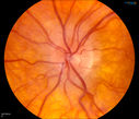
Bilateral Papilledema756 x angesehenYoung female that presents with severe frequent headaches. VA is 20/15 in both eyes. Fundus exam reveals swelling of both optic nerves.29. August 2014
Bilateral Papilledema1283 x angesehenYoung female that presents with severe frequent headaches. VA is 20/15 in both eyes. Fundus exam reveals swelling of both optic nerves.29. August 2014
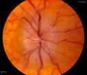
Bilateral Papilledema 1469 x angesehenYoung male presents with decreased vision in both eyes. Fundus photos reveal swollen optic nerves in both eyes.25. August 2014
Bilateral Papilledema 562 x angesehenYoung male presents with decreased vision in both eyes. Fundus photos reveal swollen optic nerves in both eyes.25. August 2014
Bilateral Papilledema 586 x angesehenYoung male presents with decreased vision in both eyes. Fundus photos reveal swollen optic nerves in both eyes.25. August 2014
Bilateral Papilledema 647 x angesehenYoung male presents with decreased vision in both eyes. Fundus photos reveal swollen optic nerves in both eyes.25. August 2014
Bilateral Optic Nerve Drusen496 x angesehen03. Juni 2014
Bilateral Optic Nerve Drusen484 x angesehen03. Juni 2014
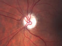
Morning Glory Syndrome, Optic Nerve1494 x angesehenPatient comes in for cataract evaluation. VA 20/60, right eye and 20/150, left eye. Fundus photo shows "morning glory syndrome" of the optic nerve in the left eye. 28. April 2014
Morning Glory Syndrome, Optic Nerve893 x angesehenPatient comes in for cataract evaluation. VA 20/60, right eye and 20/150, left eye. Fundus photo shows "morning glory syndrome" of the optic nerve in the left eye. 28. April 2014
567 x angesehen27. März 2014
501 x angesehen27. März 2014
Optic Disk Drusen Advanced Nerve Fiber Loss624 x angesehen17. März 2014
Optic Disk Drusen Advanced Nerve Fiber Loss515 x angesehen17. März 2014
Optic Disk Drusen Advanced Nerve Fiber Loss537 x angesehen17. März 2014
Optic Disk Drusen Advanced Nerve Fiber Loss462 x angesehen17. März 2014
Optic Disk Drusen Advanced Nerve Fiber Loss545 x angesehen17. März 2014
Optic Disk Drusen Advanced Nerve Fiber Loss468 x angesehen17. März 2014
Optic Disk Drusen Advanced Nerve Fiber Loss531 x angesehen17. März 2014
Optic Disk Drusen Advanced Nerve Fiber Loss504 x angesehen17. März 2014