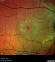
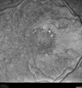
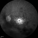
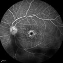
Confluent Soft Drusen with Subetinal Fluid - Right Eye533 x angesehenAsymptomatic 85 year old man. OCT shows pocket of subfoveal fluid in the center of the macula. This could be mechanical from the drusen preventing the retina from settling down. Testing did not reveal a CNVM and subsequent OCT 6 weeks later showed no change in structure.06. Februar 2017
Confluent Soft Drusen with Subetinal Fluid - Right Eye515 x angesehenAsymptomatic 85 year old man. OCT shows pocket of subfoveal fluid in the center of the macula. This could be mechanical from the drusen preventing the retina from settling down. Testing did not reveal a CNVM and subsequent OCT 6 weeks later showed no change in structure.06. Februar 2017
Confluent Soft Drusen with Subetinal Fluid - Right Eye505 x angesehenAsymptomatic 85 year old man. OCT shows pocket of subfoveal fluid in the center of the macula. This could be mechanical from the drusen preventing the retina from settling down. Testing did not reveal a CNVM and subsequent OCT 6 weeks later showed no change in structure.06. Februar 2017
Confluent Soft Drusen with Subetinal Fluid - Right Eye819 x angesehenAsymptomatic 85 year old man. OCT shows pocket of subfoveal fluid in the center of the macula. This could be mechanical from the drusen preventing the retina from settling down. Testing did not reveal a CNVM and subsequent OCT 6 weeks later showed no change in structure.06. Februar 2017
Confluent Soft Drusen with Subetinal Fluid - Right Eye648 x angesehenAsymptomatic 85 year old man. OCT shows pocket of subfoveal fluid in the center of the macula. This could be mechanical from the drusen preventing the retina from settling down. Testing did not reveal a CNVM and subsequent OCT 6 weeks later showed no change in structure.06. Februar 2017
Confluent Soft Drusen with Subetinal Fluid - Right Eye558 x angesehenAsymptomatic 85 year old man. OCT shows pocket of subfoveal fluid in the center of the macula. This could be mechanical from the drusen preventing the retina from settling down. Testing did not reveal a CNVM and subsequent OCT 6 weeks later showed no change in structure.06. Februar 2017
Confluent Soft Drusen with Subetinal Fluid - Right Eye529 x angesehenAsymptomatic 85 year old man. OCT shows pocket of subfoveal fluid in the center of the macula. This could be mechanical from the drusen preventing the retina from settling down. Testing did not reveal a CNVM and subsequent OCT 6 weeks later showed no change in structure.06. Februar 2017
Confluent Soft Drusen with Subetinal Fluid - Right Eye522 x angesehenAsymptomatic 85 year old man. OCT shows pocket of subfoveal fluid in the center of the macula. This could be mechanical from the drusen preventing the retina from settling down. Testing did not reveal a CNVM and subsequent OCT 6 weeks later showed no change in structure.06. Februar 2017
Confluent Soft Drusen with Subetinal Fluid - Right Eye544 x angesehenAsymptomatic 85 year old man. OCT shows pocket of subfoveal fluid in the center of the macula. This could be mechanical from the drusen preventing the retina from settling down. Testing did not reveal a CNVM and subsequent OCT 6 weeks later showed no change in structure.06. Februar 2017
Confluent Soft Drusen with Subetinal Fluid - Right Eye568 x angesehenAsymptomatic 85 year old man. OCT shows pocket of subfoveal fluid in the center of the macula. This could be mechanical from the drusen preventing the retina from settling down. Testing did not reveal a CNVM and subsequent OCT 6 weeks later showed no change in structure.06. Februar 2017
Confluent Soft Drusen with Subetinal Fluid - Right Eye589 x angesehenAsymptomatic 85 year old man. OCT shows pocket of subfoveal fluid in the center of the macula. This could be mechanical from the drusen preventing the retina from settling down. Testing did not reveal a CNVM and subsequent OCT 6 weeks later showed no change in structure.06. Februar 2017
Confluent Soft Drusen with Subetinal Fluid - Right Eye606 x angesehenAsymptomatic 85 year old man. OCT shows pocket of subfoveal fluid in the center of the macula. This could be mechanical from the drusen preventing the retina from settling down. Testing did not reveal a CNVM and subsequent OCT 6 weeks later showed no change in structure.06. Februar 2017
Confluent Soft Drusen with Subetinal Fluid - Right Eye615 x angesehenAsymptomatic 85 year old man. OCT shows pocket of subfoveal fluid in the center of the macula. This could be mechanical from the drusen preventing the retina from settling down. Testing did not reveal a CNVM and subsequent OCT 6 weeks later showed no change in structure.06. Februar 2017
Confluent Soft Drusen with Subetinal Fluid - Right Eye829 x angesehenAsymptomatic 85 year old man. OCT shows pocket of subfoveal fluid in the center of the macula. This could be mechanical from the drusen preventing the retina from settling down. Testing did not reveal a CNVM and subsequent OCT 6 weeks later showed no change in structure.06. Februar 2017
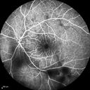
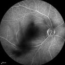
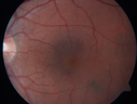
Macular Telangiectasis - visible telangiectasia on color photos462 x angesehenMulticolor photos too show pathology. This patient had 20/50 vision with pinhole to 20/2529. September 2016
Macular Telangiectasis - visible telangiectasia on color photos501 x angesehenMulticolor photos too show pathology. This patient had 20/50 vision with pinhole to 20/2529. September 2016
Macular Telangiectasis - visible telangiectasia on color photos506 x angesehenMulticolor photos too show pathology. This patient had 20/50 vision with pinhole to 20/2529. September 2016
Macular Telangiectasis - visible telangiectasia on color photos518 x angesehenMulticolor photos too show pathology. This patient had 20/50 vision with pinhole to 20/2529. September 2016
Macular Telangiectasis - visible telangiectasia on color photos472 x angesehenMulticolor photos too show pathology. This patient had 20/50 vision with pinhole to 20/2529. September 2016
Macular Telangiectasis - visible telangiectasia on color photos536 x angesehenMulticolor photos too show pathology. This patient had 20/50 vision with pinhole to 20/2529. September 2016
Macular Telangiectasis - visible telangiectasia on color photos461 x angesehenMulticolor photos too show pathology. This patient had 20/50 vision with pinhole to 20/2529. September 2016
Macular Telangiectasis - visible telangiectasia on color photos601 x angesehenMulticolor photos too show pathology. This patient had 20/50 vision with pinhole to 20/2529. September 2016
Macular Telangiectasis - visible telangiectasia on color photos529 x angesehenMulticolor photos too show pathology. This patient had 20/50 vision with pinhole to 20/2529. September 2016
Macular Telangiectasis - visible telangiectasia on color photos503 x angesehenMulticolor photos too show pathology. This patient had 20/50 vision with pinhole to 20/2529. September 2016
Macular Telangiectasis - visible telangiectasia on color photos470 x angesehenMulticolor photos too show pathology. This patient had 20/50 vision with pinhole to 20/2529. September 2016
Macular Telangiectasis - visible telangiectasia on color photos498 x angesehenMulticolor photos too show pathology. This patient had 20/50 vision with pinhole to 20/2529. September 2016
Macular Telangiectasia and dry AMD - Simulating wet AMD558 x angesehenThis patient was told different things by different retina specialists. Her case is complex because she has OCT findings of Macular Telangiectasis and dry AMD. This makes it look like she has wet AMD. Her vision is stable.29. September 2016
Macular Telangiectasia and dry AMD - Simulating wet AMD553 x angesehenThis patient was told different things by different retina specialists. Her case is complex because she has OCT findings of Macular Telangiectasis and dry AMD. This makes it look like she has wet AMD. Her vision is stable.29. September 2016
Macular Telangiectasia and dry AMD - Simulating wet AMD559 x angesehenThis patient was told different things by different retina specialists. Her case is complex because she has OCT findings of Macular Telangiectasis and dry AMD. This makes it look like she has wet AMD. Her vision is stable.29. September 2016
Macular Telangiectasia and dry AMD - Simulating wet AMD499 x angesehenThis patient was told different things by different retina specialists. Her case is complex because she has OCT findings of Macular Telangiectasis and dry AMD. This makes it look like she has wet AMD. Her vision is stable.29. September 2016
Macular Telangiectasia and dry AMD - Simulating wet AMD448 x angesehenThis patient was told different things by different retina specialists. Her case is complex because she has OCT findings of Macular Telangiectasis and dry AMD. This makes it look like she has wet AMD. Her vision is stable.29. September 2016
Macular Telangiectasia and dry AMD - Simulating wet AMD480 x angesehenThis patient was told different things by different retina specialists. Her case is complex because she has OCT findings of Macular Telangiectasis and dry AMD. This makes it look like she has wet AMD. Her vision is stable.29. September 2016
Macular Telangiectasia and dry AMD - Simulating wet AMD502 x angesehenThis patient was told different things by different retina specialists. Her case is complex because she has OCT findings of Macular Telangiectasis and dry AMD. This makes it look like she has wet AMD. Her vision is stable.29. September 2016
Macular Telangiectasia and dry AMD - Simulating wet AMD446 x angesehenThis patient was told different things by different retina specialists. Her case is complex because she has OCT findings of Macular Telangiectasis and dry AMD. This makes it look like she has wet AMD. Her vision is stable.29. September 2016
Macular Telangiectasia and dry AMD - Simulating wet AMD454 x angesehenThis patient was told different things by different retina specialists. Her case is complex because she has OCT findings of Macular Telangiectasis and dry AMD. This makes it look like she has wet AMD. Her vision is stable.29. September 2016
Macular Telangiectasia and dry AMD - Simulating wet AMD504 x angesehenThis patient was told different things by different retina specialists. Her case is complex because she has OCT findings of Macular Telangiectasis and dry AMD. This makes it look like she has wet AMD. Her vision is stable.29. September 2016
Macular Telangiectasia and dry AMD - Simulating wet AMD503 x angesehenThis patient was told different things by different retina specialists. Her case is complex because she has OCT findings of Macular Telangiectasis and dry AMD. This makes it look like she has wet AMD. Her vision is stable.29. September 2016
Macular Telangiectasia and dry AMD - Simulating wet AMD514 x angesehenThis patient was told different things by different retina specialists. Her case is complex because she has OCT findings of Macular Telangiectasis and dry AMD. This makes it look like she has wet AMD. Her vision is stable.29. September 2016
Macular Telangiectasia and dry AMD - Simulating wet AMD526 x angesehenThis patient was told different things by different retina specialists. Her case is complex because she has OCT findings of Macular Telangiectasis and dry AMD. This makes it look like she has wet AMD. Her vision is stable.29. September 2016
Macular Telangiectasia and dry AMD - Simulating wet AMD477 x angesehenThis patient was told different things by different retina specialists. Her case is complex because she has OCT findings of Macular Telangiectasis and dry AMD. This makes it look like she has wet AMD. Her vision is stable.29. September 2016
Macular Telangiectasia and dry AMD - Simulating wet AMD549 x angesehenThis patient was told different things by different retina specialists. Her case is complex because she has OCT findings of Macular Telangiectasis and dry AMD. This makes it look like she has wet AMD. Her vision is stable.29. September 2016
Macular Telangiectasia and dry AMD - Simulating wet AMD498 x angesehenThis patient was told different things by different retina specialists. Her case is complex because she has OCT findings of Macular Telangiectasis and dry AMD. This makes it look like she has wet AMD. Her vision is stable.29. September 2016
Macular Telangiectasia and dry AMD - Simulating wet AMD534 x angesehenThis patient was told different things by different retina specialists. Her case is complex because she has OCT findings of Macular Telangiectasis and dry AMD. This makes it look like she has wet AMD. Her vision is stable.29. September 2016
Macular Telangiectasia and dry AMD - Simulating wet AMD526 x angesehenThis patient was told different things by different retina specialists. Her case is complex because she has OCT findings of Macular Telangiectasis and dry AMD. This makes it look like she has wet AMD. Her vision is stable.29. September 2016
Macular Telangiectasia and dry AMD - Simulating wet AMD528 x angesehenThis patient was told different things by different retina specialists. Her case is complex because she has OCT findings of Macular Telangiectasis and dry AMD. This makes it look like she has wet AMD. Her vision is stable.29. September 2016
Macular Telangiectasia and dry AMD - Simulating wet AMD463 x angesehenThis patient was told different things by different retina specialists. Her case is complex because she has OCT findings of Macular Telangiectasis and dry AMD. This makes it look like she has wet AMD. Her vision is stable.29. September 2016
Macular Telangiectasia and dry AMD - Simulating wet AMD460 x angesehenThis patient was told different things by different retina specialists. Her case is complex because she has OCT findings of Macular Telangiectasis and dry AMD. This makes it look like she has wet AMD. Her vision is stable.29. September 2016
Macular Telangiectasia and dry AMD - Simulating wet AMD597 x angesehenThis patient was told different things by different retina specialists. Her case is complex because she has OCT findings of Macular Telangiectasis and dry AMD. This makes it look like she has wet AMD. Her vision is stable.29. September 2016
Multifocal Pattern Dystrophy484 x angesehen51 year old with normal vision - images look a lot like fundus flavimaculatus07. Juli 2016
Multifocal Pattern Dystrophy429 x angesehen51 year old with normal vision - images look a lot like fundus flavimaculatus07. Juli 2016
Multifocal Pattern Dystrophy529 x angesehen51 year old with normal vision - images look a lot like fundus flavimaculatus07. Juli 2016
Multifocal Pattern Dystrophy541 x angesehen51 year old with normal vision - images look a lot like fundus flavimaculatus07. Juli 2016
Multifocal Pattern Dystrophy554 x angesehen51 year old with normal vision - images look a lot like fundus flavimaculatus07. Juli 2016
Multifocal Pattern Dystrophy678 x angesehen51 year old with normal vision - images look a lot like fundus flavimaculatus07. Juli 2016
Multifocal Pattern Dystrophy633 x angesehen51 year old with normal vision - images look a lot like fundus flavimaculatus07. Juli 2016
Multifocal Pattern Dystrophy569 x angesehen51 year old with normal vision - images look a lot like fundus flavimaculatus07. Juli 2016
Multifocal Pattern Dystrophy990 x angesehen51 year old with normal vision - images look a lot like fundus flavimaculatus07. Juli 2016
Multifocal Pattern Dystrophy508 x angesehen51 year old with normal vision - images look a lot like fundus flavimaculatus07. Juli 2016
Multifocal Pattern Dystrophy706 x angesehen51 year old with normal vision - images look a lot like fundus flavimaculatus07. Juli 2016
Cystoid Macular Edema 10 years after Cataract Surgery510 x angesehenNo overt uveitis but the patient had aching in the eyes and responded to topical anti-inflammatory drops with recurrence of the CME on discontinuation of topical therapy07. Juli 2016
Cystoid Macular Edema 10 years after Cataract Surgery537 x angesehenNo overt uveitis but the patient had aching in the eyes and responded to topical anti-inflammatory drops with recurrence of the CME on discontinuation of topical therapy07. Juli 2016
Cystoid Macular Edema 10 years after Cataract Surgery662 x angesehenNo overt uveitis but the patient had aching in the eyes and responded to topical anti-inflammatory drops with recurrence of the CME on discontinuation of topical therapy07. Juli 2016
Cystoid Macular Edema 10 years after Cataract Surgery575 x angesehenNo overt uveitis but the patient had aching in the eyes and responded to topical anti-inflammatory drops with recurrence of the CME on discontinuation of topical therapy07. Juli 2016
Cystoid Macular Edema 10 years after Cataract Surgery574 x angesehenNo overt uveitis but the patient had aching in the eyes and responded to topical anti-inflammatory drops with recurrence of the CME on discontinuation of topical therapy07. Juli 2016
Cystoid Macular Edema 10 years after Cataract Surgery550 x angesehenNo overt uveitis but the patient had aching in the eyes and responded to topical anti-inflammatory drops with recurrence of the CME on discontinuation of topical therapy07. Juli 2016
Cystoid Macular Edema 10 years after Cataract Surgery527 x angesehenNo overt uveitis but the patient had aching in the eyes and responded to topical anti-inflammatory drops with recurrence of the CME on discontinuation of topical therapy07. Juli 2016
Cystoid Macular Edema 10 years after Cataract Surgery505 x angesehenNo overt uveitis but the patient had aching in the eyes and responded to topical anti-inflammatory drops with recurrence of the CME on discontinuation of topical therapy07. Juli 2016
Cystoid Macular Edema 10 years after Cataract Surgery502 x angesehenNo overt uveitis but the patient had aching in the eyes and responded to topical anti-inflammatory drops with recurrence of the CME on discontinuation of topical therapy07. Juli 2016
Cystoid Macular Edema 10 years after Cataract Surgery476 x angesehenNo overt uveitis but the patient had aching in the eyes and responded to topical anti-inflammatory drops with recurrence of the CME on discontinuation of topical therapy07. Juli 2016
Cystoid Macular Edema 10 years after Cataract Surgery541 x angesehenNo overt uveitis but the patient had aching in the eyes and responded to topical anti-inflammatory drops with recurrence of the CME on discontinuation of topical therapy07. Juli 2016
Cystoid Macular Edema 10 years after Cataract Surgery489 x angesehenNo overt uveitis but the patient had aching in the eyes and responded to topical anti-inflammatory drops with recurrence of the CME on discontinuation of topical therapy07. Juli 2016
Cystoid Macular Edema 10 years after Cataract Surgery432 x angesehenNo overt uveitis but the patient had aching in the eyes and responded to topical anti-inflammatory drops with recurrence of the CME on discontinuation of topical therapy07. Juli 2016
Cystoid Macular Edema 10 years after Cataract Surgery444 x angesehenNo overt uveitis but the patient had aching in the eyes and responded to topical anti-inflammatory drops with recurrence of the CME on discontinuation of topical therapy07. Juli 2016
Cystoid Macular Edema 10 years after Cataract Surgery510 x angesehenNo overt uveitis but the patient had aching in the eyes and responded to topical anti-inflammatory drops with recurrence of the CME on discontinuation of topical therapy07. Juli 2016
Cystoid Macular Edema 10 years after Cataract Surgery486 x angesehenNo overt uveitis but the patient had aching in the eyes and responded to topical anti-inflammatory drops with recurrence of the CME on discontinuation of topical therapy07. Juli 2016
Cystoid Macular Edema 10 years after Cataract Surgery553 x angesehenNo overt uveitis but the patient had aching in the eyes and responded to topical anti-inflammatory drops with recurrence of the CME on discontinuation of topical therapy07. Juli 2016
Cystoid Macular Edema 10 years after Cataract Surgery470 x angesehenNo overt uveitis but the patient had aching in the eyes and responded to topical anti-inflammatory drops with recurrence of the CME on discontinuation of topical therapy07. Juli 2016
Cystoid Macular Edema 10 years after Cataract Surgery469 x angesehenNo overt uveitis but the patient had aching in the eyes and responded to topical anti-inflammatory drops with recurrence of the CME on discontinuation of topical therapy07. Juli 2016
Cystoid Macular Edema 10 years after Cataract Surgery522 x angesehenNo overt uveitis but the patient had aching in the eyes and responded to topical anti-inflammatory drops with recurrence of the CME on discontinuation of topical therapy07. Juli 2016
Cystoid Macular Edema 10 years after Cataract Surgery444 x angesehenNo overt uveitis but the patient had aching in the eyes and responded to topical anti-inflammatory drops with recurrence of the CME on discontinuation of topical therapy07. Juli 2016
Cystoid Macular Edema 10 years after Cataract Surgery529 x angesehenNo overt uveitis but the patient had aching in the eyes and responded to topical anti-inflammatory drops with recurrence of the CME on discontinuation of topical therapy07. Juli 2016
Cystoid Macular Edema 10 years after Cataract Surgery517 x angesehenNo overt uveitis but the patient had aching in the eyes and responded to topical anti-inflammatory drops with recurrence of the CME on discontinuation of topical therapy07. Juli 2016
Cystoid Macular Edema 10 years after Cataract Surgery579 x angesehenNo overt uveitis but the patient had aching in the eyes and responded to topical anti-inflammatory drops with recurrence of the CME on discontinuation of topical therapy07. Juli 2016
Cystoid Macular Edema 10 years after Cataract Surgery522 x angesehenNo overt uveitis but the patient had aching in the eyes and responded to topical anti-inflammatory drops with recurrence of the CME on discontinuation of topical therapy07. Juli 2016
Cystoid Macular Edema 10 years after Cataract Surgery576 x angesehenNo overt uveitis but the patient had aching in the eyes and responded to topical anti-inflammatory drops with recurrence of the CME on discontinuation of topical therapy07. Juli 2016
Cystoid Macular Edema 10 years after Cataract Surgery521 x angesehenNo overt uveitis but the patient had aching in the eyes and responded to topical anti-inflammatory drops with recurrence of the CME on discontinuation of topical therapy07. Juli 2016
Cystoid Macular Edema 10 years after Cataract Surgery618 x angesehenNo overt uveitis but the patient had aching in the eyes and responded to topical anti-inflammatory drops with recurrence of the CME on discontinuation of topical therapy07. Juli 2016
hydroxychloroquine (Plaquenil) Toxicity - Taiwanese 56 year old female (Asian)529 x angesehen56 year old female on 400 mg plaquenil for 22 years presents with vision loss. VA 20/50 both eyes. Plaquenil was stopped. The vision in the left eye continued to deteriorate to 3/200. 5'2" 127 lb (6.6 mg/kg body weight)10. Mai 2016
hydroxychloroquine (Plaquenil) Toxicity - Taiwanese 56 year old female (Asian)473 x angesehen56 year old female on 400 mg plaquenil for 22 years presents with vision loss. VA 20/50 both eyes. Plaquenil was stopped. The vision in the left eye continued to deteriorate to 3/200. 5'2" 127 lb (6.6 mg/kg body weight)10. Mai 2016
hydroxychloroquine (Plaquenil) Toxicity - Taiwanese 56 year old female (Asian)478 x angesehen56 year old female on 400 mg plaquenil for 22 years presents with vision loss. VA 20/50 both eyes. Plaquenil was stopped. The vision in the left eye continued to deteriorate to 3/200. 5'2" 127 lb (6.6 mg/kg body weight)10. Mai 2016
hydroxychloroquine (Plaquenil) Toxicity - Taiwanese 56 year old female (Asian)534 x angesehen56 year old female on 400 mg plaquenil for 22 years presents with vision loss. VA 20/50 both eyes. Plaquenil was stopped. The vision in the left eye continued to deteriorate to 3/200. 5'2" 127 lb (6.6 mg/kg body weight)10. Mai 2016
Hydroxychlorquine (Plaquenil) Toxicity - Taiwanese 56 year old female (Asian)552 x angesehen56 year old female on 400 mg plaquenil for 22 years presents with vision loss. VA 20/50 both eyes. Plaquenil was stopped. The vision in the left eye continued to deteriorate to 3/200. 5'2" 127 lb (6.6 mg/kg body weight)10. Mai 2016
hydroxychloroquine (Plaquenil) Toxicity - Taiwanese 56 year old female (Asian)527 x angesehen56 year old female on 400 mg plaquenil for 22 years presents with vision loss. VA 20/50 both eyes. Plaquenil was stopped. The vision in the left eye continued to deteriorate to 3/200. 5'2" 127 lb (6.6 mg/kg body weight)10. Mai 2016
hydroxychloroquine (Plaquenil) Toxicity - Taiwanese 56 year old female (Asian)472 x angesehen56 year old female on 400 mg plaquenil for 22 years presents with vision loss. VA 20/50 both eyes. Plaquenil was stopped. The vision in the left eye continued to deteriorate to 3/200. 5'2" 127 lb (6.6 mg/kg body weight)10. Mai 2016
hydroxychloroquine (Plaquenil) Toxicity - Taiwanese 56 year old female (Asian)495 x angesehen56 year old female on 400 mg plaquenil for 22 years presents with vision loss. VA 20/50 both eyes. Plaquenil was stopped. The vision in the left eye continued to deteriorate to 3/200. 5'2" 127 lb (6.6 mg/kg body weight)10. Mai 2016
hydroxychloroquine (Plaquenil) Toxicity - Taiwanese 56 year old female (Asian)466 x angesehen56 year old female on 400 mg plaquenil for 22 years presents with vision loss. VA 20/50 both eyes. Plaquenil was stopped. The vision in the left eye continued to deteriorate to 3/200. 5'2" 127 lb (6.6 mg/kg body weight)10. Mai 2016
hydroxychloroquine (Plaquenil) Toxicity - Taiwanese 56 year old female (Asian)445 x angesehen56 year old female on 400 mg plaquenil for 22 years presents with vision loss. VA 20/50 both eyes. Plaquenil was stopped. The vision in the left eye continued to deteriorate to 3/200. 5'2" 127 lb (6.6 mg/kg body weight)10. Mai 2016
hydroxychloroquine (Plaquenil) Toxicity - Taiwanese 56 year old female (Asian)459 x angesehen56 year old female on 400 mg plaquenil for 22 years presents with vision loss. VA 20/50 both eyes. Plaquenil was stopped. The vision in the left eye continued to deteriorate to 3/200. 5'2" 127 lb (6.6 mg/kg body weight)10. Mai 2016
hydroxychloroquine (Plaquenil) Toxicity - Taiwanese 56 year old female (Asian)485 x angesehen56 year old female on 400 mg plaquenil for 22 years presents with vision loss. VA 20/50 both eyes. Plaquenil was stopped. The vision in the left eye continued to deteriorate to 3/200. 5'2" 127 lb (6.6 mg/kg body weight)10. Mai 2016
hydroxychloroquine (Plaquenil) Toxicity - Taiwanese 56 year old female (Asian)507 x angesehen56 year old female on 400 mg plaquenil for 22 years presents with vision loss. VA 20/50 both eyes. Plaquenil was stopped. The vision in the left eye continued to deteriorate to 3/200. 5'2" 127 lb (6.6 mg/kg body weight)10. Mai 2016