Last additions - Macular Disease
|

Polypoidal Choroidal Vasculopathy with Pulsating ICG polyp left eye566 viewsOct 11, 2014
|
|

Polypoidal Choroidal Vasculopathy with Pulsating ICG polyp left eye558 viewsOct 11, 2014
|
|

Polypoidal Choroidal Vasculopathy with Pulsating ICG polyp left eye740 viewsOct 11, 2014
|
|
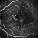
Polypoidal Choroidal Vasculopathy with Pulsating ICG polyp left eye727 viewsOct 11, 2014
|
|
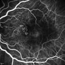
Polypoidal Choroidal Vasculopathy with Pulsating ICG polyp left eye628 viewsOct 11, 2014
|
|
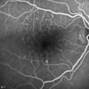
Polypoidal Choroidal Vasculopathy with Pulsating ICG polyp left eye654 viewsOct 11, 2014
|
|
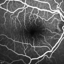
Polypoidal Choroidal Vasculopathy with Pulsating ICG polyp left eye598 viewsOct 11, 2014
|
|
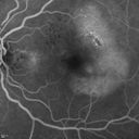
Polypoidal Choroidal Vasculopathy with Pulsating ICG polyp left eye641 viewsOct 11, 2014
|
|
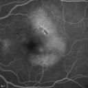
Polypoidal Choroidal Vasculopathy with Pulsating ICG polyp left eye601 viewsOct 11, 2014
|
|
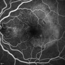
Polypoidal Choroidal Vasculopathy with Pulsating ICG polyp left eye544 viewsOct 11, 2014
|
|
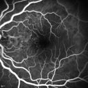
Polypoidal Choroidal Vasculopathy with Pulsating ICG polyp left eye562 viewsOct 11, 2014
|
|
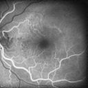
Polypoidal Choroidal Vasculopathy with Pulsating ICG polyp left eye560 viewsOct 11, 2014
|
|
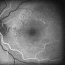
Polypoidal Choroidal Vasculopathy with Pulsating ICG polyp left eye544 viewsOct 11, 2014
|
|
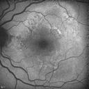
Polypoidal Choroidal Vasculopathy with Pulsating ICG polyp left eye593 viewsOct 11, 2014
|
|

Polypoidal Choroidal Vasculopathy with Pulsating ICG polyp left eye732 viewsOct 11, 2014
|
|

Polypoidal Choroidal Vasculopathy with Pulsating ICG polyp left eye677 viewsOct 11, 2014
|
|

Polypoidal Choroidal Vasculopathy with Pulsating ICG polyp left eye713 viewsOct 11, 2014
|
|

Polypoidal Choroidal Vasculopathy with Pulsating ICG polyp left eye579 viewsOct 11, 2014
|
|

Polypoidal Choroidal Vasculopathy with Pulsating ICG polyp left eye684 viewsOct 11, 2014
|
|

Polypoidal Choroidal Vasculopathy with Pulsating ICG polyp left eye642 viewsOct 11, 2014
|
|
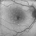
Polypoidal Choroidal Vasculopathy with Pulsating ICG polyp left eye553 viewsOct 11, 2014
|
|
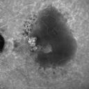
Polypoidal Choroidal Vasculopathy with Pulsating ICG polyp left eye619 viewsOct 11, 2014
|
|
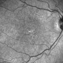
Polypoidal Choroidal Vasculopathy with Pulsating ICG polyp left eye551 viewsOct 11, 2014
|
|
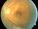
Polypoidal Choroidal Vasculopathy with Pulsating ICG polyp left eye548 viewsOct 11, 2014
|
|

Polypoidal Choroidal Vasculopathy with Pulsating ICG polyp left eye627 viewsOct 11, 2014
|
|
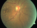
Polypoidal Choroidal Vasculopathy with Pulsating ICG polyp left eye556 viewsOct 11, 2014
|
|
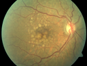
Polypoidal Choroidal Vasculopathy with Pulsating ICG polyp left eye678 viewsOct 11, 2014
|
|

Retinal Hemorrhage351 viewsFundus photo shows retinal hemorrhage located inferior, temporal to the macula in the left eye. Sep 16, 2014
|
|

Macular Pucker1282 viewsPatient comes in for followup on macular pucker, right eye. VA is 20/50 in the right eye...Fundus photo shows prominent pucker centrally pulling temporally in the right eye.Sep 16, 2014
|
|

Macular Hole Left Eye - progression from VMT - Stage II macular hole - Spontaneously Closed Macular Hole574 viewsVision has improved to 20/40 and the hole is closedSep 06, 2014
|
|

Macular Hole Left Eye - progression from VMT - Stage II macular hole - Spontaneously Closed Macular Hole630 viewsVision has improved to 20/40 and the hole is closedSep 06, 2014
|
|

Macular Hole Left Eye - progression from VMT - Stage II macular hole - Spontaneously Closed Macular Hole665 viewsVision has improved to 20/40 and the hole is closedSep 06, 2014
|
|

Macular Hole Left Eye - progression from VMT - Stage II macular hole - Spontaneously Closed Macular Hole565 viewsMacular hole is closing and vision has improved to 20/40Sep 06, 2014
|
|

Macular Hole Left Eye - progression from VMT - Stage II macular hole - Spontaneously Closed Macular Hole534 viewsMacular hole is closing and vision has improved to 20/40Sep 06, 2014
|
|

Macular Hole Left Eye - progression from VMT - Stage II macular hole - Spontaneously Closed Macular Hole551 viewsMacular hole is closing and vision has improved to 20/40Sep 06, 2014
|
|

Macular Hole Left Eye - progression from VMT - Stage II macular hole - Spontaneously Closed Macular Hole579 viewsMacular hole is closing and vision has improved to 20/40Sep 06, 2014
|
|

Macular Hole Left Eye - progression from VMT - Stage II macular hole - Spontaneously Closed Macular Hole605 viewsSep 06, 2014
|
|

Macular Hole Left Eye - progression from VMT - Stage II macular hole - Spontaneously Closed Macular Hole620 viewsSep 06, 2014
|
|

Macular Hole Left Eye - progression from VMT - Stage II macular hole - Spontaneously Closed Macular Hole532 viewsSep 06, 2014
|
|

Macular Hole Left Eye - progression from VMT - Stage II macular hole - Spontaneously Closed Macular Hole608 viewsVision has dropped to 20/50 and there is a full thickness macular holeSep 06, 2014
|
|

Macular Hole Left Eye - progression from VMT - Stage II macular hole - Spontaneously Closed Macular Hole486 viewsVision has dropped to 20/50 and there is a full thickness macular holeSep 06, 2014
|
|

Macular Hole Left Eye - progression from VMT - Stage II macular hole - Spontaneously Closed Macular Hole466 viewsPucker in fellow eye is stableSep 06, 2014
|
|

Macular Hole Left Eye - progression from VMT - Stage II macular hole - Spontaneously Closed Macular Hole490 viewsSep 06, 2014
|
|

Macular Hole Left Eye - progression from VMT - Stage II macular hole - Spontaneously Closed Macular Hole454 viewsVision has dropped to 20/50 and there is a full thickness macular holeSep 06, 2014
|
|

Macular Hole Left Eye - progression from VMT - Stage II macular hole - Spontaneously Closed Macular Hole520 viewsVision has dropped to 20/50 and there is a full thickness macular holeSep 06, 2014
|
|

Macular Hole Left Eye - progression from VMT - Stage II macular hole - Spontaneously Closed Macular Hole490 viewsSep 06, 2014
|
|

Macular Hole Left Eye - progression from VMT - Stage II macular hole - Spontaneously Closed Macular Hole531 viewsPucker False ColorSep 06, 2014
|
|

Macular Hole Left Eye - progression from VMT - Stage II macular hole - Spontaneously Closed Macular Hole524 viewsVision better left eye VMT has released the fovea (was 20/25)Sep 06, 2014
|
|

Macular Hole Left Eye - progression from VMT - Stage II macular hole - Spontaneously Closed Macular Hole537 viewsVision better left eye VMT has released the fovea (was 20/25)Sep 06, 2014
|
|

Macular Hole Left Eye - progression from VMT - Stage II macular hole - Spontaneously Closed Macular Hole481 viewsSep 06, 2014
|
|

Macular Hole Left Eye - progression from VMT - Stage II macular hole - Spontaneously Closed Macular Hole533 viewsAsymptomatic VMT OS with 20/30 VASep 06, 2014
|
|

Macular Hole Left Eye - progression from VMT - Stage II macular hole - Spontaneously Closed Macular Hole515 viewsSep 06, 2014
|
|

Macular Hole Left Eye - progression from VMT - Stage II macular hole - Spontaneously Closed Macular Hole525 viewsSep 06, 2014
|
|

Macular Hole Left Eye - progression from VMT - Stage II macular hole - Spontaneously Closed Macular Hole506 viewsSep 06, 2014
|
|

Macular Hole Left Eye - progression from VMT - Stage II macular hole - Spontaneously Closed Macular Hole545 viewsSep 06, 2014
|
|

Macular Hole Left Eye - progression from VMT - Stage II macular hole - Spontaneously Closed Macular Hole610 viewsSep 06, 2014
|
|

398 viewsJun 17, 2014
|
|
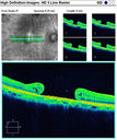
OCT Macular Hole 413 viewsMacular Hole OCTMay 15, 2014
|
|

Red Free Macular Hole 418 viewsFull Thickness Macular HoleMay 15, 2014
|
|
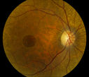
Full Thickness Macular Hole1155 viewsFull Thickness Macular HoleMay 15, 2014
|
|

440 viewsMay 03, 2014
|
|

444 viewsMay 03, 2014
|
|

408 viewsMay 03, 2014
|
|

443 viewsMay 03, 2014
|
|

623 viewsMay 03, 2014
|
|

369 viewsMay 03, 2014
|
|

401 viewsMay 03, 2014
|
|

444 viewsMay 03, 2014
|
|

454 viewsMay 03, 2014
|
|

Macular Degeneration488 viewsPatient complains of spot in the right eye. VA 20/400 in the right eye. Fundus photo shows macular degeneration changes with small layer of hemorrhage on the fovea in the right eye.Mar 28, 2014
|
|

Epiretinal Membrane1050 viewsPatient with a history of pars planitis comes in with decreased vision in the left eye. VA is 20/30 in the left eye. Fundus photos show a rather large epiretinal membrane covering the optic nerve in the left eye. Mar 27, 2014
|
|
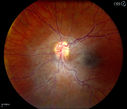
Epiretinal Membrane424 viewsPatient with a history of pars planitis comes in with decreased vision in the left eye. VA is 20/30 in the left eye. Fundus photos show a rather large epiretinal membrane covering the optic nerve in the left eye. Mar 27, 2014
|
|
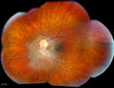
Epiretinal Membrane504 viewsPatient with a history of pars planitis comes in with decreased vision in the left eye. VA is 20/30 in the left eye. Fundus photos show a rather large epiretinal membrane covering the optic nerve in the left eye. Mar 27, 2014
|
|
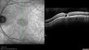
Macular Telangiectasia - Stage 5 left eye - 20/40 vision after 2 years of Avastin 741 viewsSpectral Domain OCT Line ScanMar 17, 2014
|
|
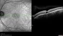
Macular Telangiectasia - Stage 5 left eye - 20/40 vision after 2 years of Avastin 507 viewsSpectral Domain OCT Line ScanMar 17, 2014
|
|
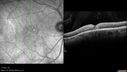
Macular Telangiectasia - Stage 3 right eye 20/40 vision591 viewsSpectral Domain OCT Line ScanMar 17, 2014
|
|
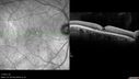
Macular Telangiectasia - Stage 3 right eye 20/40 vision538 viewsSpectral Domain OCT Line ScanMar 17, 2014
|
|
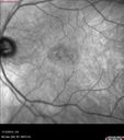
Macular Telangiectasia - Stage 5 left eye - 20/40 vision after 2 years of Avastin 466 viewsConfocal InfraRed ReflectanceMar 17, 2014
|
|
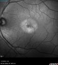
Macular Telangiectasia - Stage 5 left eye - 20/40 vision after 2 years of Avastin 500 viewsConfocal Green ReflectanceMar 17, 2014
|
|
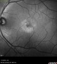
Macular Telangiectasia - Stage 5 left eye - 20/40 vision after 2 years of Avastin - Bulls Eye Blue Reflectance524 viewsConfocal Blue ReflectanceMar 17, 2014
|
|

Macular Telangiectasia - Stage 5 left eye - 20/40 vision after 2 years of Avastin 553 viewsMulticolor ImageMar 17, 2014
|
|

Macular Telangiectasia - Stage 3 right eye 20/40 vision572 viewsConfocal InfraRed ReflectanceMar 17, 2014
|
|
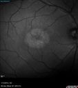
Macular Telangiectasia - Stage 3 right eye 20/40 vision530 viewsConfocal Green ReflectanceMar 17, 2014
|
|
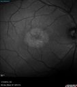
Macular Telangiectasia - Stage 3 right eye 20/40 vision - Bulls Eye Blue Reflectance535 viewsConfocal Blue ReflectanceMar 17, 2014
|
|
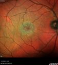
Macular Telangiectasia - Stage 3 right eye 20/40 vision558 viewsMulti-Color ImageMar 17, 2014
|
|

Vitreomacular traction - recent vision loss Right Eye - initially worse after Jetrea594 viewsFeb 18, 2014
|
|

Vitreomacular traction - recent vision loss Right Eye - initially worse after Jetrea528 viewsFeb 18, 2014
|
|

Vitreomacular traction - recent vision loss Right Eye 544 viewsFeb 18, 2014
|
|
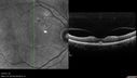
Vitreomacular traction - recent vision loss Right Eye 493 viewsFeb 18, 2014
|
|

Vitreomacular traction - recent vision loss Right Eye 558 viewsFeb 18, 2014
|
|

Vitreomacular traction - recent vision loss Right Eye 409 viewsFeb 18, 2014
|
|

Vitreomacular traction - one month post-Vitrectomy left eye - vision is back to normal - SD OCT Line Scan536 viewsFeb 18, 2014
|
|

Vitreomacular traction - recent vision loss left eye - FA527 viewsFeb 18, 2014
|
|

Vitreomacular traction - recent vision loss left eye - FA578 viewsFeb 18, 2014
|
|
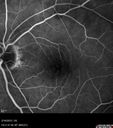
Vitreomacular traction - recent vision loss left eye - FA474 viewsFeb 18, 2014
|
|

Vitreomacular traction - recent vision loss left eye - FA522 viewsFeb 18, 2014
|
|

Vitreomacular traction - recent vision loss left eye - FA561 viewsFeb 18, 2014
|
|
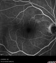
Vitreomacular traction - recent vision loss left eye - FA513 viewsFeb 18, 2014
|
|
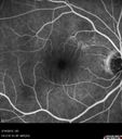
Vitreomacular traction - recent vision loss left eye - FA509 viewsFeb 18, 2014
|
|

Vitreomacular traction - recent vision loss left eye - FA586 viewsFeb 18, 2014
|
|
| 5013 files on 51 page(s) |
 |
|
|
|
|
31 | |
|
|
|
|


