Last additions - Macular Disease
|

Vitreomacular traction - recent vision loss - preop SD OCT579 viewsFeb 18, 2014
|
|

Vitreomacular traction - recent vision loss - preop SD OCT492 viewsFeb 18, 2014
|
|

Vitreomacular traction - recent vision loss - preop SD OCT574 viewsFeb 18, 2014
|
|

Vitreomacular traction - normal vision on initial presentation576 viewsFeb 18, 2014
|
|

672 viewsFeb 18, 2014
|
|

575 viewsFeb 18, 2014
|
|

633 viewsFeb 18, 2014
|
|

640 viewsFeb 18, 2014
|
|

589 viewsFeb 18, 2014
|
|

Vitreomacular traction - recent vision loss Right Eye - initially worse after Jetrea565 viewsFeb 18, 2014
|
|

Vitreomacular traction - recent vision loss Right Eye - initially worse after Jetrea then better848 viewsFeb 18, 2014
|
|
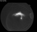
Classic Smoke Stack787 views32 yrs old male Presents: with blurry vision x 10 day's visual acuity OD CF@5ft, Note Observation was recommended. Patient return 10/ 2013 with visual improvement of 20/30 and on 11/2013 VA 20/25 stable.Jan 27, 2014
|
|

567 viewsDec 06, 2013
|
|

457 viewsDec 06, 2013
|
|
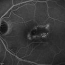
463 viewsDec 06, 2013
|
|

441 viewsDec 06, 2013
|
|

451 viewsDec 06, 2013
|
|

475 viewsDec 06, 2013
|
|

496 viewsDec 06, 2013
|
|

507 viewsDec 06, 2013
|
|

470 viewsDec 06, 2013
|
|

459 viewsDec 06, 2013
|
|

431 viewsDec 06, 2013
|
|

458 viewsDec 06, 2013
|
|

443 viewsDec 06, 2013
|
|

430 viewsDec 06, 2013
|
|

461 viewsDec 06, 2013
|
|

447 viewsDec 06, 2013
|
|

454 viewsDec 06, 2013
|
|

469 viewsDec 06, 2013
|
|

442 viewsDec 06, 2013
|
|

440 viewsDec 06, 2013
|
|

477 viewsDec 06, 2013
|
|

437 viewsDec 06, 2013
|
|

415 viewsDec 06, 2013
|
|

458 viewsDec 06, 2013
|
|

436 viewsDec 06, 2013
|
|

432 viewsDec 06, 2013
|
|

440 viewsDec 06, 2013
|
|

487 viewsDec 06, 2013
|
|

486 viewsDec 06, 2013
|
|

Cartwheel CNVM - Classic - wet AMD - evidence of possible old CSR - ICG is normal - shows nothing543 views75 y.o Decreased Vision right eye for one year. VA OD: sc20/125+2 OS 20/25. Autofluorescence and SD OCT show evidence of old serous retinal detachment superior to the fovea suggesting a history of CSR. The macular lesion on FA shows a classic cartwheel appearanceNov 01, 2013
|
|

Cartwheel CNVM - Classic - wet AMD - evidence of possible old CSR - ICG is normal - shows nothing500 views75 y.o Decreased Vision right eye for one year. VA OD: sc20/125+2 OS 20/25. Autofluorescence and SD OCT show evidence of old serous retinal detachment superior to the fovea suggesting a history of CSR. The macular lesion on FA shows a classic cartwheel appearanceNov 01, 2013
|
|

Cartwheel CNVM - Classic - wet AMD - evidence of possible old CSR - ICG is normal - shows nothing537 views75 y.o Decreased Vision right eye for one year. VA OD: sc20/125+2 OS 20/25. Autofluorescence and SD OCT show evidence of old serous retinal detachment superior to the fovea suggesting a history of CSR. The macular lesion on FA shows a classic cartwheel appearanceNov 01, 2013
|
|
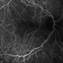
Cartwheel CNVM - Classic - wet AMD - evidence of possible old CSR - ICG is normal - shows nothing624 views75 y.o Decreased Vision right eye for one year. VA OD: sc20/125+2 OS 20/25. Autofluorescence and SD OCT show evidence of old serous retinal detachment superior to the fovea suggesting a history of CSR. The macular lesion on FA shows a classic cartwheel appearanceNov 01, 2013
|
|
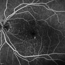
Cartwheel CNVM - Classic - wet AMD - evidence of possible old CSR - hydrophilic drusen stain early in FA570 views75 y.o Decreased Vision right eye for one year. VA OD: sc20/125+2 OS 20/25. Autofluorescence and SD OCT show evidence of old serous retinal detachment superior to the fovea suggesting a history of CSR. The macular lesion on FA shows a classic cartwheel appearanceNov 01, 2013
|
|
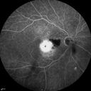
Cartwheel CNVM - Classic - wet AMD - evidence of possible old CSR - FA shows cartwheel shaped CNVM511 views75 y.o Decreased Vision right eye for one year. VA OD: sc20/125+2 OS 20/25. Autofluorescence and SD OCT show evidence of old serous retinal detachment superior to the fovea suggesting a history of CSR. The macular lesion on FA shows a classic cartwheel appearanceNov 01, 2013
|
|

Cartwheel CNVM - Classic - wet AMD - evidence of possible old CSR - FA shows cartwheel shaped CNVM694 views75 y.o Decreased Vision right eye for one year. VA OD: sc20/125+2 OS 20/25. Autofluorescence and SD OCT show evidence of old serous retinal detachment superior to the fovea suggesting a history of CSR. The macular lesion on FA shows a classic cartwheel appearanceNov 01, 2013
|
|

Cartwheel CNVM - Classic - wet AMD - evidence of possible old CSR - FA shows cartwheel shaped CNVM722 views75 y.o Decreased Vision right eye for one year. VA OD: sc20/125+2 OS 20/25. Autofluorescence and SD OCT show evidence of old serous retinal detachment superior to the fovea suggesting a history of CSR. The macular lesion on FA shows a classic cartwheel appearanceNov 01, 2013
|
|

Cartwheel CNVM - Classic - wet AMD - evidence of possible old CSR - FA shows cartwheel shaped CNVM698 views75 y.o Decreased Vision right eye for one year. VA OD: sc20/125+2 OS 20/25. Autofluorescence and SD OCT show evidence of old serous retinal detachment superior to the fovea suggesting a history of CSR. The macular lesion on FA shows a classic cartwheel appearanceNov 01, 2013
|
|

Cartwheel CNVM - Classic - wet AMD - evidence of possible old CSR569 views75 y.o Decreased Vision right eye for one year. VA OD: sc20/125+2 OS 20/25. Autofluorescence and SD OCT show evidence of old serous retinal detachment superior to the fovea suggesting a history of CSR. The macular lesion on FA shows a classic cartwheel appearanceNov 01, 2013
|
|
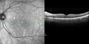
Cartwheel CNVM - Classic - wet AMD - evidence of possible old CSR543 views75 y.o Decreased Vision right eye for one year. VA OD: sc20/125+2 OS 20/25. Autofluorescence and SD OCT show evidence of old serous retinal detachment superior to the fovea suggesting a history of CSR. The macular lesion on FA shows a classic cartwheel appearanceNov 01, 2013
|
|

Cartwheel CNVM - Classic - wet AMD - evidence of possible old CSR852 views75 y.o Decreased Vision right eye for one year. VA OD: sc20/125+2 OS 20/25. Autofluorescence and SD OCT show evidence of old serous retinal detachment superior to the fovea suggesting a history of CSR. The macular lesion on FA shows a classic cartwheel appearanceNov 01, 2013
|
|

Cartwheel CNVM - Classic - wet AMD - evidence of possible old CSR - Autofluorescence shows brigh spot superior to subfoveal lesion875 views75 y.o Decreased Vision right eye for one year. VA OD: sc20/125+2 OS 20/25. Autofluorescence and SD OCT show evidence of old serous retinal detachment superior to the fovea suggesting a history of CSR. The macular lesion on FA shows a classic cartwheel appearanceNov 01, 2013
|
|

Cartwheel CNVM - Classic - wet AMD - evidence of possible old CSR769 views75 y.o Decreased Vision right eye for one year. VA OD: sc20/125+2 OS 20/25. Autofluorescence and SD OCT show evidence of old serous retinal detachment superior to the fovea suggesting a history of CSR. The macular lesion on FA shows a classic cartwheel appearanceNov 01, 2013
|
|

Cartwheel CNVM - Classic - wet AMD - evidence of possible old CSR - Fellow eye has hydrophilic Drusen771 views75 y.o Decreased Vision right eye for one year. VA OD: sc20/125+2 OS 20/25. Autofluorescence and SD OCT show evidence of old serous retinal detachment superior to the fovea suggesting a history of CSR. The macular lesion on FA shows a classic cartwheel appearanceNov 01, 2013
|
|

Cartwheel CNVM - Classic - wet AMD - evidence of possible old CSR - Subretinal hemorrhage next to subfoveal lesion1053 views75 y.o Decreased Vision right eye for one year. VA OD: sc20/125+2 OS 20/25. Autofluorescence and SD OCT show evidence of old serous retinal detachment superior to the fovea suggesting a history of CSR. The macular lesion on FA shows a classic cartwheel appearanceNov 01, 2013
|
|

Myopic Degeneration with RPE Loss1154 viewsPatient comes in for follow up for glaucoma. VA is 20/30 in both eyes. Fundus photography shows the RPE loss due to myopic degeneration, nasally, inferiority in the left eye.Oct 31, 2013
|
|

Reticular Macular Disease (Dry Macular Degeneration) - Spectral Domain Optical Coherence Tomography Line Scan843 views85 year old woman with 20/25 vision OU and reticular macular diseaseSep 25, 2013
|
|

Reticular Macular Disease (Dry Macular Degeneration) - Spectral Domain Optical Coherence Tomography Line Scan667 views85 year old woman with 20/25 vision OU and reticular macular diseaseSep 25, 2013
|
|

Reticular Macular Disease (Dry Macular Degeneration) - Spectral Domain Optical Coherence Tomography Line Scan578 views85 year old woman with 20/25 vision OU and reticular macular diseaseSep 25, 2013
|
|

Reticular Macular Disease (Dry Macular Degeneration) - Spectral Domain Optical Coherence Tomography Line Scan571 views85 year old woman with 20/25 vision OU and reticular macular diseaseSep 25, 2013
|
|

Reticular Macular Disease (Dry Macular Degeneration) - Fundus Autolfuorescence613 views85 year old woman with 20/25 vision OU and reticular macular diseaseSep 25, 2013
|
|

Reticular Macular Disease (Dry Macular Degeneration) - Fundus Autolfuorescence607 views85 year old woman with 20/25 vision OU and reticular macular diseaseSep 25, 2013
|
|

Reticular Macular Disease (Dry Macular Degeneration) - Infrared reflectance681 views85 year old woman with 20/25 vision OU and reticular macular diseaseSep 25, 2013
|
|

Reticular Macular Disease (Dry Macular Degeneration) - Infrared reflectance719 views85 year old woman with 20/25 vision OU and reticular macular diseaseSep 25, 2013
|
|

Reticular Macular Disease (Dry Macular Degeneration) - Color Photo720 views85 year old woman with 20/25 vision OU and reticular macular diseaseSep 25, 2013
|
|

Reticular Macular Disease (Dry Macular Degeneration) - Color Photo807 views85 year old woman with 20/25 vision OU and reticular macular diseaseSep 25, 2013
|
|
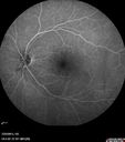
Macular Pucker 20/40 vision - minimal symptoms for 1 year Right eye555 viewsFA shows leakage from traction from pucker in the right eye - cystoid macular edemaSep 25, 2013
|
|
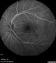
Macular Pucker 20/40 vision - minimal symptoms for 1 year Right eye486 viewsFA shows leakage from traction from pucker in the right eye - cystoid macular edemaSep 25, 2013
|
|
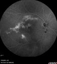
Macular Pucker 20/40 vision - minimal symptoms for 1 year Right eye481 viewsFA shows leakage from traction from pucker in the right eye - cystoid macular edemaSep 25, 2013
|
|
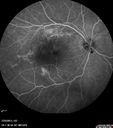
Macular Pucker 20/40 vision - minimal symptoms for 1 year Right eye468 viewsFA shows leakage from traction from pucker in the right eye - cystoid macular edemaSep 25, 2013
|
|
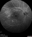
Macular Pucker 20/40 vision - minimal symptoms for 1 year Right eye487 viewsFA shows leakage from traction from pucker in the right eye - cystoid macular edemaSep 25, 2013
|
|
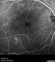
Macular Pucker 20/40 vision - minimal symptoms for 1 year Right eye486 viewsFA shows leakage from traction from pucker in the right eye - cystoid macular edemaSep 25, 2013
|
|
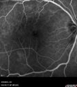
Macular Pucker 20/40 vision - minimal symptoms for 1 year Right eye538 viewsFA shows leakage from traction from pucker in the right eye - cystoid macular edemaSep 25, 2013
|
|
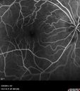
Macular Pucker 20/40 vision - minimal symptoms for 1 year Right eye512 viewsFA shows leakage from traction from pucker in the right eye - cystoid macular edemaSep 25, 2013
|
|

Macular Pucker 20/40 vision - minimal symptoms for 1 year Right eye458 viewsFA shows leakage from traction from pucker in the right eye - cystoid macular edemaSep 25, 2013
|
|
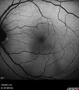
Macular Pucker 20/40 vision - minimal symptoms for 1 year Right eye466 viewsFundus AutofluorescenceSep 25, 2013
|
|
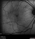
Macular Pucker 20/40 vision - minimal symptoms for 1 year Right eye514 viewsFundus AutofluorescenceSep 25, 2013
|
|
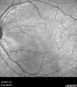
Macular Pucker 20/40 vision - minimal symptoms for 1 year Right eye488 viewsIRSep 25, 2013
|
|
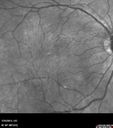
Macular Pucker 20/40 vision - minimal symptoms for 1 year Right eye468 viewsIRSep 25, 2013
|
|
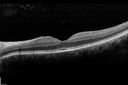
Macular Pucker 20/40 vision - minimal symptoms for 1 year Right eye619 views SD OCT line Scan Fellow Eye 20/16 Sep 25, 2013
|
|
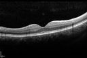
Macular Pucker 20/40 vision - minimal symptoms for 1 year Right eye592 viewsSD OCT line Scan Fellow Eye 20/16Sep 25, 2013
|
|
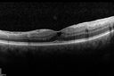
Macular Pucker 20/40 vision - minimal symptoms for 1 year Right eye594 viewsSD OCT line Scan Affected EyeSep 25, 2013
|
|
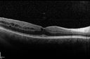
Macular Pucker 20/40 vision - minimal symptoms for 1 year Right eye518 viewsSD OCT line Scan Affected EyeSep 25, 2013
|
|
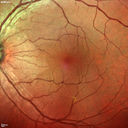
Macular Pucker 20/40 vision - minimal symptoms for 1 year Right eye616 viewsMulticolorSep 25, 2013
|
|

Macular Pucker 20/40 vision - minimal symptoms for 1 year Right eye621 viewsMulticolorSep 25, 2013
|
|
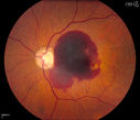
Retinal Hemorrhage 1314 viewsFundus photography shows central retinal hemorrhage. Patient was treated with Avastin Injection and will be followed up in 4-weeks.Sep 11, 2013
|
|

Myopic Maculoscheisis408 viewsFluorescence Angiography and Fundus photography shows RPE mottling and staphyloma in the right eye. VA is 20/50, right eye. No sub retinal fluid found. Sep 11, 2013
|
|

Macular Degeneration with Hemorrhage537 viewsPatient comes in with active leaking of AMD in the left eye. VA is 20/40 in the left eye. Patient was treated with Eylea and will return in 1-month for follow up.Jul 16, 2013
|
|
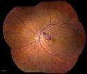
Macular Degeneration with Hemorrhage515 viewsPatient comes in with active leaking of AMD in the left eye. VA is 20/40 in the left eye. Patient was treated with Eylea and will return in 1-month for follow up.Jul 16, 2013
|
|

Outer Retinal Tubulation974 viewsPatient with a history of wet macular degeneration and glaucoma in both eyes. VA is 20/50, right eye, 20/80, left eye. Patient is treated with Eylea in both eyes. Enhanced Depth Imaging OCT reveals a small like form of a cyst which in fact isn't a cyst at all. This is called Outer Retinal Tubulation in which degenerating photo-receptors may become arranged in a circular or ovoid fashion. This is sometimes misdiagnosed as cystic changes in the retinal pigment epithelium or sub-retinal fluid. May 29, 2013
|
|

Macular Hole with Peripheral Laser Scars1180 viewsDiabetic patient comes in with flashes and floaters in both eyes. Patient's VA is 20/400, right eye and 20/25, left eye. Fundus exam presents with large macular hole with diabetic changes in the retina. Laser scars in the peripheral. May 20, 2013
|
|
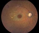
Macular Hole with Peripheral Laser Scars435 viewsDiabetic patient comes in with flashes and floaters in both eyes. Patient's VA is 20/400, right eye and 20/25, left eye. Fundus exam presents with large macular hole with diabetic changes in the retina. Laser scars in the peripheral. May 20, 2013
|
|

Idiopathic Polypoidal Choroidal Vasculopathy - Indocyanine Green Angiogram - Polyp Visible Left Eye. Branching Network Both Eye754 viewsApr 25, 2013
|
|

Idiopathic Polypoidal Choroidal Vasculopathy - Indocyanine Green Angiogram - Polyp Visible Left Eye. Branching Network Both Eye750 viewsApr 25, 2013
|
|

Idiopathic Polypoidal Choroidal Vasculopathy - Indocyanine Green Angiogram - Polyp Visible Left Eye. Branching Network Both Eye818 viewsApr 25, 2013
|
|

Asymptomatic Occult Subfoveal CNVM in Good Eye Visible on ICG617 viewsApr 25, 2013
|
|
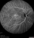
Asymptomatic Occult Subfoveal CNVM in Good Eye Visible on ICG872 viewsApr 25, 2013
|
|
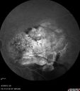
Idiopathic Polypoidal Choroidal Vasculopathy - Fluorescein Angiogram587 viewsApr 25, 2013
|
|
| 5013 files on 51 page(s) |
 |
|
|
|
|
32 | |
|
|
|
|


