Last additions - Macular Disease
|

Serous Macular Detachment Both Eyes - Wet AMD vs. CSR - 62 Year Old Man - FAF571 viewsMar 05, 2013
|
|

Serous Macular Detachment Both Eyes - Wet AMD vs. CSR - 62 Year Old Man - FAF561 viewsMar 05, 2013
|
|

Serous Macular Detachment Both Eyes - Wet AMD vs. CSR - 62 Year Old Man - IR543 viewsMar 05, 2013
|
|

Serous Macular Detachment Both Eyes - Wet AMD vs. CSR - 62 Year Old Man - IR539 viewsMar 05, 2013
|
|

Serous Macular Detachment Both Eyes - Wet AMD vs. CSR - 62 Year Old Man - Color Photo545 viewsMar 05, 2013
|
|
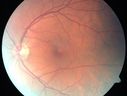
Serous Macular Detachment Both Eyes - Wet AMD vs. CSR - 62 Year Old Man - Color Photo583 viewsMar 05, 2013
|
|

Serous Macular Detachment Both Eyes - Wet AMD vs. CSR - 62 Year Old Man - Color Photo576 viewsMar 05, 2013
|
|

Wet AMD - New Small Hemorrhage Both Eyes - Mature CNVM visible on Early Indocyanine Green Angiogram827 viewsFeb 28, 2013
|
|

Wet AMD - New Small Hemorrhage Both Eyes - Mature CNVM visible on Early Indocyanine Green Angiogram757 viewsFeb 28, 2013
|
|

Wet AMD - New Small Hemorrhage Both Eyes - Mature CNVM visible on Early Indocyanine Green Angiogram588 viewsFeb 28, 2013
|
|

Wet AMD - New Small Hemorrhage Both Eyes - Mature CNVM visible on Early Indocyanine Green Angiogram721 viewsFeb 28, 2013
|
|

Wet AMD - New Small Hemorrhage Both Eyes - Mature CNVM visible on Early Indocyanine Green Angiogram706 viewsFeb 28, 2013
|
|

Wet AMD - New Small Hemorrhage Both Eyes - Mature CNVM visible on Early Indocyanine Green Angiogram630 viewsFeb 28, 2013
|
|

Wet AMD - New Small Hemorrhage Both Eyes - Mature CNVM visible on Early Indocyanine Green Angiogram577 viewsFeb 28, 2013
|
|

Wet AMD - New Small Hemorrhage Both Eyes - Mature CNVM visible on Early Indocyanine Green Angiogram612 viewsFeb 28, 2013
|
|

Wet AMD - New Small Hemorrhage Both Eyes - Mature CNVM visible on Early Indocyanine Green Angiogram657 viewsFeb 28, 2013
|
|

Wet AMD - New Small Hemorrhage Both Eyes - Mature CNVM visible on Early Indocyanine Green Angiogram658 viewsFeb 28, 2013
|
|

Wet AMD - New Small Hemorrhage Both Eyes - Mature CNVM visible on Early Indocyanine Green Angiogram630 viewsFeb 28, 2013
|
|

Wet AMD - New Small Hemorrhage Both Eyes - Mature CNVM visible on Early Indocyanine Green Angiogram653 viewsFeb 28, 2013
|
|

Wet AMD - New Small Hemorrhage Both Eyes - Mature CNVM visible on Early Indocyanine Green Angiogram668 viewsFeb 28, 2013
|
|

Wet AMD - New Small Hemorrhage Both Eyes - Mature CNVM visible on Early Indocyanine Green Angiogram668 viewsFeb 28, 2013
|
|

Wet AMD - New Small Hemorrhage Both Eyes - Mature CNVM visible on Early Indocyanine Green Angiogram671 viewsFeb 28, 2013
|
|

Wet AMD - New Small Hemorrhage Both Eyes - Mature CNVM visible on Early Indocyanine Green Angiogram666 viewsFeb 28, 2013
|
|

Wet AMD - New Small Hemorrhage Both Eyes - Mature CNVM visible on Early Indocyanine Green Angiogram687 viewsFeb 28, 2013
|
|

Psuedomacular Hole - Epiretinal Membrane - Vision unchanged 20/40 for 6 months669 viewsFeb 22, 2013
|
|
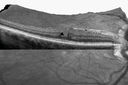
Psuedomacular Hole - Epiretinal Membrane - Vision unchanged 20/40 for 6 months512 viewsVIDEO TAKES TIME TO LOADFeb 22, 2013
|
|
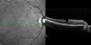
Psuedomacular Hole - Epiretinal Membrane - Vision unchanged 20/40 for 6 months692 viewsFeb 22, 2013
|
|
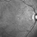
Psuedomacular Hole - Epiretinal Membrane - Vision unchanged 20/40 for 6 months621 viewsFeb 22, 2013
|
|

Asymptomatic Juxtapapillary Choroidal Neovascular Membrane - Reticular Macular Disease (Wet AMD) 582 viewsFeb 11, 2013
|
|

Asymptomatic Juxtapapillary Choroidal Neovascular Membrane - Reticular Macular Disease (Wet AMD) 501 viewsFeb 11, 2013
|
|

Asymptomatic Juxtapapillary Choroidal Neovascular Membrane - Reticular Macular Disease (Wet AMD) 559 viewsFeb 11, 2013
|
|

Asymptomatic Juxtapapillary Choroidal Neovascular Membrane - Reticular Macular Disease (Wet AMD) 546 viewsFeb 11, 2013
|
|

Asymptomatic Juxtapapillary Choroidal Neovascular Membrane - Reticular Macular Disease (Wet AMD) 504 viewsFeb 11, 2013
|
|

Asymptomatic Juxtapapillary Choroidal Neovascular Membrane - Reticular Macular Disease (Wet AMD) 513 viewsFeb 11, 2013
|
|

Asymptomatic Juxtapapillary Choroidal Neovascular Membrane - Reticular Macular Disease (Wet AMD) 514 viewsFeb 11, 2013
|
|

Asymptomatic Juxtapapillary Choroidal Neovascular Membrane - Reticular Macular Disease (Wet AMD) 567 viewsFeb 11, 2013
|
|

Asymptomatic Juxtapapillary Choroidal Neovascular Membrane - Reticular Macular Disease (Wet AMD) 558 viewsFeb 11, 2013
|
|

Asymptomatic Juxtapapillary Choroidal Neovascular Membrane - Reticular Macular Disease (Wet AMD) 591 viewsFeb 11, 2013
|
|

Asymptomatic Juxtapapillary Choroidal Neovascular Membrane - Reticular Macular Disease (Wet AMD) 538 viewsFeb 11, 2013
|
|

Asymptomatic Juxtapapillary Choroidal Neovascular Membrane - Reticular Macular Disease (Wet AMD) 593 viewsFeb 11, 2013
|
|

Asymptomatic Juxtapapillary Choroidal Neovascular Membrane - Reticular Macular Disease (Wet AMD) 561 viewsFeb 11, 2013
|
|

Asymptomatic Juxtapapillary Choroidal Neovascular Membrane - Reticular Macular Disease (Wet AMD) 601 viewsFeb 11, 2013
|
|

Asymptomatic Juxtapapillary Choroidal Neovascular Membrane - Reticular Macular Disease (Wet AMD) 506 viewsFeb 11, 2013
|
|

Asymptomatic Juxtapapillary Choroidal Neovascular Membrane - Reticular Macular Disease (Wet AMD) 515 viewsFeb 11, 2013
|
|

Asymptomatic Juxtapapillary Choroidal Neovascular Membrane - Reticular Macular Disease (Wet AMD) 756 viewsFeb 11, 2013
|
|

Asymptomatic Juxtapapillary Choroidal Neovascular Membrane - Reticular Macular Disease (Wet AMD) 576 viewsFeb 11, 2013
|
|

Asymptomatic Juxtapapillary Choroidal Neovascular Membrane - Reticular Macular Disease (Wet AMD) 568 viewsFeb 11, 2013
|
|

Asymptomatic Juxtapapillary Choroidal Neovascular Membrane - Reticular Macular Disease (Wet AMD) 580 viewsFeb 11, 2013
|
|

Asymptomatic Juxtapapillary Choroidal Neovascular Membrane - Reticular Macular Disease (Wet AMD) - FAF620 viewsFeb 11, 2013
|
|

Asymptomatic Juxtapapillary Choroidal Neovascular Membrane - Reticular Macular Disease (Wet AMD) - FAF796 viewsFeb 11, 2013
|
|

Asymptomatic Juxtapapillary Choroidal Neovascular Membrane - Reticular Macular Disease (Wet AMD) - Heidelberg Color766 viewsFeb 11, 2013
|
|

Asymptomatic Juxtapapillary Choroidal Neovascular Membrane - Reticular Macular Disease (Wet AMD) - Heidelberg Color706 viewsFeb 11, 2013
|
|

Asymptomatic Juxtapapillary Choroidal Neovascular Membrane - Reticular Macular Disease (Wet AMD) - Heidelberg Color751 viewsFeb 11, 2013
|
|

Central Serous Retinopathy 25 year old professional baseball player - Atypical - Choroidal Hypo-perfusion?664 viewsFeb 01, 2013
|
|

Central Serous Retinopathy 25 year old professional baseball player - Atypical - Choroidal Hypo-perfusion?500 viewsFeb 01, 2013
|
|

Central Serous Retinopathy 25 year old professional baseball player - Atypical - Choroidal Hypo-perfusion?530 viewsFeb 01, 2013
|
|

Central Serous Retinopathy 25 year old professional baseball player - Atypical - Choroidal Hypo-perfusion?481 viewsFeb 01, 2013
|
|

Central Serous Retinopathy 25 year old professional baseball player - Atypical - Choroidal Hypo-perfusion?490 viewsFeb 01, 2013
|
|

Central Serous Retinopathy 25 year old professional baseball player - Atypical - Choroidal Hypo-perfusion?482 viewsFeb 01, 2013
|
|

Central Serous Retinopathy 25 year old professional baseball player - Atypical - Choroidal Hypo-perfusion?516 viewsFeb 01, 2013
|
|

Central Serous Retinopathy 25 year old professional baseball player - Atypical - Choroidal Hypo-perfusion?523 viewsFeb 01, 2013
|
|

Central Serous Retinopathy 25 year old professional baseball player - Atypical - Choroidal Hypo-perfusion?446 viewsFeb 01, 2013
|
|

Central Serous Retinopathy 25 year old professional baseball player - Atypical - Choroidal Hypo-perfusion?444 viewsFeb 01, 2013
|
|

Central Serous Retinopathy 25 year old professional baseball player - Atypical - Choroidal Hypo-perfusion?440 viewsFeb 01, 2013
|
|

Central Serous Retinopathy 25 year old professional baseball player - Atypical - Choroidal Hypo-perfusion?602 viewsFeb 01, 2013
|
|

Central Serous Retinopathy 25 year old professional baseball player - Atypical - Choroidal Hypo-perfusion?550 viewsFeb 01, 2013
|
|

Central Serous Retinopathy 25 year old professional baseball player - Atypical - Choroidal Hypo-perfusion?558 viewsFeb 01, 2013
|
|

Central Serous Retinopathy 25 year old professional baseball player - Atypical - Choroidal Hypo-perfusion?550 viewsFeb 01, 2013
|
|

Central Serous Retinopathy 25 year old professional baseball player - Atypical - Choroidal Hypo-perfusion?559 viewsFeb 01, 2013
|
|

Central Serous Retinopathy 25 year old professional baseball player - Atypical - Choroidal Hypo-perfusion?569 viewsFeb 01, 2013
|
|

Central Serous Retinopathy 25 year old professional baseball player - Atypical - Choroidal Hypo-perfusion?474 viewsFeb 01, 2013
|
|

Central Serous Retinopathy 25 year old professional baseball player - Atypical - Choroidal Hypo-perfusion?Central Serous Retinopathy 25 year old professional baseball player - Atypical - Choroidal Hypo-perfusion?482 viewsFeb 01, 2013
|
|

Central Serous Retinopathy 25 year old professional baseball player - Atypical - Choroidal Hypo-perfusion?497 viewsFeb 01, 2013
|
|

Central Serous Retinopathy 25 year old professional baseball player - Atypical - Choroidal Hypo-perfusion?481 viewsFeb 01, 2013
|
|

Central Serous Retinopathy 25 year old professional baseball player - Atypical - Choroidal Hypo-perfusion?500 viewsFeb 01, 2013
|
|

Central Serous Retinopathy 25 year old professional baseball player - Atypical - Choroidal Hypo-perfusion?501 viewsFeb 01, 2013
|
|

Central Serous Retinopathy 25 year old professional baseball player - Atypical - Choroidal Hypo-perfusion?491 viewsFeb 01, 2013
|
|

Central Serous Retinopathy 25 year old professional baseball player - Atypical - Choroidal Hypo-perfusion?506 viewsFeb 01, 2013
|
|

Central Serous Retinopathy 25 year old professional baseball player - Atypical - Choroidal Hypo-perfusion?512 viewsFeb 01, 2013
|
|

Central Serous Retinopathy 25 year old professional baseball player - Atypical - Choroidal Hypo-perfusion?567 viewsFeb 01, 2013
|
|

Central Serous Retinopathy 25 year old professional baseball player - Atypical - Choroidal Hypo-perfusion?567 viewsFeb 01, 2013
|
|

Central Serous Retinopathy 25 year old professional baseball player - Atypical - Choroidal Hypo-perfusion?514 viewsFeb 01, 2013
|
|

Central Serous Retinopathy 25 year old professional baseball player - Atypical - Choroidal Hypo-perfusion?537 viewsFeb 01, 2013
|
|

Central Serous Retinopathy 25 year old professional baseball player - Atypical - Choroidal Hypo-perfusion?516 viewsFeb 01, 2013
|
|

Central Serous Retinopathy 25 year old professional baseball player - Atypical - Choroidal Hypo-perfusion?589 viewsFeb 01, 2013
|
|

Central Serous Retinopathy 25 year old professional baseball player - Atypical - Choroidal Hypo-perfusion?638 viewsFeb 01, 2013
|
|

Central Serous Retinopathy494 viewsPatient comes in with blurred vision. Patient also had a lung transplant. VA was 20/150 with pinhole correction to 20/60 in the right eye. Fundus exam in the right eye shows a great amount of sub retinal fluid over the macula. Will monitor to see if it goes away on its own, if not then will consider laser treatment. Jan 22, 2013
|
|

Central Serous Retinopathy350 viewsPatient comes in with blurred vision. Patient also had a lung transplant. VA was 20/150 with pinhole correction to 20/60 in the right eye. Fundus exam in the right eye shows a great amount of sub retinal fluid over the macula. Will monitor to see if it goes away on its own, if not then will consider laser treatment. Jan 22, 2013
|
|

Central Serous Retinopathy350 viewsPatient comes in with blurred vision. Patient also had a lung transplant. VA was 20/150 with pinhole correction to 20/60 in the right eye. Fundus exam in the right eye shows a great amount of sub retinal fluid over the macula. Will monitor to see if it goes away on its own, if not then will consider laser treatment. Jan 22, 2013
|
|

Central Serous Retinopathy369 viewsPatient comes in with blurred vision. Patient also had a lung transplant. VA was 20/150 with pinhole correction to 20/60 in the right eye. Fundus exam in the right eye shows a great amount of sub retinal fluid over the macula. Will monitor to see if it goes away on its own, if not then will consider laser treatment. Jan 22, 2013
|
|

Central Serous Retinopathy359 viewsPatient comes in with blurred vision. Patient also had a lung transplant. VA was 20/150 with pinhole correction to 20/60 in the right eye. Fundus exam in the right eye shows a great amount of sub retinal fluid over the macula. Will monitor to see if it goes away on its own, if not then will consider laser treatment. Jan 22, 2013
|
|

Central Serous Retinopathy342 viewsPatient comes in with blurred vision. Patient also had a lung transplant. VA was 20/150 with pinhole correction to 20/60 in the right eye. Fundus exam in the right eye shows a great amount of sub retinal fluid over the macula. Will monitor to see if it goes away on its own, if not then will consider laser treatment. Jan 22, 2013
|
|

Central Serous Retinopathy370 viewsPatient comes in with blurred vision. Patient also had a lung transplant. VA was 20/150 with pinhole correction to 20/60 in the right eye. Fundus exam in the right eye shows a great amount of sub retinal fluid over the macula. Will monitor to see if it goes away on its own, if not then will consider laser treatment. Jan 22, 2013
|
|

Central Serous Retinopathy353 viewsPatient comes in with blurred vision. Patient also had a lung transplant. VA was 20/150 with pinhole correction to 20/60 in the right eye. Fundus exam in the right eye shows a great amount of sub retinal fluid over the macula. Will monitor to see if it goes away on its own, if not then will consider laser treatment. Jan 22, 2013
|
|

Central Serous Retinopathy381 viewsPatient comes in with blurred vision. Patient also had a lung transplant. VA was 20/150 with pinhole correction to 20/60 in the right eye. Fundus exam in the right eye shows a great amount of sub retinal fluid over the macula. Will monitor to see if it goes away on its own, if not then will consider laser treatment. Jan 22, 2013
|
|

Central Serous Retinopathy361 viewsPatient comes in with blurred vision. Patient also had a lung transplant. VA was 20/150 with pinhole correction to 20/60 in the right eye. Fundus exam in the right eye shows a great amount of sub retinal fluid over the macula. Will monitor to see if it goes away on its own, if not then will consider laser treatment. Jan 22, 2013
|
|

Central Serous Retinopathy448 viewsPatient comes in with blurred vision. Patient also had a lung transplant. VA was 20/150 with pinhole correction to 20/60 in the right eye. Fundus exam in the right eye shows a great amount of sub retinal fluid over the macula. Will monitor to see if it goes away on its own, if not then will consider laser treatment. Jan 22, 2013
|
|

Central Serous Retinopathy490 viewsPatient comes in with blurred vision. Patient also had a lung transplant. VA was 20/150 with pinhole correction to 20/60 in the right eye. Fundus exam in the right eye shows a great amount of sub retinal fluid over the macula. Will monitor to see if it goes away on its own, if not then will consider laser treatment. Jan 22, 2013
|
|

Central Serous Retinopathy470 viewsPatient comes in with blurred vision. Patient also had a lung transplant. VA was 20/150 with pinhole correction to 20/60 in the right eye. Fundus exam in the right eye shows a great amount of sub retinal fluid over the macula. Will monitor to see if it goes away on its own, if not then will consider laser treatment. Jan 22, 2013
|
|

Central Serous Retinopathy515 viewsPatient comes in with blurred vision. Patient also had a lung transplant. VA was 20/150 with pinhole correction to 20/60 in the right eye. Fundus exam in the right eye shows a great amount of sub retinal fluid over the macula. Will monitor to see if it goes away on its own, if not then will consider laser treatment. Jan 22, 2013
|
|
| 5013 files on 51 page(s) |
 |
|
|
|
|
34 | |
|
|
|
|


