Last additions - Macular Disease
|

HD OCT of Macular Hole with Operculum702 viewsJul 11, 2012
|
|

HD OCT Image of Operculum1070 viewsJul 11, 2012
|
|

HD Grayscale OCT of Macular Hole706 viewsJul 11, 2012
|
|

HD OCT of Macular Hole with Operculum1040 viewsJul 11, 2012
|
|
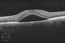
HD Grayscale OCT of Central Serous Retinopathy1113 viewsJul 11, 2012
|
|

Macular Telangiectasis (Group 2a Juxtafoveal Telangiectasis) Decreased Fundus Autofluorescence1182 views63-year-old woman has juxtafoveal retinal telangiectasis in both eyes. She notices her vision a little worse with more distortion and change over the last six months.
VISUAL ACUITY: OD 20/40, OS 20/40.Jun 28, 2012
|
|

Macular Telangiectasis (Group 2a Juxtafoveal Telangiectasis) Decreased Fundus Autofluorescence1353 views63-year-old woman has juxtafoveal retinal telangiectasis in both eyes. She notices her vision a little worse with more distortion and change over the last six months.
VISUAL ACUITY: OD 20/40, OS 20/40.Jun 28, 2012
|
|

Macular Telangiectasis (Group 2a Juxtafoveal Telangiectasis) IR709 views63-year-old woman has juxtafoveal retinal telangiectasis in both eyes. She notices her vision a little worse with more distortion and change over the last six months.
VISUAL ACUITY: OD 20/40, OS 20/40.Jun 28, 2012
|
|

Macular Telangiectasis (Group 2a Juxtafoveal Telangiectasis) IR699 views63-year-old woman has juxtafoveal retinal telangiectasis in both eyes. She notices her vision a little worse with more distortion and change over the last six months.
VISUAL ACUITY: OD 20/40, OS 20/40.Jun 28, 2012
|
|

Macular Telangiectasis (Group 2a Juxtafoveal Telangiectasis) Decreased Fundus Autofluorescence778 views63-year-old woman has juxtafoveal retinal telangiectasis in both eyes. She notices her vision a little worse with more distortion and change over the last six months.
VISUAL ACUITY: OD 20/40, OS 20/40.Jun 28, 2012
|
|

Macular Telangiectasis (Group 2a Juxtafoveal Telangiectasis) Decreased Fundus Autofluorescence988 views63-year-old woman has juxtafoveal retinal telangiectasis in both eyes. She notices her vision a little worse with more distortion and change over the last six months.
VISUAL ACUITY: OD 20/40, OS 20/40.Jun 28, 2012
|
|

Post-Retinal Detachment Repair Occult Subfoveal CNVM568 viewsThis pleasant 56-year-old man has had a complex retinal detachment repair in the right eye. I most recently removed his silicone oil 3 months ago. His vision now is the best it has ever been.
VISUAL ACUITY: OD 4/200Jun 28, 2012
|
|
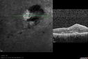
Post-Retinal Detachment Repair Occult Subfoveal CNVM561 viewsThis pleasant 56-year-old man has had a complex retinal detachment repair in the right eye. I most recently removed his silicone oil 3 months ago. His vision now is the best it has ever been.
VISUAL ACUITY: OD 4/200Jun 28, 2012
|
|

Post-Retinal Detachment Repair Occult Subfoveal CNVM586 viewsThis pleasant 56-year-old man has had a complex retinal detachment repair in the right eye. I most recently removed his silicone oil 3 months ago. His vision now is the best it has ever been.
VISUAL ACUITY: OD 4/200Jun 28, 2012
|
|
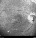
Post-Retinal Detachment Repair Occult Subfoveal CNVM495 viewsThis pleasant 56-year-old man has had a complex retinal detachment repair in the right eye. I most recently removed his silicone oil 3 months ago. His vision now is the best it has ever been.
VISUAL ACUITY: OD 4/200Jun 28, 2012
|
|

Post-Retinal Detachment Repair Occult Subfoveal CNVM487 viewsThis pleasant 56-year-old man has had a complex retinal detachment repair in the right eye. I most recently removed his silicone oil 3 months ago. His vision now is the best it has ever been.
VISUAL ACUITY: OD 4/200Jun 28, 2012
|
|

Post-Retinal Detachment Repair Occult Subfoveal CNVM476 viewsThis pleasant 56-year-old man has had a complex retinal detachment repair in the right eye. I most recently removed his silicone oil 3 months ago. His vision now is the best it has ever been.
VISUAL ACUITY: OD 4/200Jun 28, 2012
|
|

Post-Retinal Detachment Repair Occult Subfoveal CNVM469 viewsThis pleasant 56-year-old man has had a complex retinal detachment repair in the right eye. I most recently removed his silicone oil 3 months ago. His vision now is the best it has ever been.
VISUAL ACUITY: OD 4/200Jun 28, 2012
|
|
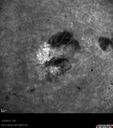
Post-Retinal Detachment Repair Occult Subfoveal CNVM504 viewsThis pleasant 56-year-old man has had a complex retinal detachment repair in the right eye. I most recently removed his silicone oil 3 months ago. His vision now is the best it has ever been.
VISUAL ACUITY: OD 4/200Jun 28, 2012
|
|

Post-Retinal Detachment Repair Occult Subfoveal CNVM476 viewsThis pleasant 56-year-old man has had a complex retinal detachment repair in the right eye. I most recently removed his silicone oil 3 months ago. His vision now is the best it has ever been.
VISUAL ACUITY: OD 4/200Jun 28, 2012
|
|

Post-Retinal Detachment Repair Occult Subfoveal CNVM521 viewsThis pleasant 56-year-old man has had a complex retinal detachment repair in the right eye. I most recently removed his silicone oil 3 months ago. His vision now is the best it has ever been.
VISUAL ACUITY: OD 4/200Jun 28, 2012
|
|
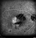
Post-Retinal Detachment Repair Occult Subfoveal CNVM599 viewsThis pleasant 56-year-old man has had a complex retinal detachment repair in the right eye. I most recently removed his silicone oil 3 months ago. His vision now is the best it has ever been.
VISUAL ACUITY: OD 4/200Jun 28, 2012
|
|

Coats' Disease1367 views11 year old male seen for trauma to OS from a volleyball. After examination, it was determined the pt had Coat's Disease. VA 20/20 OD 20/60 OS. FA confirmed the diagnosis.May 25, 2012
|
|

Wet Age-related Macular Degeneration (AMD) Radiating Chorioretinal Folds from Contracting Macular Scar598 views93-year-old man has wet age-related macular degeneration in his left eye with a subfoveal pigment epithelial detachment.
Vision OS is 20/40. There are striking radiating chorioretinal folds emanating off of the scar, which suggests it is contracting.May 22, 2012
|
|

Wet Age-related Macular Degeneration (AMD) Radiating Chorioretinal Folds from Contracting Macular Scar606 views93-year-old man has wet age-related macular degeneration in his left eye with a subfoveal pigment epithelial detachment.
Vision OS is 20/40. There are striking radiating chorioretinal folds emanating off of the scar, which suggests it is contracting.May 22, 2012
|
|

Wet Age-related Macular Degeneration (AMD) Radiating Chorioretinal Folds from Contracting Macular Scar616 views93-year-old man has wet age-related macular degeneration in his left eye with a subfoveal pigment epithelial detachment.
Vision OS is 20/40. There are striking radiating chorioretinal folds emanating off of the scar, which suggests it is contracting.May 22, 2012
|
|

Wet Age-related Macular Degeneration (AMD) Radiating Chorioretinal Folds from Contracting Macular Scar578 views93-year-old man has wet age-related macular degeneration in his left eye with a subfoveal pigment epithelial detachment.
Vision OS is 20/40. There are striking radiating chorioretinal folds emanating off of the scar, which suggests it is contracting.May 22, 2012
|
|

Wet Age-related Macular Degeneration (AMD) Radiating Chorioretinal Folds from Contracting Macular Scar698 views93-year-old man has wet age-related macular degeneration in his left eye with a subfoveal pigment epithelial detachment.
Vision OS is 20/40. There are striking radiating chorioretinal folds emanating off of the scar, which suggests it is contracting.May 22, 2012
|
|

Wet Age-related Macular Degeneration (AMD) Radiating Chorioretinal Folds from Contracting Macular Scar632 views93-year-old man has wet age-related macular degeneration in his left eye with a subfoveal pigment epithelial detachment.
Vision OS is 20/40. There are striking radiating chorioretinal folds emanating off of the scar, which suggests it is contracting.May 22, 2012
|
|

Wet Age-related Macular Degeneration (AMD) Radiating Chorioretinal Folds from Contracting Macular Scar775 views93-year-old man has wet age-related macular degeneration in his left eye with a subfoveal pigment epithelial detachment.
Vision OS is 20/40. There are striking radiating chorioretinal folds emanating off of the scar, which suggests it is contracting.May 22, 2012
|
|

Acute Central Serous Retinopathy - CSR - Steroid Induced (probably) - Good Vision - 6 months later - vision Better - Normal Photo776 views42-year-old man has central serous retinopathy in the right eye with serous retinal detachment. 6 month follow-up Vision is better.
VISUAL ACUITY: OD 20/20.
May 22, 2012
|
|

Acute Central Serous Retinopathy - CSR - Steroid Induced (probably) - Good Vision - 6 months later - vision Better - FAF shows small abnormality834 views42-year-old man has central serous retinopathy in the right eye with serous retinal detachment. 6 month follow-up Vision is better.
VISUAL ACUITY: OD 20/20. May 22, 2012
|
|

Acute Central Serous Retinopathy - CSR - Steroid Induced (probably) - Good Vision - 6 months later - vision Better INFRARED SHOWS EXACT VISION DEFECT785 views42-year-old man has central serous retinopathy in the right eye with serous retinal detachment. 6 month follow-up Vision is better.OD 20/20. The patient looked at this infra-red image and said "That's exactly what I see!"May 22, 2012
|
|

Acute Central Serous Retinopathy - CSR - Steroid Induced (probably) - Good Vision - 6 months later - vision Better - OCT Extended Depth Imaging - Choroidal Congestion646 views42-year-old man has central serous retinopathy in the right eye with serous retinal detachment. 6 month follow-up Vision is better.
VISUAL ACUITY: OD 20/20. May 22, 2012
|
|

Acute Central Serous Retinopathy - CSR - Steroid Induced (probably) - Good Vision - 6 months later - vision Better640 views42-year-old man has central serous retinopathy in the right eye with serous retinal detachment. 6 month follow-up Vision is better.OD 20/20. The patient looked at this infra-red image and said "That's exactly what I see!"
VISUAL ACUITY: OD 20/20. May 22, 2012
|
|

Acute Central Serous Retinopathy - CSR - Steroid Induced (probably) - Good Vision - 6 months later - vision Better668 views42-year-old man has central serous retinopathy in the right eye with serous retinal detachment. 6 month follow-up Vision is better.
VISUAL ACUITY: OD 20/20. May 22, 2012
|
|

Acute Central Serous Retinopathy - CSR - Steroid Induced (probably) - Good Vision - ICG Fellow Eye672 views42-year-old man was seen in the office on October 5, 2011. He had noticed starting in August after a course of antibiotic and steroids, that he developed new spots in his vision in the right eye. He may have had an episode like this sometime in the past. He did take steroids a few years ago and his vision did change at that time, but then returned.
VISUAL ACUITY: OD 20/32, OS 20/32. The OCT scan of the right eye confirms subretinal fluid.May 22, 2012
|
|

Acute Central Serous Retinopathy - CSR - Steroid Induced (probably) - Good Vision - ICG Fellow Eye604 views42-year-old man was seen in the office on October 5, 2011. He had noticed starting in August after a course of antibiotic and steroids, that he developed new spots in his vision in the right eye. He may have had an episode like this sometime in the past. He did take steroids a few years ago and his vision did change at that time, but then returned.
VISUAL ACUITY: OD 20/32, OS 20/32. The OCT scan of the right eye confirms subretinal fluid.May 22, 2012
|
|

Acute Central Serous Retinopathy - CSR - Steroid Induced (probably) - Good Vision - ICG Fellow Eye617 views42-year-old man was seen in the office on October 5, 2011. He had noticed starting in August after a course of antibiotic and steroids, that he developed new spots in his vision in the right eye. He may have had an episode like this sometime in the past. He did take steroids a few years ago and his vision did change at that time, but then returned.
VISUAL ACUITY: OD 20/32, OS 20/32. The OCT scan of the right eye confirms subretinal fluid.May 22, 2012
|
|

Acute Central Serous Retinopathy - CSR - Steroid Induced (probably) - Good Vision - FA Fellow Eye517 views42-year-old man was seen in the office on October 5, 2011. He had noticed starting in August after a course of antibiotic and steroids, that he developed new spots in his vision in the right eye. He may have had an episode like this sometime in the past. He did take steroids a few years ago and his vision did change at that time, but then returned.
VISUAL ACUITY: OD 20/32, OS 20/32. The OCT scan of the right eye confirms subretinal fluid.May 22, 2012
|
|

Acute Central Serous Retinopathy - CSR - Steroid Induced (probably) - Good Vision - FA Fellow Eye512 views42-year-old man was seen in the office on October 5, 2011. He had noticed starting in August after a course of antibiotic and steroids, that he developed new spots in his vision in the right eye. He may have had an episode like this sometime in the past. He did take steroids a few years ago and his vision did change at that time, but then returned.
VISUAL ACUITY: OD 20/32, OS 20/32. The OCT scan of the right eye confirms subretinal fluid.May 22, 2012
|
|

Acute Central Serous Retinopathy - CSR - Steroid Induced (probably) - Good Vision - FA Fellow Eye520 views42-year-old man was seen in the office on October 5, 2011. He had noticed starting in August after a course of antibiotic and steroids, that he developed new spots in his vision in the right eye. He may have had an episode like this sometime in the past. He did take steroids a few years ago and his vision did change at that time, but then returned.
VISUAL ACUITY: OD 20/32, OS 20/32. The OCT scan of the right eye confirms subretinal fluid.May 22, 2012
|
|

Acute Central Serous Retinopathy - CSR - Steroid Induced (probably) - Good Vision - ICG Affected Eye - Choroidal Leakage523 views42-year-old man was seen in the office on October 5, 2011. He had noticed starting in August after a course of antibiotic and steroids, that he developed new spots in his vision in the right eye. He may have had an episode like this sometime in the past. He did take steroids a few years ago and his vision did change at that time, but then returned.
VISUAL ACUITY: OD 20/32, OS 20/32. The OCT scan of the right eye confirms subretinal fluid.May 22, 2012
|
|

Acute Central Serous Retinopathy - CSR - Steroid Induced (probably) - Good Vision - ICG Affected Eye - Choroidal Leakage508 views42-year-old man was seen in the office on October 5, 2011. He had noticed starting in August after a course of antibiotic and steroids, that he developed new spots in his vision in the right eye. He may have had an episode like this sometime in the past. He did take steroids a few years ago and his vision did change at that time, but then returned.
VISUAL ACUITY: OD 20/32, OS 20/32. The OCT scan of the right eye confirms subretinal fluid.May 22, 2012
|
|

Acute Central Serous Retinopathy - CSR - Steroid Induced (probably) - Good Vision - ICG Affected Eye - Choroidal Leakage540 views42-year-old man was seen in the office on October 5, 2011. He had noticed starting in August after a course of antibiotic and steroids, that he developed new spots in his vision in the right eye. He may have had an episode like this sometime in the past. He did take steroids a few years ago and his vision did change at that time, but then returned.
VISUAL ACUITY: OD 20/32, OS 20/32. The OCT scan of the right eye confirms subretinal fluid.May 22, 2012
|
|

Acute Central Serous Retinopathy - CSR - Steroid Induced (probably) - Good Vision - ICG Affected Eye - Choroidal Leakage699 views42-year-old man was seen in the office on October 5, 2011. He had noticed starting in August after a course of antibiotic and steroids, that he developed new spots in his vision in the right eye. He may have had an episode like this sometime in the past. He did take steroids a few years ago and his vision did change at that time, but then returned.
VISUAL ACUITY: OD 20/32, OS 20/32. The OCT scan of the right eye confirms subretinal fluid.May 22, 2012
|
|

Acute Central Serous Retinopathy - CSR - Steroid Induced (probably) - Good Vision - FA Affected Eye548 views42-year-old man was seen in the office on October 5, 2011. He had noticed starting in August after a course of antibiotic and steroids, that he developed new spots in his vision in the right eye. He may have had an episode like this sometime in the past. He did take steroids a few years ago and his vision did change at that time, but then returned.
VISUAL ACUITY: OD 20/32, OS 20/32. The OCT scan of the right eye confirms subretinal fluid.May 22, 2012
|
|

Acute Central Serous Retinopathy - CSR - Steroid Induced (probably) - Good Vision - FA Affected Eye542 views42-year-old man was seen in the office on October 5, 2011. He had noticed starting in August after a course of antibiotic and steroids, that he developed new spots in his vision in the right eye. He may have had an episode like this sometime in the past. He did take steroids a few years ago and his vision did change at that time, but then returned.
VISUAL ACUITY: OD 20/32, OS 20/32. The OCT scan of the right eye confirms subretinal fluid.May 22, 2012
|
|

Acute Central Serous Retinopathy - CSR - Steroid Induced (probably) - Good Vision - FA Affected Eye680 views42-year-old man was seen in the office on October 5, 2011. He had noticed starting in August after a course of antibiotic and steroids, that he developed new spots in his vision in the right eye. He may have had an episode like this sometime in the past. He did take steroids a few years ago and his vision did change at that time, but then returned.
VISUAL ACUITY: OD 20/32, OS 20/32. The OCT scan of the right eye confirms subretinal fluid.May 22, 2012
|
|

Acute Central Serous Retinopathy - CSR - Steroid Induced (probably) - Good Vision 550 views42-year-old man was seen in the office on October 5, 2011. He had noticed starting in August after a course of antibiotic and steroids, that he developed new spots in his vision in the right eye. He may have had an episode like this sometime in the past. He did take steroids a few years ago and his vision did change at that time, but then returned.
VISUAL ACUITY: OD 20/32, OS 20/32. The OCT scan of the right eye confirms subretinal fluid.May 22, 2012
|
|

Acute Central Serous Retinopathy - CSR - Steroid Induced (probably) - Good Vision 676 views42-year-old man was seen in the office on October 5, 2011. He had noticed starting in August after a course of antibiotic and steroids, that he developed new spots in his vision in the right eye. He may have had an episode like this sometime in the past. He did take steroids a few years ago and his vision did change at that time, but then returned.
VISUAL ACUITY: OD 20/32, OS 20/32. The OCT scan of the right eye confirms subretinal fluid.May 22, 2012
|
|

Acute Central Serous Retinopathy - CSR - Steroid Induced (probably) - Good Vision 565 views42-year-old man was seen in the office on October 5, 2011. He had noticed starting in August after a course of antibiotic and steroids, that he developed new spots in his vision in the right eye. He may have had an episode like this sometime in the past. He did take steroids a few years ago and his vision did change at that time, but then returned.
VISUAL ACUITY: OD 20/32, OS 20/32. The OCT scan of the right eye confirms subretinal fluid.May 22, 2012
|
|

570 views42-year-old man was seen in the office on October 5, 2011. He had noticed starting in August after a course of antibiotic and steroids, that he developed new spots in his vision in the right eye. He may have had an episode like this sometime in the past. He did take steroids a few years ago and his vision did change at that time, but then returned.
VISUAL ACUITY: OD 20/32, OS 20/32. The OCT scan of the right eye confirms subretinal fluid.May 22, 2012
|
|

561 views42-year-old man was seen in the office on October 5, 2011. He had noticed starting in August after a course of antibiotic and steroids, that he developed new spots in his vision in the right eye. He may have had an episode like this sometime in the past. He did take steroids a few years ago and his vision did change at that time, but then returned.
VISUAL ACUITY: OD 20/32, OS 20/32. The OCT scan of the right eye confirms subretinal fluid.May 22, 2012
|
|

Acute Central Serous Retinopathy - CSR - Steroid Induced (probably) - Good Vision 687 views42-year-old man was seen in the office on October 5, 2011. He had noticed starting in August after a course of antibiotic and steroids, that he developed new spots in his vision in the right eye. He may have had an episode like this sometime in the past. He did take steroids a few years ago and his vision did change at that time, but then returned.
VISUAL ACUITY: OD 20/32, OS 20/32. The OCT scan of the right eye confirms subretinal fluid.May 22, 2012
|
|

Acute Central Serous Retinopathy - CSR - Steroid Induced (probably) - Good Vision - Fundus Autofluorescence633 views42-year-old man was seen in the office on October 5, 2011. He had noticed starting in August after a course of antibiotic and steroids, that he developed new spots in his vision in the right eye. He may have had an episode like this sometime in the past. He did take steroids a few years ago and his vision did change at that time, but then returned.
VISUAL ACUITY: OD 20/32, OS 20/32. The OCT scan of the right eye confirms subretinal fluid.May 22, 2012
|
|

Acute Central Serous Retinopathy - CSR - Steroid Induced (probably) - Good Vision - Fundus HyperAutofluorescence Serous Retinal Detachment901 views42-year-old man was seen in the office on October 5, 2011. He had noticed starting in August after a course of antibiotic and steroids, that he developed new spots in his vision in the right eye. He may have had an episode like this sometime in the past. He did take steroids a few years ago and his vision did change at that time, but then returned.
VISUAL ACUITY: OD 20/32, OS 20/32. The OCT scan of the right eye confirms subretinal fluid.May 22, 2012
|
|
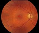
Macular Hole612 viewsMay 10, 2012
|
|

OCT of Macular Hole with Operculum636 viewsOCT of macular hole in 3-D with Tractional maculopathy. Operculum is presentMay 08, 2012
|
|

Solar Retinopathy1402 viewsFoveal defect due to sun staringMay 08, 2012
|
|

578 viewsMay 07, 2012
|
|
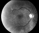
548 viewsMay 07, 2012
|
|
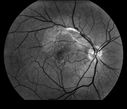
504 viewsMay 07, 2012
|
|

479 viewsMay 07, 2012
|
|

404 viewsMay 07, 2012
|
|

469 viewsMay 07, 2012
|
|

439 viewsMay 07, 2012
|
|

438 viewsMay 07, 2012
|
|

468 viewsMay 07, 2012
|
|

430 viewsMay 07, 2012
|
|

445 viewsMay 07, 2012
|
|

465 viewsMay 07, 2012
|
|

500 viewsMay 07, 2012
|
|
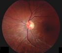
474 viewsMay 07, 2012
|
|

470 viewsMay 07, 2012
|
|
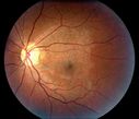
531 viewsMay 07, 2012
|
|

Pseudoxanthoma Elasticum with CNV540 viewsMay 07, 2012
|
|

568 viewsMay 07, 2012
|
|

616 viewsMay 07, 2012
|
|

Epiretinal Membrane with Pseudohole518 viewsEpiretinal Membrane with Pseudohole
James L. Perron, C.R.A.May 04, 2012
|
|

801 views87 year old male seen for neovascular macular degeneration. Patient had huge disciform scar and lipid.Apr 25, 2012
|
|

506 views87 year old male seen for neovascular macular degeneration. Patient had huge disciform scar and lipid.Apr 25, 2012
|
|

343 views75 year old male seen for ARMD. Pt developed massive hemorrhage with lipid and macular edema.Apr 25, 2012
|
|

420 views75 year old male seen for ARMD. Pt developed massive hemorrhage with lipid and macular edema.Apr 25, 2012
|
|

654 viewsMar 28, 2012
|
|

669 views74-year-old woman
OD 20/25, OS 20/32Mar 20, 2012
|
|

Calcified Macular Drusen - Age Related Macular Degeneration - Dry723 views74-year-old woman
OD 20/25, OS 20/32Mar 20, 2012
|
|

Calcified Macular Drusen - Age Related Macular Degeneration - Dry749 views74-year-old woman
OD 20/25, OS 20/32Mar 20, 2012
|
|

Calcified Macular Drusen - Age Related Macular Degeneration - Dry725 views74-year-old woman
OD 20/25, OS 20/32Mar 20, 2012
|
|

Calcified Macular Drusen - Age Related Macular Degeneration - Dry754 views74-year-old woman
OD 20/25, OS 20/32Mar 20, 2012
|
|

Calcified Macular Drusen - Age Related Macular Degeneration - Dry811 views74-year-old woman
OD 20/25, OS 20/32Mar 20, 2012
|
|

Angioid Streaks - Peau D'Orange - Pseudoxanthoma Elasticum - Asymptomatic Extrafoveal Choroidal Neovascular Membrane586 views60-year-old woman has pseudoxanthoma elasticum since 2008. Her sister has the same problem and lost vision in both eyes. She had a problem with blood vessel growth in the left eye for which she had injections of Avastin in August, September, and October of 2011 (5 months ago). Subsequent to that, the macula has been dry and you have just been watching her carefully. She is in Florida for a short time but will ultimately be spending nine months out of the year here. Both of her eyes have good vision at Mar 20, 2012
|
|

Angioid Streaks - Peau D'Orange - Pseudoxanthoma Elasticum - Asymptomatic Extrafoveal Choroidal Neovascular Membrane545 views60-year-old woman has pseudoxanthoma elasticum since 2008. Her sister has the same problem and lost vision in both eyes. She had a problem with blood vessel growth in the left eye for which she had injections of Avastin in August, September, and October of 2011 (5 months ago). Subsequent to that, the macula has been dry and you have just been watching her carefully. She is in Florida for a short time but will ultimately be spending nine months out of the year here. Both of her eyes have good vision at Mar 20, 2012
|
|

Angioid Streaks - Peau D'Orange - Pseudoxanthoma Elasticum - Asymptomatic Extrafoveal Choroidal Neovascular Membrane513 views60-year-old woman has pseudoxanthoma elasticum since 2008. Her sister has the same problem and lost vision in both eyes. She had a problem with blood vessel growth in the left eye for which she had injections of Avastin in August, September, and October of 2011 (5 months ago). Subsequent to that, the macula has been dry and you have just been watching her carefully. She is in Florida for a short time but will ultimately be spending nine months out of the year here. Both of her eyes have good vision at Mar 20, 2012
|
|

Angioid Streaks - Peau D'Orange - Pseudoxanthoma Elasticum - Asymptomatic Extrafoveal Choroidal Neovascular Membrane547 views60-year-old woman has pseudoxanthoma elasticum since 2008. Her sister has the same problem and lost vision in both eyes. She had a problem with blood vessel growth in the left eye for which she had injections of Avastin in August, September, and October of 2011 (5 months ago). Subsequent to that, the macula has been dry and you have just been watching her carefully. She is in Florida for a short time but will ultimately be spending nine months out of the year here. Both of her eyes have good vision at Mar 20, 2012
|
|

Angioid Streaks - Peau D'Orange - Pseudoxanthoma Elasticum - Asymptomatic Extrafoveal Choroidal Neovascular Membrane697 views60-year-old woman has pseudoxanthoma elasticum since 2008. Her sister has the same problem and lost vision in both eyes. She had a problem with blood vessel growth in the left eye for which she had injections of Avastin in August, September, and October of 2011 (5 months ago). Subsequent to that, the macula has been dry and you have just been watching her carefully. She is in Florida for a short time but will ultimately be spending nine months out of the year here. Both of her eyes have good vision at Mar 20, 2012
|
|

Angioid Streaks - Peau D'Orange - Pseudoxanthoma Elasticum - Asymptomatic Extrafoveal Choroidal Neovascular Membrane646 views60-year-old woman has pseudoxanthoma elasticum since 2008. Her sister has the same problem and lost vision in both eyes. She had a problem with blood vessel growth in the left eye for which she had injections of Avastin in August, September, and October of 2011 (5 months ago). Subsequent to that, the macula has been dry and you have just been watching her carefully. She is in Florida for a short time but will ultimately be spending nine months out of the year here. Both of her eyes have good vision at Mar 20, 2012
|
|

Angioid Streaks - Peau D'Orange - Pseudoxanthoma Elasticum - Asymptomatic Extrafoveal Choroidal Neovascular Membrane800 views60-year-old woman has pseudoxanthoma elasticum since 2008. Her sister has the same problem and lost vision in both eyes. She had a problem with blood vessel growth in the left eye for which she had injections of Avastin in August, September, and October of 2011 (5 months ago). Subsequent to that, the macula has been dry and you have just been watching her carefully. She is in Florida for a short time but will ultimately be spending nine months out of the year here. Both of her eyes have good vision at Mar 20, 2012
|
|

Angioid Streaks - Peau D'Orange - Pseudoxanthoma Elasticum - Asymptomatic Extrafoveal Choroidal Neovascular Membrane636 views60-year-old woman has pseudoxanthoma elasticum since 2008. Her sister has the same problem and lost vision in both eyes. She had a problem with blood vessel growth in the left eye for which she had injections of Avastin in August, September, and October of 2011 (5 months ago). Subsequent to that, the macula has been dry and you have just been watching her carefully. She is in Florida for a short time but will ultimately be spending nine months out of the year here. Both of her eyes have good vision at Mar 20, 2012
|
|

Angioid Streaks - Peau D'Orange - Pseudoxanthoma Elasticum - Asymptomatic Extrafoveal Choroidal Neovascular Membrane1255 views60-year-old woman has pseudoxanthoma elasticum since 2008. Her sister has the same problem and lost vision in both eyes. She had a problem with blood vessel growth in the left eye for which she had injections of Avastin in August, September, and October of 2011 (5 months ago). Subsequent to that, the macula has been dry and you have just been watching her carefully. She is in Florida for a short time but will ultimately be spending nine months out of the year here. Both of her eyes have good vision at Mar 20, 2012
|
|
| 5013 files on 51 page(s) |
 |
|
|
|
|
36 | |
|
|
|
|


