Last additions - Macular Disease
|

Angioid Streaks - Peau D'Orange - Pseudoxanthoma Elasticum - Asymptomatic Extrafoveal Choroidal Neovascular Membrane871 views60-year-old woman has pseudoxanthoma elasticum since 2008. Her sister has the same problem and lost vision in both eyes. She had a problem with blood vessel growth in the left eye for which she had injections of Avastin in August, September, and October of 2011 (5 months ago). Subsequent to that, the macula has been dry and you have just been watching her carefully. She is in Florida for a short time but will ultimately be spending nine months out of the year here. Both of her eyes have good vision at Mar 20, 2012
|
|

Angioid Streaks - Peau D'Orange - Pseudoxanthoma Elasticum - Asymptomatic Extrafoveal Choroidal Neovascular Membrane707 views60-year-old woman has pseudoxanthoma elasticum since 2008. Her sister has the same problem and lost vision in both eyes. She had a problem with blood vessel growth in the left eye for which she had injections of Avastin in August, September, and October of 2011 (5 months ago). Subsequent to that, the macula has been dry and you have just been watching her carefully. She is in Florida for a short time but will ultimately be spending nine months out of the year here. Both of her eyes have good vision at Mar 20, 2012
|
|

Angioid Streaks - Peau D'Orange - Pseudoxanthoma Elasticum - Asymptomatic Extrafoveal Choroidal Neovascular Membrane779 views60-year-old woman has pseudoxanthoma elasticum since 2008. Her sister has the same problem and lost vision in both eyes. She had a problem with blood vessel growth in the left eye for which she had injections of Avastin in August, September, and October of 2011 (5 months ago). Subsequent to that, the macula has been dry and you have just been watching her carefully. She is in Florida for a short time but will ultimately be spending nine months out of the year here. Both of her eyes have good vision at Mar 20, 2012
|
|

Angioid Streaks - Peau D'Orange - Pseudoxanthoma Elasticum - Asymptomatic Extrafoveal Choroidal Neovascular Membrane607 views60-year-old woman has pseudoxanthoma elasticum since 2008. Her sister has the same problem and lost vision in both eyes. She had a problem with blood vessel growth in the left eye for which she had injections of Avastin in August, September, and October of 2011 (5 months ago). Subsequent to that, the macula has been dry and you have just been watching her carefully. She is in Florida for a short time but will ultimately be spending nine months out of the year here. Both of her eyes have good vision at Mar 20, 2012
|
|

Angioid Streaks - Peau D'Orange - Pseudoxanthoma Elasticum - Asymptomatic Extrafoveal Choroidal Neovascular Membrane676 views60-year-old woman has pseudoxanthoma elasticum since 2008. Her sister has the same problem and lost vision in both eyes. She had a problem with blood vessel growth in the left eye for which she had injections of Avastin in August, September, and October of 2011 (5 months ago). Subsequent to that, the macula has been dry and you have just been watching her carefully. She is in Florida for a short time but will ultimately be spending nine months out of the year here. Both of her eyes have good vision at Mar 20, 2012
|
|

Angioid Streaks - Peau D'Orange - Pseudoxanthoma Elasticum - Asymptomatic Extrafoveal Choroidal Neovascular Membrane866 views60-year-old woman has pseudoxanthoma elasticum since 2008. Her sister has the same problem and lost vision in both eyes. She had a problem with blood vessel growth in the left eye for which she had injections of Avastin in August, September, and October of 2011 (5 months ago). Subsequent to that, the macula has been dry and you have just been watching her carefully. She is in Florida for a short time but will ultimately be spending nine months out of the year here. Both of her eyes have good vision at Mar 20, 2012
|
|

Angioid Streaks - Peau D'Orange - Pseudoxanthoma Elasticum - Asymptomatic Extrafoveal Choroidal Neovascular Membrane876 views60-year-old woman has pseudoxanthoma elasticum since 2008. Her sister has the same problem and lost vision in both eyes. She had a problem with blood vessel growth in the left eye for which she had injections of Avastin in August, September, and October of 2011 (5 months ago). Subsequent to that, the macula has been dry and you have just been watching her carefully. She is in Florida for a short time but will ultimately be spending nine months out of the year here. Both of her eyes have good vision at Mar 20, 2012
|
|

Angioid Streaks - Peau D'Orange - Pseudoxanthoma Elasticum - Asymptomatic Extrafoveal Choroidal Neovascular Membrane1084 views60-year-old woman has pseudoxanthoma elasticum since 2008. Her sister has the same problem and lost vision in both eyes. She had a problem with blood vessel growth in the left eye for which she had injections of Avastin in August, September, and October of 2011 (5 months ago). Subsequent to that, the macula has been dry and you have just been watching her carefully. She is in Florida for a short time but will ultimately be spending nine months out of the year here. Both of her eyes have good vision at Mar 20, 2012
|
|

Angioid Streaks - Peau D'Orange - Pseudoxanthoma Elasticum - Asymptomatic Extrafoveal Choroidal Neovascular Membrane1037 views60-year-old woman has pseudoxanthoma elasticum since 2008. Her sister has the same problem and lost vision in both eyes. She had a problem with blood vessel growth in the left eye for which she had injections of Avastin in August, September, and October of 2011 (5 months ago). Subsequent to that, the macula has been dry and you have just been watching her carefully. She is in Florida for a short time but will ultimately be spending nine months out of the year here. Both of her eyes have good vision at Mar 20, 2012
|
|

Angioid Streaks - Peau D'Orange - Pseudoxanthoma Elasticum - Asymptomatic Extrafoveal Choroidal Neovascular Membrane762 views60-year-old woman has pseudoxanthoma elasticum since 2008. Her sister has the same problem and lost vision in both eyes. She had a problem with blood vessel growth in the left eye for which she had injections of Avastin in August, September, and October of 2011 (5 months ago). Subsequent to that, the macula has been dry and you have just been watching her carefully. She is in Florida for a short time but will ultimately be spending nine months out of the year here. Both of her eyes have good vision at Mar 20, 2012
|
|

Angioid Streaks - Peau D'Orange - Pseudoxanthoma Elasticum - Asymptomatic Extrafoveal Choroidal Neovascular Membrane1042 views60-year-old woman has pseudoxanthoma elasticum since 2008. Her sister has the same problem and lost vision in both eyes. She had a problem with blood vessel growth in the left eye for which she had injections of Avastin in August, September, and October of 2011 (5 months ago). Subsequent to that, the macula has been dry and you have just been watching her carefully. She is in Florida for a short time but will ultimately be spending nine months out of the year here. Both of her eyes have good vision at Mar 20, 2012
|
|

Angioid Streaks - Peau D'Orange - Pseudoxanthoma Elasticum - Asymptomatic Extrafoveal Choroidal Neovascular Membrane1138 views60-year-old woman has pseudoxanthoma elasticum since 2008. Her sister has the same problem and lost vision in both eyes. She had a problem with blood vessel growth in the left eye for which she had injections of Avastin in August, September, and October of 2011 (5 months ago). Subsequent to that, the macula has been dry and you have just been watching her carefully. She is in Florida for a short time but will ultimately be spending nine months out of the year here. Both of her eyes have good vision at Mar 20, 2012
|
|

Macular Hole Closed - Nerve Fiber Layer Defects Visible after Internal Limiting Membrane Peel - Vision 20/60582 views53-year-old woman had vitrectomy for a stage IV macular hole in the left eye on June 1st. Her vision has improved some but still has a bump in it.
VISUAL ACUITY: Vision OS is 20/80, PH is 20/60Mar 20, 2012
|
|

Macular Hole Closed - Nerve Fiber Layer Defects Visible after Internal Limiting Membrane Peel - Vision 20/60611 views53-year-old woman had vitrectomy for a stage IV macular hole in the left eye on June 1st. Her vision has improved some but still has a bump in it.
VISUAL ACUITY: Vision OS is 20/80, PH is 20/60Mar 20, 2012
|
|

Macular Hole - Stage 4580 views53-year-old woman was seen in the office on 5/20/2011. She had cataract surgery in June of 2009. She noticed the vision in the left eye was starting to get foggy some time around the middle of April (1 month ago).
VISUAL ACUITY: Vision OD is 20/20, OS is 20/160Mar 20, 2012
|
|

Macular Hole - Stage 4581 views53-year-old woman was seen in the office on 5/20/2011. She had cataract surgery in June of 2009. She noticed the vision in the left eye was starting to get foggy some time around the middle of April (1 month ago).
VISUAL ACUITY: Vision OD is 20/20, OS is 20/160Mar 20, 2012
|
|

ANGIOID STREAKS2095 viewsA 29 year old woman was referred to the ophthalmologist for an eye exam since the dermatologist had noted “chicken-skin†papules in the neck skin. PAD showed for pseudoxathoma elasticum typical pathology. The eye exam disclosed angioid streaks, asymptomatic so far.
Magnus Gjötterberg MD
International membrer 1440
S:t Erik Eye Hospital
SE-11282
Stockholm
Sweden
magnusgjotterberg@hem.utfors.se
magnus.gjotterberg@sankterik.se.
Photographer/technician
Malin Langhals
Mar 16, 2012
|
|

Angioid Streaks and Treated CNVM1237 viewsMaculopathy associated to angioid streaks in a 45 years old patient, man,
treated for related CNV
Feb 29, 2012
|
|
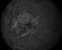
Angioid Streaks and Treated CNVM703 viewsMaculopathy associated to angioid streaks in a 45 years old patient, man,
treated for related CNV
Feb 29, 2012
|
|

Small vessel vasculitis causing cystoid macular edema - right eye - late frame fluorescein angiography 423 views36 year old female with no significant past medical history noted painless, progressive loss of vision over one month. BCVA was 20/100 in her right eye. Dilated fundus exam, OCT and FA identified small vessel vasculitis associated with cystoid macular edema. Left eye was also involved with less marked findings.Feb 28, 2012
|
|
Small vessel vasculitis causing cystoid macular edema - right eye - early frame fluorescein angiography 438 views36 year old female with no significant past medical history noted painless, progressive loss of vision over one month. BCVA was 20/100 in her right eye. Dilated fundus exam, OCT and FA identified small vessel vasculitis associated with cystoid macular edema. Left eye was also involved with less marked findings.Feb 28, 2012
|
|

Equatorial Drusen558 views76-year-old man with equatorial drusen
VISUAL ACUITY: Vision OD is 20/40, OS is 20/25. Feb 28, 2012
|
|
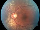
Equatorial Drusen782 views76-year-old man with equatorial drusen
VISUAL ACUITY: Vision OD is 20/40, OS is 20/25. Feb 28, 2012
|
|

Equatorial Drusen764 views76-year-old man with equatorial drusen
VISUAL ACUITY: Vision OD is 20/40, OS is 20/25. Feb 28, 2012
|
|

Equatorial Drusen831 views76-year-old man with equatorial drusen
VISUAL ACUITY: Vision OD is 20/40, OS is 20/25. Feb 28, 2012
|
|

Hyperpigmentation of the Retinal Pigment Epithelium Right Eye (Treated wet AMD OS)575 views85-year-old man has wet age-related macular degeneration in the left eye treated for one year most recently one year ago. OD is 20/30, OS is 20/60Feb 28, 2012
|
|

Hyperpigmentation of the Retinal Pigment Epithelium Right Eye (Treated wet AMD OS)559 views85-year-old man has wet age-related macular degeneration in the left eye treated for one year most recently one year ago. OD is 20/30, OS is 20/60Feb 28, 2012
|
|

Hyperpigmentation of the Retinal Pigment Epithelium Right Eye (Treated wet AMD OS)539 views85-year-old man has wet age-related macular degeneration in the left eye treated for one year most recently one year ago. OD is 20/30, OS is 20/60Feb 28, 2012
|
|

Hyperpigmentation of the Retinal Pigment Epithelium Right Eye (Treated wet AMD OS)661 views85-year-old man has wet age-related macular degeneration in the left eye treated for one year most recently one year ago. OD is 20/30, OS is 20/60Feb 28, 2012
|
|

Hyperpigmentation of the Retinal Pigment Epithelium Right Eye (Treated wet AMD OS)735 views85-year-old man has wet age-related macular degeneration in the left eye treated for one year most recently one year ago. OD is 20/30, OS is 20/60Feb 28, 2012
|
|

Hyperpigmentation of the Retinal Pigment Epithelium Right Eye (Treated wet AMD OS)833 views85-year-old man has wet age-related macular degeneration in the left eye treated for one year most recently one year ago. OD is 20/30, OS is 20/60Feb 28, 2012
|
|

Hyperpigmentation of the Retinal Pigment Epithelium Right Eye (Treated wet AMD OS)838 views85-year-old man has wet age-related macular degeneration in the left eye treated for one year most recently one year ago. OD is 20/30, OS is 20/60Feb 28, 2012
|
|

Hyperpigmentation of the Retinal Pigment Epithelium Right Eye (Treated wet AMD OS)1308 views85-year-old man has wet age-related macular degeneration in the left eye treated for one year most recently one year ago. OD is 20/30, OS is 20/60Feb 28, 2012
|
|

Hyperpigmentation of the Retinal Pigment Epithelium Right Eye (Treated wet AMD OS)1203 views85-year-old man has wet age-related macular degeneration in the left eye treated for one year most recently one year ago. OD is 20/30, OS is 20/60Feb 28, 2012
|
|

Cuticular Drusen (Basal Laminar Drusen) and Bilateral Chronic Subfoveal Fluid (Serous Retinal Detachment) unresponsive to monthly Avastin - 15 minute ICG Angiogram Left Eye756 views66-year-old man with bilateral subfoveal fluid unresponsive to one year of monthly avastin therapy.
OD 20/160, Pinhole 20/80. OS 20/63, Pinhole 20/50. OCT scan shows subretinal fluid with normal choroidal thickness. Fluorescein angiogram shows cuticular/basal laminar drusen. ICG shows no foci of choriocapillaris leakage.
Feb 15, 2012
|
|

Cuticular Drusen (Basal Laminar Drusen) and Bilateral Chronic Subfoveal Fluid (Serous Retinal Detachment) unresponsive to monthly Avastin - ICG OS684 views66-year-old man with bilateral subfoveal fluid unresponsive to one year of monthly avastin therapy.
OD 20/160, Pinhole 20/80. OS 20/63, Pinhole 20/50. OCT scan shows subretinal fluid with normal choroidal thickness. Fluorescein angiogram shows cuticular/basal laminar drusen. ICG shows no foci of choriocapillaris leakage.
Feb 15, 2012
|
|

Cuticular Drusen (Basal Laminar Drusen) and Bilateral Chronic Subfoveal Fluid (Serous Retinal Detachment) unresponsive to monthly Avastin - Late FA OS689 views66-year-old man with bilateral subfoveal fluid unresponsive to one year of monthly avastin therapy.
OD 20/160, Pinhole 20/80. OS 20/63, Pinhole 20/50. OCT scan shows subretinal fluid with normal choroidal thickness. Fluorescein angiogram shows cuticular/basal laminar drusen. ICG shows no foci of choriocapillaris leakage.
Feb 15, 2012
|
|

Cuticular Drusen (Basal Laminar Drusen) and Bilateral Chronic Subfoveal Fluid (Serous Retinal Detachment) unresponsive to monthly Avastin - Early ICG OS549 views66-year-old man with bilateral subfoveal fluid unresponsive to one year of monthly avastin therapy.
OD 20/160, Pinhole 20/80. OS 20/63, Pinhole 20/50. OCT scan shows subretinal fluid with normal choroidal thickness. Fluorescein angiogram shows cuticular/basal laminar drusen. ICG shows no foci of choriocapillaris leakage.
Feb 15, 2012
|
|

Cuticular Drusen (Basal Laminar Drusen) and Bilateral Chronic Subfoveal Fluid (Serous Retinal Detachment) unresponsive to monthly Avastin FA OS525 views66-year-old man with bilateral subfoveal fluid unresponsive to one year of monthly avastin therapy.
OD 20/160, Pinhole 20/80. OS 20/63, Pinhole 20/50. OCT scan shows subretinal fluid with normal choroidal thickness. Fluorescein angiogram shows cuticular/basal laminar drusen. ICG shows no foci of choriocapillaris leakage.
Feb 15, 2012
|
|

Cuticular Drusen (Basal Laminar Drusen) and Bilateral Chronic Subfoveal Fluid (Serous Retinal Detachment) unresponsive to monthly Avastin Very Early ICG OS466 views66-year-old man with bilateral subfoveal fluid unresponsive to one year of monthly avastin therapy.
OD 20/160, Pinhole 20/80. OS 20/63, Pinhole 20/50. OCT scan shows subretinal fluid with normal choroidal thickness. Fluorescein angiogram shows cuticular/basal laminar drusen. ICG shows no foci of choriocapillaris leakage.
Feb 15, 2012
|
|

Cuticular Drusen (Basal Laminar Drusen) and Bilateral Chronic Subfoveal Fluid (Serous Retinal Detachment) unresponsive to monthly Avastin FA OS463 views66-year-old man with bilateral subfoveal fluid unresponsive to one year of monthly avastin therapy.
OD 20/160, Pinhole 20/80. OS 20/63, Pinhole 20/50. OCT scan shows subretinal fluid with normal choroidal thickness. Fluorescein angiogram shows cuticular/basal laminar drusen. ICG shows no foci of choriocapillaris leakage.
Feb 15, 2012
|
|

Cuticular Drusen (Basal Laminar Drusen) and Bilateral Chronic Subfoveal Fluid (Serous Retinal Detachment) unresponsive to monthly Avastin ICG OS460 views66-year-old man with bilateral subfoveal fluid unresponsive to one year of monthly avastin therapy.
OD 20/160, Pinhole 20/80. OS 20/63, Pinhole 20/50. OCT scan shows subretinal fluid with normal choroidal thickness. Fluorescein angiogram shows cuticular/basal laminar drusen. ICG shows no foci of choriocapillaris leakage.
Feb 15, 2012
|
|

Cuticular Drusen (Basal Laminar Drusen) and Bilateral Chronic Subfoveal Fluid (Serous Retinal Detachment) unresponsive to monthly Avastin Early FA OS631 views66-year-old man with bilateral subfoveal fluid unresponsive to one year of monthly avastin therapy.
OD 20/160, Pinhole 20/80. OS 20/63, Pinhole 20/50. OCT scan shows subretinal fluid with normal choroidal thickness. Fluorescein angiogram shows cuticular/basal laminar drusen. ICG shows no foci of choriocapillaris leakage.
Feb 15, 2012
|
|

Cuticular Drusen (Basal Laminar Drusen) and Bilateral Chronic Subfoveal Fluid (Serous Retinal Detachment) unresponsive to monthly Avastin 15 Minute ICG OD547 views66-year-old man with bilateral subfoveal fluid unresponsive to one year of monthly avastin therapy.
OD 20/160, Pinhole 20/80. OS 20/63, Pinhole 20/50. OCT scan shows subretinal fluid with normal choroidal thickness. Fluorescein angiogram shows cuticular/basal laminar drusen. ICG shows no foci of choriocapillaris leakage.
Feb 15, 2012
|
|

Cuticular Drusen (Basal Laminar Drusen) and Bilateral Chronic Subfoveal Fluid (Serous Retinal Detachment) unresponsive to monthly Avastin 5 minute ICG OD629 views66-year-old man with bilateral subfoveal fluid unresponsive to one year of monthly avastin therapy.
OD 20/160, Pinhole 20/80. OS 20/63, Pinhole 20/50. OCT scan shows subretinal fluid with normal choroidal thickness. Fluorescein angiogram shows cuticular/basal laminar drusen. ICG shows no foci of choriocapillaris leakage.
Feb 15, 2012
|
|

Cuticular Drusen (Basal Laminar Drusen) and Bilateral Chronic Subfoveal Fluid (Serous Retinal Detachment) unresponsive to monthly Avastin Late FA OD631 views66-year-old man with bilateral subfoveal fluid unresponsive to one year of monthly avastin therapy.
OD 20/160, Pinhole 20/80. OS 20/63, Pinhole 20/50. OCT scan shows subretinal fluid with normal choroidal thickness. Fluorescein angiogram shows cuticular/basal laminar drusen. ICG shows no foci of choriocapillaris leakage.
Feb 15, 2012
|
|

Cuticular Drusen (Basal Laminar Drusen) and Bilateral Chronic Subfoveal Fluid (Serous Retinal Detachment) unresponsive to monthly Avastin Early ICG OD610 views66-year-old man with bilateral subfoveal fluid unresponsive to one year of monthly avastin therapy.
OD 20/160, Pinhole 20/80. OS 20/63, Pinhole 20/50. OCT scan shows subretinal fluid with normal choroidal thickness. Fluorescein angiogram shows cuticular/basal laminar drusen. ICG shows no foci of choriocapillaris leakage.
Feb 15, 2012
|
|

Cuticular Drusen (Basal Laminar Drusen) and Bilateral Chronic Subfoveal Fluid (Serous Retinal Detachment) unresponsive to monthly Avastin Early FA OD681 views66-year-old man with bilateral subfoveal fluid unresponsive to one year of monthly avastin therapy.
OD 20/160, Pinhole 20/80. OS 20/63, Pinhole 20/50. OCT scan shows subretinal fluid with normal choroidal thickness. Fluorescein angiogram shows cuticular/basal laminar drusen. ICG shows no foci of choriocapillaris leakage.
Feb 15, 2012
|
|

Cuticular Drusen (Basal Laminar Drusen) and Bilateral Chronic Subfoveal Fluid (Serous Retinal Detachment) unresponsive to monthly Avastin Very Early ICG OD509 views66-year-old man with bilateral subfoveal fluid unresponsive to one year of monthly avastin therapy.
OD 20/160, Pinhole 20/80. OS 20/63, Pinhole 20/50. OCT scan shows subretinal fluid with normal choroidal thickness. Fluorescein angiogram shows cuticular/basal laminar drusen. ICG shows no foci of choriocapillaris leakage.
Feb 15, 2012
|
|

Cuticular Drusen (Basal Laminar Drusen) and Bilateral Chronic Subfoveal Fluid (Serous Retinal Detachment) unresponsive to monthly Avastin Early FA OD580 views66-year-old man with bilateral subfoveal fluid unresponsive to one year of monthly avastin therapy.
OD 20/160, Pinhole 20/80. OS 20/63, Pinhole 20/50. OCT scan shows subretinal fluid with normal choroidal thickness. Fluorescein angiogram shows cuticular/basal laminar drusen. ICG shows no foci of choriocapillaris leakage.
Feb 15, 2012
|
|

Cuticular Drusen (Basal Laminar Drusen) and Bilateral Chronic Subfoveal Fluid (Serous Retinal Detachment) unresponsive to monthly Avastin Very Early ICG OD564 views66-year-old man with bilateral subfoveal fluid unresponsive to one year of monthly avastin therapy.
OD 20/160, Pinhole 20/80. OS 20/63, Pinhole 20/50. OCT scan shows subretinal fluid with normal choroidal thickness. Fluorescein angiogram shows cuticular/basal laminar drusen. ICG shows no foci of choriocapillaris leakage.
Feb 15, 2012
|
|

Cuticular Drusen (Basal Laminar Drusen) and Bilateral Chronic Subfoveal Fluid (Serous Retinal Detachment) unresponsive to monthly Avastin Early FA OD538 views66-year-old man with bilateral subfoveal fluid unresponsive to one year of monthly avastin therapy.
OD 20/160, Pinhole 20/80. OS 20/63, Pinhole 20/50. OCT scan shows subretinal fluid with normal choroidal thickness. Fluorescein angiogram shows cuticular/basal laminar drusen. ICG shows no foci of choriocapillaris leakage.
Feb 15, 2012
|
|

Cuticular Drusen (Basal Laminar Drusen) and Bilateral Chronic Subfoveal Fluid (Serous Retinal Detachment) unresponsive to monthly Avastin - Fill ICG FA OD533 views66-year-old man with bilateral subfoveal fluid unresponsive to one year of monthly avastin therapy.
OD 20/160, Pinhole 20/80. OS 20/63, Pinhole 20/50. OCT scan shows subretinal fluid with normal choroidal thickness. Fluorescein angiogram shows cuticular/basal laminar drusen. ICG shows no foci of choriocapillaris leakage.
Feb 15, 2012
|
|

Cuticular Drusen (Basal Laminar Drusen) and Bilateral Chronic Subfoveal Fluid (Serous Retinal Detachment) unresponsive to monthly Avastin - Fill ICG FA OD467 views66-year-old man with bilateral subfoveal fluid unresponsive to one year of monthly avastin therapy.
OD 20/160, Pinhole 20/80. OS 20/63, Pinhole 20/50. OCT scan shows subretinal fluid with normal choroidal thickness. Fluorescein angiogram shows cuticular/basal laminar drusen. ICG shows no foci of choriocapillaris leakage.
Feb 15, 2012
|
|

Cuticular Drusen (Basal Laminar Drusen) and Bilateral Chronic Subfoveal Fluid (Serous Retinal Detachment) unresponsive to monthly Avastin - Fill ICG FA OD530 views66-year-old man with bilateral subfoveal fluid unresponsive to one year of monthly avastin therapy.
OD 20/160, Pinhole 20/80. OS 20/63, Pinhole 20/50. OCT scan shows subretinal fluid with normal choroidal thickness. Fluorescein angiogram shows cuticular/basal laminar drusen. ICG shows no foci of choriocapillaris leakage.
Feb 15, 2012
|
|

Cuticular Drusen (Basal Laminar Drusen) and Bilateral Chronic Subfoveal Fluid (Serous Retinal Detachment) unresponsive to monthly Avastin - Fill ICG FA OD507 views66-year-old man with bilateral subfoveal fluid unresponsive to one year of monthly avastin therapy.
OD 20/160, Pinhole 20/80. OS 20/63, Pinhole 20/50. OCT scan shows subretinal fluid with normal choroidal thickness. Fluorescein angiogram shows cuticular/basal laminar drusen. ICG shows no foci of choriocapillaris leakage.
Feb 15, 2012
|
|

Cuticular Drusen (Basal Laminar Drusen) and Bilateral Chronic Subfoveal Fluid (Serous Retinal Detachment) unresponsive to monthly Avastin - Fill ICG FA OD522 views66-year-old man with bilateral subfoveal fluid unresponsive to one year of monthly avastin therapy.
OD 20/160, Pinhole 20/80. OS 20/63, Pinhole 20/50. OCT scan shows subretinal fluid with normal choroidal thickness. Fluorescein angiogram shows cuticular/basal laminar drusen. ICG shows no foci of choriocapillaris leakage.
Feb 15, 2012
|
|

Cuticular Drusen (Basal Laminar Drusen) and Bilateral Chronic Subfoveal Fluid (Serous Retinal Detachment) unresponsive to monthly Avastin - Fill ICG FA OD599 views66-year-old man with bilateral subfoveal fluid unresponsive to one year of monthly avastin therapy.
OD 20/160, Pinhole 20/80. OS 20/63, Pinhole 20/50. OCT scan shows subretinal fluid with normal choroidal thickness. Fluorescein angiogram shows cuticular/basal laminar drusen. ICG shows no foci of choriocapillaris leakage.
Feb 15, 2012
|
|

Cuticular Drusen (Basal Laminar Drusen) and Bilateral Chronic Subfoveal Fluid (Serous Retinal Detachment) unresponsive to monthly Avastin - Fundus Autofluorescence OS632 views66-year-old man with bilateral subfoveal fluid unresponsive to one year of monthly avastin therapy.
OD 20/160, Pinhole 20/80. OS 20/63, Pinhole 20/50. OCT scan shows subretinal fluid with normal choroidal thickness. Fluorescein angiogram shows cuticular/basal laminar drusen. ICG shows no foci of choriocapillaris leakage.
Feb 15, 2012
|
|

Cuticular Drusen (Basal Laminar Drusen) and Bilateral Chronic Subfoveal Fluid (Serous Retinal Detachment) unresponsive to monthly Avastin - Fundus Autofluorescence OD883 views66-year-old man with bilateral subfoveal fluid unresponsive to one year of monthly avastin therapy.
OD 20/160, Pinhole 20/80. OS 20/63, Pinhole 20/50. OCT scan shows subretinal fluid with normal choroidal thickness. Fluorescein angiogram shows cuticular/basal laminar drusen. ICG shows no foci of choriocapillaris leakage.
Feb 15, 2012
|
|

Cuticular Drusen (Basal Laminar Drusen) and Bilateral Chronic Subfoveal Fluid (Serous Retinal Detachment) unresponsive to monthly Avastin - Infrared OS661 views66-year-old man with bilateral subfoveal fluid unresponsive to one year of monthly avastin therapy.
OD 20/160, Pinhole 20/80. OS 20/63, Pinhole 20/50. OCT scan shows subretinal fluid with normal choroidal thickness. Fluorescein angiogram shows cuticular/basal laminar drusen. ICG shows no foci of choriocapillaris leakage.
Feb 15, 2012
|
|

Cuticular Drusen (Basal Laminar Drusen) and Bilateral Chronic Subfoveal Fluid (Serous Retinal Detachment) unresponsive to monthly Avastin Infrared OD556 views66-year-old man with bilateral subfoveal fluid unresponsive to one year of monthly avastin therapy.
OD 20/160, Pinhole 20/80. OS 20/63, Pinhole 20/50. OCT scan shows subretinal fluid with normal choroidal thickness. Fluorescein angiogram shows cuticular/basal laminar drusen. ICG shows no foci of choriocapillaris leakage.
Feb 15, 2012
|
|

Cuticular Drusen (Basal Laminar Drusen) and Bilateral Chronic Subfoveal Fluid (Serous Retinal Detachment) unresponsive to monthly Avastin OCT OS VIDEO TAKES A LONG LONG TIME TO LOAD633 views66-year-old man with bilateral subfoveal fluid unresponsive to one year of monthly avastin therapy.
OD 20/160, Pinhole 20/80. OS 20/63, Pinhole 20/50. OCT scan shows subretinal fluid with normal choroidal thickness. Fluorescein angiogram shows cuticular/basal laminar drusen. ICG shows no foci of choriocapillaris leakage.
Feb 15, 2012
|
|

Cuticular Drusen (Basal Laminar Drusen) and Bilateral Chronic Subfoveal Fluid (Serous Retinal Detachment) unresponsive to monthly Avastin OCT OD VIDEO TAKES A LONG LONG TIME TO LOAD626 views66-year-old man with bilateral subfoveal fluid unresponsive to one year of monthly avastin therapy.
OD 20/160, Pinhole 20/80. OS 20/63, Pinhole 20/50. OCT scan shows subretinal fluid with normal choroidal thickness. Fluorescein angiogram shows cuticular/basal laminar drusen. ICG shows no foci of choriocapillaris leakage.
Feb 15, 2012
|
|

Cuticular Drusen (Basal Laminar Drusen) and Bilateral Chronic Subfoveal Fluid (Serous Retinal Detachment) unresponsive to monthly Avastin OCT OS929 views66-year-old man with bilateral subfoveal fluid unresponsive to one year of monthly avastin therapy.
OD 20/160, Pinhole 20/80. OS 20/63, Pinhole 20/50. OCT scan shows subretinal fluid with normal choroidal thickness. Fluorescein angiogram shows cuticular/basal laminar drusen. ICG shows no foci of choriocapillaris leakage.
Feb 15, 2012
|
|
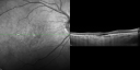
Cuticular Drusen (Basal Laminar Drusen) and Bilateral Chronic Subfoveal Fluid (Serous Retinal Detachment) unresponsive to monthly Avastin OCT OD916 views66-year-old man with bilateral subfoveal fluid unresponsive to one year of monthly avastin therapy.
OD 20/160, Pinhole 20/80. OS 20/63, Pinhole 20/50. OCT scan shows subretinal fluid with normal choroidal thickness. Fluorescein angiogram shows cuticular/basal laminar drusen. ICG shows no foci of choriocapillaris leakage.
Feb 15, 2012
|
|

Cuticular Drusen (Basal Laminar Drusen) and Bilateral Chronic Subfoveal Fluid (Serous Retinal Detachment) unresponsive to monthly Avastin OCT OS EDI1231 views66-year-old man with bilateral subfoveal fluid unresponsive to one year of monthly avastin therapy.
OD 20/160, Pinhole 20/80. OS 20/63, Pinhole 20/50. OCT scan shows subretinal fluid with normal choroidal thickness. Fluorescein angiogram shows cuticular/basal laminar drusen. ICG shows no foci of choriocapillaris leakage.
Feb 15, 2012
|
|

Cuticular Drusen (Basal Laminar Drusen) and Bilateral Chronic Subfoveal Fluid (Serous Retinal Detachment) unresponsive to monthly Avastin OCT OD EDI1038 views66-year-old man with bilateral subfoveal fluid unresponsive to one year of monthly avastin therapy.
OD 20/160, Pinhole 20/80. OS 20/63, Pinhole 20/50. OCT scan shows subretinal fluid with normal choroidal thickness. Fluorescein angiogram shows cuticular/basal laminar drusen. ICG shows no foci of choriocapillaris leakage.
Feb 15, 2012
|
|

Cuticular Drusen (Basal Laminar Drusen) and Bilateral Chronic Subfoveal Fluid (Serous Retinal Detachment) unresponsive to monthly Avastin 947 views66-year-old man with bilateral subfoveal fluid unresponsive to one year of monthly avastin therapy.
OD 20/160, Pinhole 20/80. OS 20/63, Pinhole 20/50. OCT scan shows subretinal fluid with normal choroidal thickness. Fluorescein angiogram shows cuticular/basal laminar drusen. ICG shows no foci of choriocapillaris leakage.
Feb 15, 2012
|
|

Cuticular Drusen (Basal Laminar Drusen) and Bilateral Chronic Subfoveal Fluid (Serous Retinal Detachment) unresponsive to monthly Avastin 760 views66-year-old man with bilateral subfoveal fluid unresponsive to one year of monthly avastin therapy.
OD 20/160, Pinhole 20/80. OS 20/63, Pinhole 20/50. OCT scan shows subretinal fluid with normal choroidal thickness. Fluorescein angiogram shows cuticular/basal laminar drusen. ICG shows no foci of choriocapillaris leakage.
Feb 15, 2012
|
|

Cuticular Drusen (Basal Laminar Drusen) and Bilateral Chronic Subfoveal Fluid (Serous Retinal Detachment) unresponsive to monthly Avastin 712 views66-year-old man with bilateral subfoveal fluid unresponsive to one year of monthly avastin therapy.
OD 20/160, Pinhole 20/80. OS 20/63, Pinhole 20/50. OCT scan shows subretinal fluid with normal choroidal thickness. Fluorescein angiogram shows cuticular/basal laminar drusen. ICG shows no foci of choriocapillaris leakage.
Feb 15, 2012
|
|
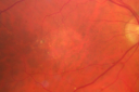
Cuticular Drusen (Basal Laminar Drusen) and Bilateral Chronic Subfoveal Fluid (Serous Retinal Detachment) unresponsive to monthly Avastin 943 views66-year-old man with bilateral subfoveal fluid unresponsive to one year of monthly avastin therapy.
OD 20/160, Pinhole 20/80. OS 20/63, Pinhole 20/50. OCT scan shows subretinal fluid with normal choroidal thickness. Fluorescein angiogram shows cuticular/basal laminar drusen. ICG shows no foci of choriocapillaris leakage.
Feb 15, 2012
|
|
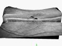
Group 2a Juxtafoveal Telangiectasis - MacTel - Stage 5 left eye - Macular Hole Right eye554 views76-year-old woman has juxtafoveal retinal telangiectasis. She had Stage five neovascularization in the left eye I treated with injections between September of 2007 and October of 2008 with Avastin (3 years ago). OCT scan today shows macular hole right eye. VISUAL ACUITY: OD 20/400, OS 20/40. Feb 03, 2012
|
|
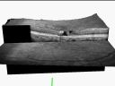
Group 2a Juxtafoveal Telangiectasis - MacTel - Stage 5 left eye - Macular Hole Right eye544 views76-year-old woman has juxtafoveal retinal telangiectasis. She had Stage five neovascularization in the left eye I treated with injections between September of 2007 and October of 2008 with Avastin (3 years ago). OCT scan today shows macular hole right eye. VISUAL ACUITY: OD 20/400, OS 20/40. Feb 03, 2012
|
|
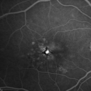
Group 2a Juxtafoveal Telangiectasis - MacTel - Stage 5 left eye - Macular Hole Right eye470 views76-year-old woman has juxtafoveal retinal telangiectasis. She had Stage five neovascularization in the left eye I treated with injections between September of 2007 and October of 2008 with Avastin (3 years ago). OCT scan today shows macular hole right eye. VISUAL ACUITY: OD 20/400, OS 20/40. Feb 03, 2012
|
|
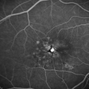
Group 2a Juxtafoveal Telangiectasis - MacTel - Stage 5 left eye - Macular Hole Right eye494 views76-year-old woman has juxtafoveal retinal telangiectasis. She had Stage five neovascularization in the left eye I treated with injections between September of 2007 and October of 2008 with Avastin (3 years ago). OCT scan today shows macular hole right eye. VISUAL ACUITY: OD 20/400, OS 20/40. Feb 03, 2012
|
|
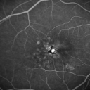
Group 2a Juxtafoveal Telangiectasis - MacTel - Stage 5 left eye - Macular Hole Right eye484 views76-year-old woman has juxtafoveal retinal telangiectasis. She had Stage five neovascularization in the left eye I treated with injections between September of 2007 and October of 2008 with Avastin (3 years ago). OCT scan today shows macular hole right eye. VISUAL ACUITY: OD 20/400, OS 20/40. Feb 03, 2012
|
|
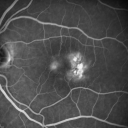
Group 2a Juxtafoveal Telangiectasis - MacTel - Stage 5 left eye - Macular Hole Right eye508 views76-year-old woman has juxtafoveal retinal telangiectasis. She had Stage five neovascularization in the left eye I treated with injections between September of 2007 and October of 2008 with Avastin (3 years ago). OCT scan today shows macular hole right eye. VISUAL ACUITY: OD 20/400, OS 20/40. Feb 03, 2012
|
|
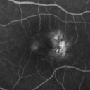
Group 2a Juxtafoveal Telangiectasis - MacTel - Stage 5 left eye - Macular Hole Right eye573 views76-year-old woman has juxtafoveal retinal telangiectasis. She had Stage five neovascularization in the left eye I treated with injections between September of 2007 and October of 2008 with Avastin (3 years ago). OCT scan today shows macular hole right eye. VISUAL ACUITY: OD 20/400, OS 20/40. Feb 03, 2012
|
|
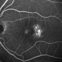
Group 2a Juxtafoveal Telangiectasis - MacTel - Stage 5 left eye - Macular Hole Right eye510 views76-year-old woman has juxtafoveal retinal telangiectasis. She had Stage five neovascularization in the left eye I treated with injections between September of 2007 and October of 2008 with Avastin (3 years ago). OCT scan today shows macular hole right eye. VISUAL ACUITY: OD 20/400, OS 20/40. Feb 03, 2012
|
|
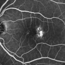
Group 2a Juxtafoveal Telangiectasis - MacTel - Stage 5 left eye - Macular Hole Right eye553 views76-year-old woman has juxtafoveal retinal telangiectasis. She had Stage five neovascularization in the left eye I treated with injections between September of 2007 and October of 2008 with Avastin (3 years ago). OCT scan today shows macular hole right eye. VISUAL ACUITY: OD 20/400, OS 20/40. Feb 03, 2012
|
|
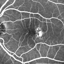
Group 2a Juxtafoveal Telangiectasis - MacTel - Stage 5 left eye - Macular Hole Right eye541 views76-year-old woman has juxtafoveal retinal telangiectasis. She had Stage five neovascularization in the left eye I treated with injections between September of 2007 and October of 2008 with Avastin (3 years ago). OCT scan today shows macular hole right eye. VISUAL ACUITY: OD 20/400, OS 20/40. Feb 03, 2012
|
|
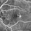
Group 2a Juxtafoveal Telangiectasis - MacTel - Stage 5 left eye - Macular Hole Right eye544 views76-year-old woman has juxtafoveal retinal telangiectasis. She had Stage five neovascularization in the left eye I treated with injections between September of 2007 and October of 2008 with Avastin (3 years ago). OCT scan today shows macular hole right eye. VISUAL ACUITY: OD 20/400, OS 20/40. Feb 03, 2012
|
|

Group 2a Juxtafoveal Telangiectasis - MacTel - Stage 5 left eye - Macular Hole Right eye604 views76-year-old woman has juxtafoveal retinal telangiectasis. She had Stage five neovascularization in the left eye I treated with injections between September of 2007 and October of 2008 with Avastin (3 years ago). OCT scan today shows macular hole right eye. VISUAL ACUITY: OD 20/400, OS 20/40. Feb 03, 2012
|
|

Group 2a Juxtafoveal Telangiectasis - MacTel - Stage 5 left eye - Macular Hole Right eye483 views76-year-old woman has juxtafoveal retinal telangiectasis. She had Stage five neovascularization in the left eye I treated with injections between September of 2007 and October of 2008 with Avastin (3 years ago). OCT scan today shows macular hole right eye. VISUAL ACUITY: OD 20/400, OS 20/40. Feb 03, 2012
|
|
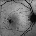
Group 2a Juxtafoveal Telangiectasis - MacTel - Stage 5 left eye - Macular Hole Right eye736 views76-year-old woman has juxtafoveal retinal telangiectasis. She had Stage five neovascularization in the left eye I treated with injections between September of 2007 and October of 2008 with Avastin (3 years ago). OCT scan today shows macular hole right eye. VISUAL ACUITY: OD 20/400, OS 20/40. Feb 03, 2012
|
|

Pattern Dystrophy - Adult Vitelliform - 95 Year Old Man with Good Vision962 views95-year-old man in excellent health and he doesn’t notice a vision problem. OD 20/32, OS 20/80Feb 03, 2012
|
|

Pattern Dystrophy - Adult Vitelliform - 95 Year Old Man with Good Vision664 views95-year-old man in excellent health and he doesn’t notice a vision problem. OD 20/32, OS 20/80Feb 03, 2012
|
|

Pattern Dystrophy - Adult Vitelliform - 95 Year Old Man with Good Vision678 views95-year-old man in excellent health and he doesn’t notice a vision problem. OD 20/32, OS 20/80Feb 03, 2012
|
|

Pattern Dystrophy - Adult Vitelliform - 95 Year Old Man with Good Vision604 views95-year-old man in excellent health and he doesn’t notice a vision problem. OD 20/32, OS 20/80Feb 03, 2012
|
|

Pattern Dystrophy - Adult Vitelliform - 95 Year Old Man with Good Vision771 views95-year-old man in excellent health and he doesn’t notice a vision problem. OD 20/32, OS 20/80Feb 03, 2012
|
|

Pattern Dystrophy - Adult Vitelliform - 95 Year Old Man with Good Vision639 views95-year-old man in excellent health and he doesn’t notice a vision problem. OD 20/32, OS 20/80Feb 03, 2012
|
|
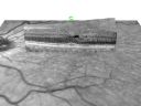
Juxtafoveal Retinal Telangiectasia (Telangiectasis) - MacTel - Striking Gray Ring in Macula663 views59-year-old woman decreasing vision in the right eye for a few years. She has had diabetes for five years. OD 20/100, Pinhole 20/40. OS 20/20Feb 02, 2012
|
|

Juxtafoveal Retinal Telangiectasia (Telangiectasis) - MacTel - Striking Gray Ring in Macula - VIDEO TAKES A LONG TIME TO LOAD612 views59-year-old woman decreasing vision in the right eye for a few years. She has had diabetes for five years. OD 20/100, Pinhole 20/40. OS 20/20Feb 02, 2012
|
|
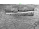
Juxtafoveal Retinal Telangiectasia (Telangiectasis) - MacTel - Striking Gray Ring in Macula - VIDEO TAKES A LONG TIME TO LOAD598 views59-year-old woman decreasing vision in the right eye for a few years. She has had diabetes for five years. OD 20/100, Pinhole 20/40. OS 20/20Feb 02, 2012
|
|
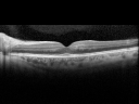
Juxtafoveal Retinal Telangiectasia (Telangiectasis) - MacTel - Striking Gray Ring in Macula - SDOCT599 views59-year-old woman decreasing vision in the right eye for a few years. She has had diabetes for five years. OD 20/100, Pinhole 20/40. OS 20/20Feb 02, 2012
|
|
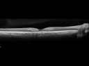
Juxtafoveal Retinal Telangiectasia (Telangiectasis) - MacTel - Striking Gray Ring in Macula - SDOCT614 views59-year-old woman decreasing vision in the right eye for a few years. She has had diabetes for five years. OD 20/100, Pinhole 20/40. OS 20/20Feb 02, 2012
|
|
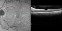
Juxtafoveal Retinal Telangiectasia (Telangiectasis) - MacTel - Striking Gray Ring in Macula - SDOCT666 views59-year-old woman decreasing vision in the right eye for a few years. She has had diabetes for five years. OD 20/100, Pinhole 20/40. OS 20/20Feb 02, 2012
|
|

Juxtafoveal Retinal Telangiectasia (Telangiectasis) - MacTel - Striking Gray Ring in Macula - SDOCT667 views59-year-old woman decreasing vision in the right eye for a few years. She has had diabetes for five years. OD 20/100, Pinhole 20/40. OS 20/20Feb 02, 2012
|
|
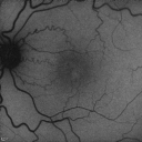
Juxtafoveal Retinal Telangiectasia (Telangiectasis) - MacTel - Striking Gray Ring in Macula - Funuds Autofluorescence762 views59-year-old woman decreasing vision in the right eye for a few years. She has had diabetes for five years. OD 20/100, Pinhole 20/40. OS 20/20Feb 02, 2012
|
|
| 5013 files on 51 page(s) |
 |
|
|
|
|
37 | |
|
|
|
|


