Last additions - Macular Disease
|

Basal Laminar Drusen - Cuticular Drusen - 19 Year Old with Lupus Nephritis693 views19-year-old. She has a history of developing severe systemic lupus erythematosus in 2001. She was in renal failure and had biopsy proven Class 2 lupus nephritis. She is currently on Plaquenil 400 mg for six years, as well as Prednisolone orally, every other day. OD 20/20, OS 20/25.Oct 27, 2010
|
|

Basal Laminar Drusen - Cuticular Drusen - 19 Year Old with Lupus Nephritis728 views19-year-old. She has a history of developing severe systemic lupus erythematosus in 2001. She was in renal failure and had biopsy proven Class 2 lupus nephritis. She is currently on Plaquenil 400 mg for six years, as well as Prednisolone orally, every other day. OD 20/20, OS 20/25.Oct 27, 2010
|
|

Basal Laminar Drusen - Cuticular Drusen - 19 Year Old with Lupus Nephritis736 views19-year-old. She has a history of developing severe systemic lupus erythematosus in 2001. She was in renal failure and had biopsy proven Class 2 lupus nephritis. She is currently on Plaquenil 400 mg for six years, as well as Prednisolone orally, every other day. OD 20/20, OS 20/25.Oct 27, 2010
|
|

Basal Laminar Drusen - Cuticular Drusen - 19 Year Old with Lupus Nephritis881 views19-year-old. She has a history of developing severe systemic lupus erythematosus in 2001. She was in renal failure and had biopsy proven Class 2 lupus nephritis. She is currently on Plaquenil 400 mg for six years, as well as Prednisolone orally, every other day. OD 20/20, OS 20/25.Oct 27, 2010
|
|

Basal Laminar Drusen - Cuticular Drusen - 19 Year Old with Lupus Nephritis711 views19-year-old. She has a history of developing severe systemic lupus erythematosus in 2001. She was in renal failure and had biopsy proven Class 2 lupus nephritis. She is currently on Plaquenil 400 mg for six years, as well as Prednisolone orally, every other day. OD 20/20, OS 20/25.Oct 27, 2010
|
|

Macular Pucker573 viewsThis pleasant 59-year-old man was seen in the office on 7/9/2010. He had new onset floaters about a year ago. Subsequent to that his vision was not quite right in the right eye and then he noticed, starting about three weeks ago, substantial change in the vision. In the morning he sees what he calls a small gray circle with a ring of fire around it and then subsequent to that the vision becomes more normal but not quite right. His vision in the left eye is fine. Vision OD is 20/50, OS is 20/16. Oct 25, 2010
|
|

Macular Pucker507 views. This pleasant 59-year-old man had vitrectomy for a macular pucker in the right eye in July. His vision is gradually improving.
VISUAL ACUITY: Vision OD is 20/30. IOP: OD 22. There is 2+ nuclear sclerosis.
EXTENDED OPHTHALMOSCOPY:
OD: Vertical C/D ratio is 0.5. The macula is smooth and the retina is attached.
OCT SCAN: The OCT scan shows an average central foveal thickness of 349 microns, which compares favorably to the pre-op thickness of 482 microns.
Oct 25, 2010
|
|

Macular Pucker566 views. This pleasant 59-year-old man had vitrectomy for a macular pucker in the right eye in July. His vision is gradually improving.
VISUAL ACUITY: Vision OD is 20/30. IOP: OD 22. There is 2+ nuclear sclerosis.
EXTENDED OPHTHALMOSCOPY:
OD: Vertical C/D ratio is 0.5. The macula is smooth and the retina is attached.
OCT SCAN: The OCT scan shows an average central foveal thickness of 349 microns, which compares favorably to the pre-op thickness of 482 microns.
Oct 25, 2010
|
|

Macular Pucker657 viewsThis pleasant 59-year-old man was seen in the office on 7/9/2010. He had new onset floaters about a year ago. Subsequent to that his vision was not quite right in the right eye and then he noticed, starting about three weeks ago, substantial change in the vision. In the morning he sees what he calls a small gray circle with a ring of fire around it and then subsequent to that the vision becomes more normal but not quite right. His vision in the left eye is fine. Vision OD is 20/50, OS is 20/16. Oct 25, 2010
|
|

Central Serous Retinopathy769 viewsOCT scan showing serous RDOct 24, 2010
|
|

Central Serous Retinopathy756 viewsFundus photo showing serous RDOct 24, 2010
|
|

Central Serous Retinopathy755 viewsEarly FAOct 24, 2010
|
|
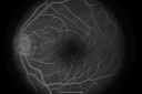
Central Serous Retinopathy735 viewsLater FAOct 24, 2010
|
|

Central Serous Retinopathy832 viewsRed Free photoOct 24, 2010
|
|

Macular Pucker888 viewsFundus photo with OCT superimposed, 20/100 macular puckerOct 22, 2010
|
|
|
| 5015 files on 51 page(s) |
 |
|
|
|
|
|
|
|
|
51 |
|


