neueste Dateien - Retinal Vascular Disease associated with Systemic Disease
|

Microaneursyms and Macular Edema Not Diabetic482 x angesehen85 year old man has been tested about 2-3 times and is always negative for diabetes20. Dezember 2018
|
|

Microaneursyms and Macular Edema Not Diabetic482 x angesehen85 year old man has been tested about 2-3 times and is always negative for diabetes20. Dezember 2018
|
|

Microaneursyms and Macular Edema Not Diabetic384 x angesehen85 year old man has been tested about 2-3 times and is always negative for diabetes20. Dezember 2018
|
|

Microaneursyms and Macular Edema Not Diabetic476 x angesehen85 year old man has been tested about 2-3 times and is always negative for diabetes20. Dezember 2018
|
|

Microaneursyms and Macular Edema Not Diabetic759 x angesehen85 year old man has been tested about 2-3 times and is always negative for diabetes20. Dezember 2018
|
|

Microaneursyms and Macular Edema Not Diabetic428 x angesehen85 year old man has been tested about 2-3 times and is always negative for diabetes20. Dezember 2018
|
|

Microaneursyms and Macular Edema Not Diabetic443 x angesehen85 year old man has been tested about 2-3 times and is always negative for diabetes20. Dezember 2018
|
|
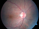
Microaneursyms and Macular Edema Not Diabetic453 x angesehen85 year old man has been tested about 2-3 times and is always negative for diabetes20. Dezember 2018
|
|
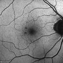
Microaneursyms and Macular Edema Not Diabetic422 x angesehen85 year old man has been tested about 2-3 times and is always negative for diabetes20. Dezember 2018
|
|

Microaneursyms and Macular Edema Not Diabetic396 x angesehen85 year old man has been tested about 2-3 times and is always negative for diabetes20. Dezember 2018
|
|

Microaneursyms and Macular Edema Not Diabetic416 x angesehen85 year old man has been tested about 2-3 times and is always negative for diabetes20. Dezember 2018
|
|

Microaneursyms and Macular Edema Not Diabetic413 x angesehen85 year old man has been tested about 2-3 times and is always negative for diabetes20. Dezember 2018
|
|

Microaneursyms and Macular Edema Not Diabetic490 x angesehen85 year old man has been tested about 2-3 times and is always negative for diabetes20. Dezember 2018
|
|

Anemic Retinopathy783 x angesehenMyelodysplasia - HgB 8 and very low platelet count05. November 2018
|
|

Anemic Retinopathy548 x angesehenMyelodysplasia - HgB 8 and very low platelet count05. November 2018
|
|

Anemic Retinopathy763 x angesehenMyelodysplasia - HgB 8 and very low platelet count05. November 2018
|
|
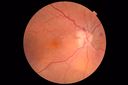
Anemic Retinopathy687 x angesehenMyelodysplasia - HgB 8 and very low platelet count05. November 2018
|
|

Parafoveal Microaneurysm - Not Diabetic503 x angesehen88 year old man asymptomatic with parafoveal microaneurysm26. Februar 2018
|
|

Parafoveal Microaneurysm - Not Diabetic496 x angesehen88 year old man asymptomatic with parafoveal microaneurysm26. Februar 2018
|
|
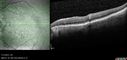
Parafoveal Microaneurysm - Not Diabetic457 x angesehen88 year old man asymptomatic with parafoveal microaneurysm26. Februar 2018
|
|

Foveal Hypoplasia in 23 year old patient with ROP457 x angesehen11. Januar 2018
|
|

Foveal Hypoplasia in 23 year old patient with ROP432 x angesehen11. Januar 2018
|
|

Foveal Hypoplasia in adult with ROP - good Vision429 x angesehen26 year old female Born at 1 lb 15 oz and had cryo for ROP at birth.
VA OD: Dcc20/25
VA OS: Dcc20/20
SD OCT shows foveal hypoplasia
The SD OCT scan shows incursion of one or more of the plexiform layers into the center of the fovea.11. Januar 2018
|
|

Foveal Hypoplasia in adult with ROP - good Vision431 x angesehen26 year old female Born at 1 lb 15 oz and had cryo for ROP at birth.
VA OD: Dcc20/25
VA OS: Dcc20/20
SD OCT shows foveal hypoplasia
The SD OCT scan shows incursion of one or more of the plexiform layers into the center of the fovea.11. Januar 2018
|
|

Foveal Hypoplasia in adult with ROP - good Vision508 x angesehen26 year old female Born at 1 lb 15 oz and had cryo for ROP at birth.
VA OD: Dcc20/25
VA OS: Dcc20/20
SD OCT shows foveal hypoplasia
The SD OCT scan shows incursion of one or more of the plexiform layers into the center of the fovea.11. Januar 2018
|
|

Foveal Hypoplasia in adult with ROP - good Vision451 x angesehen26 year old female Born at 1 lb 15 oz and had cryo for ROP at birth.
VA OD: Dcc20/25
VA OS: Dcc20/20
SD OCT shows foveal hypoplasia
The SD OCT scan shows incursion of one or more of the plexiform layers into the center of the fovea.11. Januar 2018
|
|

Foveal Hypoplasia in adult with ROP - good Vision499 x angesehen26 year old female Born at 1 lb 15 oz and had cryo for ROP at birth.
VA OD: Dcc20/25
VA OS: Dcc20/20
SD OCT shows foveal hypoplasia
The SD OCT scan shows incursion of one or more of the plexiform layers into the center of the fovea.11. Januar 2018
|
|

Foveal Hypoplasia in adult with ROP - good Vision446 x angesehen26 year old female Born at 1 lb 15 oz and had cryo for ROP at birth.
VA OD: Dcc20/25
VA OS: Dcc20/20
SD OCT shows foveal hypoplasia
The SD OCT scan shows incursion of one or more of the plexiform layers into the center of the fovea.11. Januar 2018
|
|

Foveal Hypoplasia in adult with ROP - good Vision445 x angesehen26 year old female Born at 1 lb 15 oz and had cryo for ROP at birth.
VA OD: Dcc20/25
VA OS: Dcc20/20
SD OCT shows foveal hypoplasia
The SD OCT scan shows incursion of one or more of the plexiform layers into the center of the fovea.11. Januar 2018
|
|

Foveal Hypoplasia in adult with ROP - good Vision425 x angesehen26 year old female Born at 1 lb 15 oz and had cryo for ROP at birth.
VA OD: Dcc20/25
VA OS: Dcc20/20
SD OCT shows foveal hypoplasia
The SD OCT scan shows incursion of one or more of the plexiform layers into the center of the fovea.11. Januar 2018
|
|

Cotton Wool Spot at 6 Weeks Pregnant460 x angesehen 31 y/o white female who is 4-6 weeks pregnant, no medical problems. Saw new shadow12. Oktober 2017
|
|

Orbital Cellulitis - Optic nerve compression with ocular ischemia482 x angesehen81 year old woman with constricted eye movement, orbital cellulitis and severe vision loss in the right eye (LP). She has whitening of the retina and the OCT shows ischemic changes as well as subretinal fluid and blood. The vision in this eye declined to NLP and the eye remained ischemic despite control of her infection.06. Mai 2016
|
|
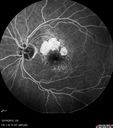
Orbital Cellulitis - Optic nerve compression with ocular ischemia557 x angesehen81 year old woman with constricted eye movement, orbital cellulitis and severe vision loss in the right eye (LP). She has whitening of the retina and the OCT shows ischemic changes as well as subretinal fluid and blood. The vision in this eye declined to NLP and the eye remained ischemic despite control of her infection.06. Mai 2016
|
|

Orbital Cellulitis - Optic nerve compression with ocular ischemia521 x angesehen81 year old woman with constricted eye movement, orbital cellulitis and severe vision loss in the right eye (LP). She has whitening of the retina and the OCT shows ischemic changes as well as subretinal fluid and blood. The vision in this eye declined to NLP and the eye remained ischemic despite control of her infection.06. Mai 2016
|
|

Orbital Cellulitis - Optic nerve compression with ocular ischemia508 x angesehen81 year old woman with constricted eye movement, orbital cellulitis and severe vision loss in the right eye (LP). She has whitening of the retina and the OCT shows ischemic changes as well as subretinal fluid and blood. The vision in this eye declined to NLP and the eye remained ischemic despite control of her infection.06. Mai 2016
|
|

Orbital Cellulitis - Optic nerve compression with ocular ischemia514 x angesehen81 year old woman with constricted eye movement, orbital cellulitis and severe vision loss in the right eye (LP). She has whitening of the retina and the OCT shows ischemic changes as well as subretinal fluid and blood. The vision in this eye declined to NLP and the eye remained ischemic despite control of her infection.06. Mai 2016
|
|

Orbital Cellulitis - Optic nerve compression with ocular ischemia524 x angesehen81 year old woman with constricted eye movement, orbital cellulitis and severe vision loss in the right eye (LP). She has whitening of the retina and the OCT shows ischemic changes as well as subretinal fluid and blood. The vision in this eye declined to NLP and the eye remained ischemic despite control of her infection.06. Mai 2016
|
|

Orbital Cellulitis - Optic nerve compression with ocular ischemia565 x angesehen81 year old woman with constricted eye movement, orbital cellulitis and severe vision loss in the right eye (LP). She has whitening of the retina and the OCT shows ischemic changes as well as subretinal fluid and blood. The vision in this eye declined to NLP and the eye remained ischemic despite control of her infection.06. Mai 2016
|
|

Orbital Cellulitis - Optic nerve compression with ocular ischemia530 x angesehen81 year old woman with constricted eye movement, orbital cellulitis and severe vision loss in the right eye (LP). She has whitening of the retina and the OCT shows ischemic changes as well as subretinal fluid and blood. The vision in this eye declined to NLP and the eye remained ischemic despite control of her infection.06. Mai 2016
|
|

Orbital Cellulitis - Optic nerve compression with ocular ischemia494 x angesehen81 year old woman with constricted eye movement, orbital cellulitis and severe vision loss in the right eye (LP). She has whitening of the retina and the OCT shows ischemic changes as well as subretinal fluid and blood. The vision in this eye declined to NLP and the eye remained ischemic despite control of her infection.06. Mai 2016
|
|
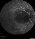
Orbital Cellulitis - Optic nerve compression with ocular ischemia499 x angesehen81 year old woman with constricted eye movement, orbital cellulitis and severe vision loss in the right eye (LP). She has whitening of the retina and the OCT shows ischemic changes as well as subretinal fluid and blood. The vision in this eye declined to NLP and the eye remained ischemic despite control of her infection.06. Mai 2016
|
|

Orbital Cellulitis - Optic nerve compression with ocular ischemia484 x angesehen81 year old woman with constricted eye movement, orbital cellulitis and severe vision loss in the right eye (LP). She has whitening of the retina and the OCT shows ischemic changes as well as subretinal fluid and blood. The vision in this eye declined to NLP and the eye remained ischemic despite control of her infection.06. Mai 2016
|
|

Orbital Cellulitis - Optic nerve compression with ocular ischemia530 x angesehen81 year old woman with constricted eye movement, orbital cellulitis and severe vision loss in the right eye (LP). She has whitening of the retina and the OCT shows ischemic changes as well as subretinal fluid and blood. The vision in this eye declined to NLP and the eye remained ischemic despite control of her infection.06. Mai 2016
|
|

Orbital Cellulitis - Optic nerve compression with ocular ischemia497 x angesehen81 year old woman with constricted eye movement, orbital cellulitis and severe vision loss in the right eye (LP). She has whitening of the retina and the OCT shows ischemic changes as well as subretinal fluid and blood. The vision in this eye declined to NLP and the eye remained ischemic despite control of her infection.06. Mai 2016
|
|

Orbital Cellulitis - Optic nerve compression with ocular ischemia522 x angesehen81 year old woman with constricted eye movement, orbital cellulitis and severe vision loss in the right eye (LP). She has whitening of the retina and the OCT shows ischemic changes as well as subretinal fluid and blood. The vision in this eye declined to NLP and the eye remained ischemic despite control of her infection.06. Mai 2016
|
|
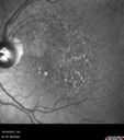
Orbital Cellulitis - Optic nerve compression with ocular ischemia497 x angesehen81 year old woman with constricted eye movement, orbital cellulitis and severe vision loss in the right eye (LP). She has whitening of the retina and the OCT shows ischemic changes as well as subretinal fluid and blood. The vision in this eye declined to NLP and the eye remained ischemic despite control of her infection.06. Mai 2016
|
|

Orbital Cellulitis - Optic nerve compression with ocular ischemia556 x angesehen81 year old woman with constricted eye movement, orbital cellulitis and severe vision loss in the right eye (LP). She has whitening of the retina and the OCT shows ischemic changes as well as subretinal fluid and blood. The vision in this eye declined to NLP and the eye remained ischemic despite control of her infection.06. Mai 2016
|
|

Orbital Cellulitis - Optic nerve compression with ocular ischemia579 x angesehen81 year old woman with constricted eye movement, orbital cellulitis and severe vision loss in the right eye (LP). She has whitening of the retina and the OCT shows ischemic changes as well as subretinal fluid and blood. The vision in this eye declined to NLP and the eye remained ischemic despite control of her infection.06. Mai 2016
|
|

Orbital Cellulitis - Optic nerve compression with ocular ischemia584 x angesehen81 year old woman with constricted eye movement, orbital cellulitis and severe vision loss in the right eye (LP). She has whitening of the retina and the OCT shows ischemic changes as well as subretinal fluid and blood. The vision in this eye declined to NLP and the eye remained ischemic despite control of her infection.06. Mai 2016
|
|

Orbital Cellulitis - Optic nerve compression with ocular ischemia618 x angesehen81 year old woman with constricted eye movement, orbital cellulitis and severe vision loss in the right eye (LP). She has whitening of the retina and the OCT shows ischemic changes as well as subretinal fluid and blood. The vision in this eye declined to NLP and the eye remained ischemic despite control of her infection.06. Mai 2016
|
|

Central retinal artery and vein occlusion - Protein C Deficiency 33 year old male536 x angesehen33 year old male with vision loss for 3 hours. VA 20/12 right eye, 5/200 left eye. He has a protein C deficiency. The left eye recovered vision in about 1 month to 20/16. FA shows very slow recirculation time.06. Mai 2016
|
|

Central retinal artery and vein occlusion - Protein C Deficiency 33 year old male469 x angesehen33 year old male with vision loss for 3 hours. VA 20/12 right eye, 5/200 left eye. He has a protein C deficiency. The left eye recovered vision in about 1 month to 20/16. FA shows very slow recirculation time.06. Mai 2016
|
|

Central retinal artery and vein occlusion - Protein C Deficiency 33 year old male525 x angesehen33 year old male with vision loss for 3 hours. VA 20/12 right eye, 5/200 left eye. He has a protein C deficiency. The left eye recovered vision in about 1 month to 20/16. FA shows very slow recirculation time.06. Mai 2016
|
|

Central retinal artery and vein occlusion - Protein C Deficiency 33 year old male525 x angesehen33 year old male with vision loss for 3 hours. VA 20/12 right eye, 5/200 left eye. He has a protein C deficiency. The left eye recovered vision in about 1 month to 20/16. FA shows very slow recirculation time.06. Mai 2016
|
|

Central retinal artery and vein occlusion - Protein C Deficiency 33 year old male490 x angesehen33 year old male with vision loss for 3 hours. VA 20/12 right eye, 5/200 left eye. He has a protein C deficiency. The left eye recovered vision in about 1 month to 20/16. FA shows very slow recirculation time.06. Mai 2016
|
|

Central retinal artery and vein occlusion - Protein C Deficiency 33 year old male463 x angesehen33 year old male with vision loss for 3 hours. VA 20/12 right eye, 5/200 left eye. He has a protein C deficiency. The left eye recovered vision in about 1 month to 20/16. FA shows very slow recirculation time.06. Mai 2016
|
|
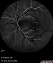
Central retinal artery and vein occlusion - Protein C Deficiency 33 year old male502 x angesehen33 year old male with vision loss for 3 hours. VA 20/12 right eye, 5/200 left eye. He has a protein C deficiency. The left eye recovered vision in about 1 month to 20/16. FA shows very slow recirculation time.06. Mai 2016
|
|

Central retinal artery and vein occlusion - Protein C Deficiency 33 year old male480 x angesehen33 year old male with vision loss for 3 hours. VA 20/12 right eye, 5/200 left eye. He has a protein C deficiency. The left eye recovered vision in about 1 month to 20/16. FA shows very slow recirculation time.06. Mai 2016
|
|

Central retinal artery and vein occlusion - Protein C Deficiency 33 year old male486 x angesehen33 year old male with vision loss for 3 hours. VA 20/12 right eye, 5/200 left eye. He has a protein C deficiency. The left eye recovered vision in about 1 month to 20/16. FA shows very slow recirculation time.06. Mai 2016
|
|

Central retinal artery and vein occlusion - Protein C Deficiency 33 year old male453 x angesehen33 year old male with vision loss for 3 hours. VA 20/12 right eye, 5/200 left eye. He has a protein C deficiency. The left eye recovered vision in about 1 month to 20/16. FA shows very slow recirculation time.06. Mai 2016
|
|

Central retinal artery and vein occlusion - Protein C Deficiency 33 year old male454 x angesehen33 year old male with vision loss for 3 hours. VA 20/12 right eye, 5/200 left eye. He has a protein C deficiency. The left eye recovered vision in about 1 month to 20/16. FA shows very slow recirculation time.06. Mai 2016
|
|

Central retinal artery and vein occlusion - Protein C Deficiency 33 year old male471 x angesehen33 year old male with vision loss for 3 hours. VA 20/12 right eye, 5/200 left eye. He has a protein C deficiency. The left eye recovered vision in about 1 month to 20/16. FA shows very slow recirculation time.06. Mai 2016
|
|

Central retinal artery and vein occlusion - Protein C Deficiency 33 year old male544 x angesehen33 year old male with vision loss for 3 hours. VA 20/12 right eye, 5/200 left eye. He has a protein C deficiency. The left eye recovered vision in about 1 month to 20/16. FA shows very slow recirculation time.06. Mai 2016
|
|

Central retinal artery and vein occlusion - Protein C Deficiency 33 year old male511 x angesehen33 year old male with vision loss for 3 hours. VA 20/12 right eye, 5/200 left eye. He has a protein C deficiency. The left eye recovered vision in about 1 month to 20/16. FA shows very slow recirculation time.06. Mai 2016
|
|

Central retinal artery and vein occlusion - Protein C Deficiency 33 year old male491 x angesehen33 year old male with vision loss for 3 hours. VA 20/12 right eye, 5/200 left eye. He has a protein C deficiency. The left eye recovered vision in about 1 month to 20/16. FA shows very slow recirculation time.06. Mai 2016
|
|

Central retinal artery and vein occlusion - Protein C Deficiency 33 year old male521 x angesehen33 year old male with vision loss for 3 hours. VA 20/12 right eye, 5/200 left eye. He has a protein C deficiency. The left eye recovered vision in about 1 month to 20/16. FA shows very slow recirculation time.06. Mai 2016
|
|
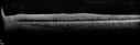
Central retinal artery and vein occlusion - Protein C Deficiency 33 year old male461 x angesehen33 year old male with vision loss for 3 hours. VA 20/12 right eye, 5/200 left eye. He has a protein C deficiency. The left eye recovered vision in about 1 month to 20/16. FA shows very slow recirculation time.06. Mai 2016
|
|

Central retinal artery and vein occlusion - Protein C Deficiency 33 year old male505 x angesehen33 year old male with vision loss for 3 hours. VA 20/12 right eye, 5/200 left eye. He has a protein C deficiency. The left eye recovered vision in about 1 month to 20/16. FA shows very slow recirculation time.06. Mai 2016
|
|

Central retinal artery and vein occlusion - Protein C Deficiency 33 year old male473 x angesehen33 year old male with vision loss for 3 hours. VA 20/12 right eye, 5/200 left eye. He has a protein C deficiency. The left eye recovered vision in about 1 month to 20/16. FA shows very slow recirculation time.06. Mai 2016
|
|
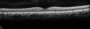
Central retinal artery and vein occlusion - Protein C Deficiency 33 year old male511 x angesehen33 year old male with vision loss for 3 hours. VA 20/12 right eye, 5/200 left eye. He has a protein C deficiency. The left eye recovered vision in about 1 month to 20/16. FA shows very slow recirculation time.06. Mai 2016
|
|

Central retinal artery and vein occlusion - Protein C Deficiency 33 year old male513 x angesehen33 year old male with vision loss for 3 hours. VA 20/12 right eye, 5/200 left eye. He has a protein C deficiency. The left eye recovered vision in about 1 month to 20/16. FA shows very slow recirculation time.06. Mai 2016
|
|
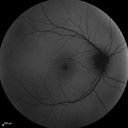
Central retinal artery and vein occlusion - Protein C Deficiency 33 year old male504 x angesehen33 year old male with vision loss for 3 hours. VA 20/12 right eye, 5/200 left eye. He has a protein C deficiency. The left eye recovered vision in about 1 month to 20/16. FA shows very slow recirculation time.06. Mai 2016
|
|

Central retinal artery and vein occlusion - Protein C Deficiency 33 year old male582 x angesehen33 year old male with vision loss for 3 hours. VA 20/12 right eye, 5/200 left eye. He has a protein C deficiency. The left eye recovered vision in about 1 month to 20/16. FA shows very slow recirculation time.06. Mai 2016
|
|

Central retinal artery and vein occlusion - Protein C Deficiency 33 year old male529 x angesehen33 year old male with vision loss for 3 hours. VA 20/12 right eye, 5/200 left eye. He has a protein C deficiency. The left eye recovered vision in about 1 month to 20/16. FA shows very slow recirculation time.06. Mai 2016
|
|

Central retinal artery and vein occlusion - Protein C Deficiency 33 year old male561 x angesehen33 year old male with vision loss for 3 hours. VA 20/12 right eye, 5/200 left eye. He has a protein C deficiency. The left eye recovered vision in about 1 month to 20/16. FA shows very slow recirculation time.06. Mai 2016
|
|

Central retinal artery and vein occlusion - Protein C Deficiency 33 year old male630 x angesehen33 year old male with vision loss for 3 hours. VA 20/12 right eye, 5/200 left eye. He has a protein C deficiency. The left eye recovered vision in about 1 month to 20/16. FA shows very slow recirculation time.06. Mai 2016
|
|

Central retinal artery and vein occlusion - Protein C Deficiency 33 year old male450 x angesehen33 year old male with vision loss for 3 hours. VA 20/12 right eye, 5/200 left eye. He has a protein C deficiency. The left eye recovered vision in about 1 month to 20/16. FA shows very slow recirculation time.06. Mai 2016
|
|

Hypertensive Retinopathy - BP 240 / 130 mmHg - Grade 4503 x angesehen71 year old woman who claims to be asymptomatic with decreased vision, macular edema, cotton wool spots, subretinal fluid and optic nerve edema06. Mai 2016
|
|

Hypertensive Retinopathy - BP 240 / 130 mmHg - Grade 4470 x angesehen71 year old woman who claims to be asymptomatic with decreased vision, macular edema, cotton wool spots, subretinal fluid and optic nerve edema06. Mai 2016
|
|

Hypertensive Retinopathy - BP 240 / 130 mmHg - Grade 4428 x angesehen71 year old woman who claims to be asymptomatic with decreased vision, macular edema, cotton wool spots, subretinal fluid and optic nerve edema06. Mai 2016
|
|

Hypertensive Retinopathy - BP 240 / 130 mmHg - Grade 4498 x angesehen71 year old woman who claims to be asymptomatic with decreased vision, macular edema, cotton wool spots, subretinal fluid and optic nerve edema06. Mai 2016
|
|

Hypertensive Retinopathy - BP 240 / 130 mmHg - Grade 4522 x angesehen71 year old woman who claims to be asymptomatic with decreased vision, macular edema, cotton wool spots, subretinal fluid and optic nerve edema06. Mai 2016
|
|

Hypertensive Retinopathy - BP 240 / 130 mmHg - Grade 4595 x angesehen71 year old woman who claims to be asymptomatic with decreased vision, macular edema, cotton wool spots, subretinal fluid and optic nerve edema06. Mai 2016
|
|

Hypertensive Retinopathy - BP 240 / 130 mmHg - Grade 4568 x angesehen71 year old woman who claims to be asymptomatic with decreased vision, macular edema, cotton wool spots, subretinal fluid and optic nerve edema06. Mai 2016
|
|

Hypertensive Retinopathy - BP 240 / 130 mmHg - Grade 4640 x angesehen71 year old woman who claims to be asymptomatic with decreased vision, macular edema, cotton wool spots, subretinal fluid and optic nerve edema06. Mai 2016
|
|

Hypertensive Retinopathy - BP 240 / 130 mmHg - Grade 4660 x angesehen71 year old woman who claims to be asymptomatic with decreased vision, macular edema, cotton wool spots, subretinal fluid and optic nerve edema06. Mai 2016
|
|

Hypertensive Retinopathy - BP 240 / 130 mmHg - Grade 4545 x angesehen71 year old woman who claims to be asymptomatic with decreased vision, macular edema, cotton wool spots, subretinal fluid and optic nerve edema06. Mai 2016
|
|

Trousseau Sign - CRVO and Pancreatic Cancer617 x angesehenLeft eye shows CRVO with severe edema. The right eye has multiple small hemorrhages. Some of this is probably anemic retinopathy and some from hypercoaguability of cancer. The patient has pancreatic cancer.07. März 2016
|
|

Trousseau Sign - CRVO and Pancreatic Cancer499 x angesehenLeft eye shows CRVO with severe edema. The right eye has multiple small hemorrhages. Some of this is probably anemic retinopathy and some from hypercoaguability of cancer. The patient has pancreatic cancer.07. März 2016
|
|

Trousseau Sign - CRVO and Pancreatic Cancer503 x angesehenLeft eye shows CRVO with severe edema. The right eye has multiple small hemorrhages. Some of this is probably anemic retinopathy and some from hypercoaguability of cancer. The patient has pancreatic cancer.07. März 2016
|
|

Trousseau Sign - CRVO and Pancreatic Cancer629 x angesehenLeft eye shows CRVO with severe edema. The right eye has multiple small hemorrhages. Some of this is probably anemic retinopathy and some from hypercoaguability of cancer. The patient has pancreatic cancer.07. März 2016
|
|

Trousseau Sign - CRVO and Pancreatic Cancer603 x angesehenLeft eye shows CRVO with severe edema. The right eye has multiple small hemorrhages. Some of this is probably anemic retinopathy and some from hypercoaguability of cancer. The patient has pancreatic cancer.07. März 2016
|
|

Trousseau Sign - CRVO and Pancreatic Cancer659 x angesehenLeft eye shows CRVO with severe edema. The right eye has multiple small hemorrhages. Some of this is probably anemic retinopathy and some from hypercoaguability of cancer. The patient has pancreatic cancer.07. März 2016
|
|
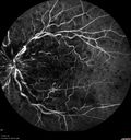
Trousseau Sign - CRVO and Pancreatic Cancer570 x angesehenLeft eye shows CRVO with severe edema. The right eye has multiple small hemorrhages. Some of this is probably anemic retinopathy and some from hypercoaguability of cancer. The patient has pancreatic cancer.07. März 2016
|
|

Trousseau Sign - CRVO and Pancreatic Cancer518 x angesehenLeft eye shows CRVO with severe edema. The right eye has multiple small hemorrhages. Some of this is probably anemic retinopathy and some from hypercoaguability of cancer. The patient has pancreatic cancer.07. März 2016
|
|

Trousseau Sign - CRVO and Pancreatic Cancer634 x angesehenLeft eye shows CRVO with severe edema. The right eye has multiple small hemorrhages. Some of this is probably anemic retinopathy and some from hypercoaguability of cancer. The patient has pancreatic cancer.07. März 2016
|
|

Trousseau Sign - CRVO and Pancreatic Cancer580 x angesehenLeft eye shows CRVO with severe edema. The right eye has multiple small hemorrhages. Some of this is probably anemic retinopathy and some from hypercoaguability of cancer. The patient has pancreatic cancer.07. März 2016
|
|
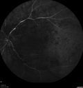
Trousseau Sign - CRVO and Pancreatic Cancer545 x angesehenLeft eye shows CRVO with severe edema. The right eye has multiple small hemorrhages. Some of this is probably anemic retinopathy and some from hypercoaguability of cancer. The patient has pancreatic cancer.07. März 2016
|
|

Trousseau Sign - CRVO and Pancreatic Cancer623 x angesehenLeft eye shows CRVO with severe edema. The right eye has multiple small hemorrhages. Some of this is probably anemic retinopathy and some from hypercoaguability of cancer. The patient has pancreatic cancer.07. März 2016
|
|

Trousseau Sign - CRVO and Pancreatic Cancer637 x angesehenLeft eye shows CRVO with severe edema. The right eye has multiple small hemorrhages. Some of this is probably anemic retinopathy and some from hypercoaguability of cancer. The patient has pancreatic cancer.07. März 2016
|
|
| 844 Dateien auf 9 Seite(n) |
 |
|
4 | |
|
|
|
|


