Last additions - Posterior Uveitis - Chorioretinitis
|

Unilteral Acute Idiopathic Maculopathy - Initial Presentation479 viewsFeb 02, 2018
|
|
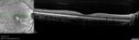
470 viewsFeb 02, 2018
|
|

475 viewsFeb 02, 2018
|
|

432 viewsFeb 02, 2018
|
|

461 viewsFeb 02, 2018
|
|

463 viewsFeb 02, 2018
|
|

440 viewsFeb 02, 2018
|
|

433 viewsFeb 02, 2018
|
|

427 viewsFeb 02, 2018
|
|

421 viewsFeb 02, 2018
|
|

444 viewsFeb 02, 2018
|
|

419 viewsFeb 02, 2018
|
|
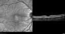
415 viewsFeb 02, 2018
|
|

455 viewsFeb 02, 2018
|
|

487 viewsFeb 02, 2018
|
|

Sarcoidosis Multifocal Choroiditis542 views66 year old man with dense cataracts and recurrent uveitis. Images show multifocal choroidal granulomas from sarcoidosis more in the right eye than the left eye.Jan 29, 2018
|
|

Sarcoidosis Multifocal Choroiditis573 views66 year old man with dense cataracts and recurrent uveitis. Images show multifocal choroidal granulomas from sarcoidosis more in the right eye than the left eye.Jan 29, 2018
|
|

Sarcoidosis Multifocal Choroiditis529 views66 year old man with dense cataracts and recurrent uveitis. Images show multifocal choroidal granulomas from sarcoidosis more in the right eye than the left eye.Jan 29, 2018
|
|

Sarcoidosis Multifocal Choroiditis518 views66 year old man with dense cataracts and recurrent uveitis. Images show multifocal choroidal granulomas from sarcoidosis more in the right eye than the left eye.Jan 29, 2018
|
|

Sarcoidosis Multifocal Choroiditis528 views66 year old man with dense cataracts and recurrent uveitis. Images show multifocal choroidal granulomas from sarcoidosis more in the right eye than the left eye.Jan 29, 2018
|
|

Sarcoidosis Multifocal Choroiditis513 views66 year old man with dense cataracts and recurrent uveitis. Images show multifocal choroidal granulomas from sarcoidosis more in the right eye than the left eye.Jan 29, 2018
|
|

Sarcoidosis Multifocal Choroiditis567 views66 year old man with dense cataracts and recurrent uveitis. Images show multifocal choroidal granulomas from sarcoidosis more in the right eye than the left eye.Jan 29, 2018
|
|

Sarcoidosis Multifocal Choroiditis466 views66 year old man with dense cataracts and recurrent uveitis. Images show multifocal choroidal granulomas from sarcoidosis more in the right eye than the left eye.Jan 29, 2018
|
|

Sarcoidosis Multifocal Choroiditis469 views66 year old man with dense cataracts and recurrent uveitis. Images show multifocal choroidal granulomas from sarcoidosis more in the right eye than the left eye.Jan 29, 2018
|
|

Sarcoidosis Multifocal Choroiditis461 views66 year old man with dense cataracts and recurrent uveitis. Images show multifocal choroidal granulomas from sarcoidosis more in the right eye than the left eye.Jan 29, 2018
|
|
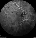
Sarcoidosis Multifocal Choroiditis543 views66 year old man with dense cataracts and recurrent uveitis. Images show multifocal choroidal granulomas from sarcoidosis more in the right eye than the left eye.Jan 29, 2018
|
|

Sarcoidosis Multifocal Choroiditis469 views66 year old man with dense cataracts and recurrent uveitis. Images show multifocal choroidal granulomas from sarcoidosis more in the right eye than the left eye.Jan 29, 2018
|
|

Sarcoidosis Multifocal Choroiditis491 views66 year old man with dense cataracts and recurrent uveitis. Images show multifocal choroidal granulomas from sarcoidosis more in the right eye than the left eye.Jan 29, 2018
|
|
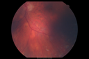
Sarcoidosis Multifocal Choroiditis483 views66 year old man with dense cataracts and recurrent uveitis. Images show multifocal choroidal granulomas from sarcoidosis more in the right eye than the left eye.Jan 29, 2018
|
|
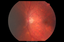
Sarcoidosis Multifocal Choroiditis512 views66 year old man with dense cataracts and recurrent uveitis. Images show multifocal choroidal granulomas from sarcoidosis more in the right eye than the left eye.Jan 29, 2018
|
|

Sarcoidosis Multifocal Choroiditis491 views66 year old man with dense cataracts and recurrent uveitis. Images show multifocal choroidal granulomas from sarcoidosis more in the right eye than the left eye.Jan 29, 2018
|
|
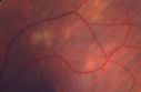
Sarcoidosis Multifocal Choroiditis521 views66 year old man with dense cataracts and recurrent uveitis. Images show multifocal choroidal granulomas from sarcoidosis more in the right eye than the left eye.Jan 29, 2018
|
|

Sarcoidosis Multifocal Choroiditis3721 views66 year old man with dense cataracts and recurrent uveitis. Images show multifocal choroidal granulomas from sarcoidosis more in the right eye than the left eye.Jan 29, 2018
|
|

Sarcoidosis Multifocal Choroiditis606 views66 year old man with dense cataracts and recurrent uveitis. Images show multifocal choroidal granulomas from sarcoidosis more in the right eye than the left eye.Jan 29, 2018
|
|

Sarcoidosis Multifocal Choroiditis475 views66 year old man with dense cataracts and recurrent uveitis. Images show multifocal choroidal granulomas from sarcoidosis more in the right eye than the left eye.Jan 29, 2018
|
|

Sarcoidosis Multifocal Choroiditis563 views66 year old man with dense cataracts and recurrent uveitis. Images show multifocal choroidal granulomas from sarcoidosis more in the right eye than the left eye.Jan 29, 2018
|
|

Sarcoidosis Multifocal Choroiditis624 views66 year old man with dense cataracts and recurrent uveitis. Images show multifocal choroidal granulomas from sarcoidosis more in the right eye than the left eye.Jan 29, 2018
|
|

Toxoplasmosis - Recurrent - Moderate Vitritis634 views32 year old woman with 20/80 vision and moderately painful red eye in the left eye only. The left eye had some redness about 10 years ago. She is a native of Sierra LeoneJan 05, 2018
|
|

Toxoplasmosis - Recurrent - Moderate Vitritis534 views32 year old woman with 20/80 vision and moderately painful red eye in the left eye only. The left eye had some redness about 10 years ago. She is a native of Sierra LeoneJan 05, 2018
|
|

Toxoplasmosis - Recurrent - Moderate Vitritis526 views32 year old woman with 20/80 vision and moderately painful red eye in the left eye only. The left eye had some redness about 10 years ago. She is a native of Sierra LeoneJan 05, 2018
|
|

Toxoplasmosis - Recurrent - Moderate Vitritis605 views32 year old woman with 20/80 vision and moderately painful red eye in the left eye only. The left eye had some redness about 10 years ago. She is a native of Sierra LeoneJan 05, 2018
|
|

Toxoplasmosis - Recurrent - Moderate Vitritis527 views32 year old woman with 20/80 vision and moderately painful red eye in the left eye only. The left eye had some redness about 10 years ago. She is a native of Sierra LeoneJan 05, 2018
|
|

Toxoplasmosis - Recurrent - Moderate Vitritis523 views32 year old woman with 20/80 vision and moderately painful red eye in the left eye only. The left eye had some redness about 10 years ago. She is a native of Sierra LeoneJan 05, 2018
|
|

Toxoplasmosis - Recurrent - Moderate Vitritis470 views32 year old woman with 20/80 vision and moderately painful red eye in the left eye only. The left eye had some redness about 10 years ago. She is a native of Sierra LeoneJan 05, 2018
|
|

Toxoplasmosis - Recurrent - Moderate Vitritis583 views32 year old woman with 20/80 vision and moderately painful red eye in the left eye only. The left eye had some redness about 10 years ago. She is a native of Sierra LeoneJan 05, 2018
|
|

AMN 3 years later594 viewsShe still sees the blind spot one she looks for it. Nov 03, 2017
|
|

AMN 3 years later626 viewsShe still sees the blind spot one she looks for it. Nov 03, 2017
|
|

Multifocal Choroiditis - Linear Scars - Schlaegel lines530 views66 year old woman with macular pucker right eye. Images show typical linear peripheral scars for MFC or POHS - this is unilateral.Oct 25, 2017
|
|

Multifocal Choroiditis - Linear Scars - Schlaegel lines518 views66 year old woman with macular pucker right eye. Images show typical linear peripheral scars for MFC or POHS - this is unilateral.Oct 25, 2017
|
|

Multifocal Choroiditis - Linear Scars - Schlaegel lines699 views66 year old woman with macular pucker right eye. Images show typical linear peripheral scars for MFC or POHS - this is unilateral.Oct 25, 2017
|
|
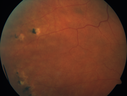
Multifocal Choroiditis - Linear Scars - Schlaegel lines534 views66 year old woman with macular pucker right eye. Images show typical linear peripheral scars for MFC or POHS - this is unilateral.Oct 25, 2017
|
|
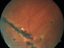
Multifocal Choroiditis - Linear Scars - Schlaegel lines690 views66 year old woman with macular pucker right eye. Images show typical linear peripheral scars for MFC or POHS - this is unilateral.Oct 25, 2017
|
|

Multifocal Choroiditis - Linear Scars - Schlaegel lines527 views66 year old woman with macular pucker right eye. Images show typical linear peripheral scars for MFC or POHS - this is unilateral.Oct 25, 2017
|
|

Multifocal Choroiditis - Linear Scars - Schlaegel lines494 views66 year old woman with macular pucker right eye. Images show typical linear peripheral scars for MFC or POHS - this is unilateral.Oct 25, 2017
|
|

Multifocal Choroiditis - Linear Scars - Schlaegel lines612 views66 year old woman with macular pucker right eye. Images show typical linear peripheral scars for MFC or POHS - this is unilateral.Oct 25, 2017
|
|

Multifocal Choroiditis - Linear Scars - Schlaegel lines596 views66 year old woman with macular pucker right eye. Images show typical linear peripheral scars for MFC or POHS - this is unilateral.Oct 25, 2017
|
|

Multifocal Choroiditis - Linear Scars - Schlaegel lines751 views66 year old woman with macular pucker right eye. Images show typical linear peripheral scars for MFC or POHS - this is unilateral.Oct 25, 2017
|
|

Multifocal Choroiditis - Linear Scars - Schlaegel lines511 views66 year old woman with macular pucker right eye. Images show typical linear peripheral scars for MFC or POHS - this is unilateral.Oct 25, 2017
|
|

Multifocal Choroiditis - Linear Scars - Schlaegel lines499 views66 year old woman with macular pucker right eye. Images show typical linear peripheral scars for MFC or POHS - this is unilateral.Oct 25, 2017
|
|

Neuroretinitis - NOT cat scratch - 5 year old489 viewsPresented with nerve swelling, serous RD and uveitis. Ultimately responded to steroids and methotrexate. ANA is positive. All other extensive testing negative including MRI worried about MSJun 02, 2017
|
|

Neuroretinitis - NOT cat scratch - 5 year old447 viewsPresented with nerve swelling, serous RD and uveitis. Ultimately responded to steroids and methotrexate. ANA is positive. All other extensive testing negative including MRI worried about MSJun 02, 2017
|
|

Neuroretinitis - NOT cat scratch - 5 year old440 viewsPresented with nerve swelling, serous RD and uveitis. Ultimately responded to steroids and methotrexate. ANA is positive. All other extensive testing negative including MRI worried about MSJun 02, 2017
|
|

Neuroretinitis - NOT cat scratch - 5 year old421 viewsPresented with nerve swelling, serous RD and uveitis. Ultimately responded to steroids and methotrexate. ANA is positive. All other extensive testing negative including MRI worried about MSJun 02, 2017
|
|

Neuroretinitis - NOT cat scratch - 5 year old454 viewsPresented with nerve swelling, serous RD and uveitis. Ultimately responded to steroids and methotrexate. ANA is positive. All other extensive testing negative including MRI worried about MSJun 02, 2017
|
|

Neuroretinitis - NOT cat scratch - 5 year old419 viewsPresented with nerve swelling, serous RD and uveitis. Ultimately responded to steroids and methotrexate. ANA is positive. All other extensive testing negative including MRI worried about MSJun 02, 2017
|
|

Neuroretinitis - NOT cat scratch - 5 year old438 viewsPresented with nerve swelling, serous RD and uveitis. Ultimately responded to steroids and methotrexate. ANA is positive. All other extensive testing negative including MRI worried about MSJun 02, 2017
|
|
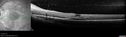
Neuroretinitis - NOT cat scratch - 5 year old493 viewsPresented with nerve swelling, serous RD and uveitis. Ultimately responded to steroids and methotrexate. ANA is positive. All other extensive testing negative including MRI worried about MSJun 02, 2017
|
|

Recurrent MEWDS Left Eye - Teenage Girl635 viewsPatient presented in 2014 (first images) 2 months following vision loss left eye. Vision went from 20/60 - 20/20. OCT showed outer retinal irregularities which normalized. Then 3 years later in 2017 she had full blown MEWDS. She presented acutely and all the images are typical. Both episodes where in the same time of year, about 3 months after her flu shot for sports.May 05, 2017
|
|
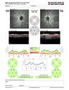
Recurrent MEWDS Left Eye - Teenage Girl487 viewsPatient presented in 2014 (first images) 2 months following vision loss left eye. Vision went from 20/60 - 20/20. OCT showed outer retinal irregularities which normalized. Then 3 years later in 2017 she had full blown MEWDS. She presented acutely and all the images are typical. Both episodes where in the same time of year, about 3 months after her flu shot for sports.May 05, 2017
|
|

Recurrent MEWDS Left Eye - Teenage Girl524 viewsPatient presented in 2014 (first images) 2 months following vision loss left eye. Vision went from 20/60 - 20/20. OCT showed outer retinal irregularities which normalized. Then 3 years later in 2017 she had full blown MEWDS. She presented acutely and all the images are typical. Both episodes where in the same time of year, about 3 months after her flu shot for sports.May 05, 2017
|
|

Recurrent MEWDS Left Eye - Teenage Girl535 viewsPatient presented in 2014 (first images) 2 months following vision loss left eye. Vision went from 20/60 - 20/20. OCT showed outer retinal irregularities which normalized. Then 3 years later in 2017 she had full blown MEWDS. She presented acutely and all the images are typical. Both episodes where in the same time of year, about 3 months after her flu shot for sports.May 05, 2017
|
|

Recurrent MEWDS Left Eye - Teenage Girl445 viewsPatient presented in 2014 (first images) 2 months following vision loss left eye. Vision went from 20/60 - 20/20. OCT showed outer retinal irregularities which normalized. Then 3 years later in 2017 she had full blown MEWDS. She presented acutely and all the images are typical. Both episodes where in the same time of year, about 3 months after her flu shot for sports.May 05, 2017
|
|

Recurrent MEWDS Left Eye - Teenage Girl550 viewsPatient presented in 2014 (first images) 2 months following vision loss left eye. Vision went from 20/60 - 20/20. OCT showed outer retinal irregularities which normalized. Then 3 years later in 2017 she had full blown MEWDS. She presented acutely and all the images are typical. Both episodes where in the same time of year, about 3 months after her flu shot for sports.May 05, 2017
|
|

Recurrent MEWDS Left Eye - Teenage Girl479 viewsPatient presented in 2014 (first images) 2 months following vision loss left eye. Vision went from 20/60 - 20/20. OCT showed outer retinal irregularities which normalized. Then 3 years later in 2017 she had full blown MEWDS. She presented acutely and all the images are typical. Both episodes where in the same time of year, about 3 months after her flu shot for sports.May 05, 2017
|
|

Recurrent MEWDS Left Eye - Teenage Girl498 viewsPatient presented in 2014 (first images) 2 months following vision loss left eye. Vision went from 20/60 - 20/20. OCT showed outer retinal irregularities which normalized. Then 3 years later in 2017 she had full blown MEWDS. She presented acutely and all the images are typical. Both episodes where in the same time of year, about 3 months after her flu shot for sports.May 05, 2017
|
|

Recurrent MEWDS Left Eye - Teenage Girl534 viewsPatient presented in 2014 (first images) 2 months following vision loss left eye. Vision went from 20/60 - 20/20. OCT showed outer retinal irregularities which normalized. Then 3 years later in 2017 she had full blown MEWDS. She presented acutely and all the images are typical. Both episodes where in the same time of year, about 3 months after her flu shot for sports.May 05, 2017
|
|

Recurrent MEWDS Left Eye - Teenage Girl485 viewsPatient presented in 2014 (first images) 2 months following vision loss left eye. Vision went from 20/60 - 20/20. OCT showed outer retinal irregularities which normalized. Then 3 years later in 2017 she had full blown MEWDS. She presented acutely and all the images are typical. Both episodes where in the same time of year, about 3 months after her flu shot for sports.May 05, 2017
|
|
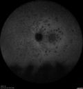
Recurrent MEWDS Left Eye - Teenage Girl536 viewsPatient presented in 2014 (first images) 2 months following vision loss left eye. Vision went from 20/60 - 20/20. OCT showed outer retinal irregularities which normalized. Then 3 years later in 2017 she had full blown MEWDS. She presented acutely and all the images are typical. Both episodes where in the same time of year, about 3 months after her flu shot for sports.May 05, 2017
|
|

Recurrent MEWDS Left Eye - Teenage Girl500 viewsPatient presented in 2014 (first images) 2 months following vision loss left eye. Vision went from 20/60 - 20/20. OCT showed outer retinal irregularities which normalized. Then 3 years later in 2017 she had full blown MEWDS. She presented acutely and all the images are typical. Both episodes where in the same time of year, about 3 months after her flu shot for sports.May 05, 2017
|
|
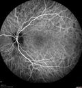
Recurrent MEWDS Left Eye - Teenage Girl524 viewsPatient presented in 2014 (first images) 2 months following vision loss left eye. Vision went from 20/60 - 20/20. OCT showed outer retinal irregularities which normalized. Then 3 years later in 2017 she had full blown MEWDS. She presented acutely and all the images are typical. Both episodes where in the same time of year, about 3 months after her flu shot for sports.May 05, 2017
|
|
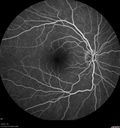
Recurrent MEWDS Left Eye - Teenage Girl507 viewsPatient presented in 2014 (first images) 2 months following vision loss left eye. Vision went from 20/60 - 20/20. OCT showed outer retinal irregularities which normalized. Then 3 years later in 2017 she had full blown MEWDS. She presented acutely and all the images are typical. Both episodes where in the same time of year, about 3 months after her flu shot for sports.May 05, 2017
|
|

Recurrent MEWDS Left Eye - Teenage Girl542 viewsPatient presented in 2014 (first images) 2 months following vision loss left eye. Vision went from 20/60 - 20/20. OCT showed outer retinal irregularities which normalized. Then 3 years later in 2017 she had full blown MEWDS. She presented acutely and all the images are typical. Both episodes where in the same time of year, about 3 months after her flu shot for sports.May 05, 2017
|
|

Recurrent MEWDS Left Eye - Teenage Girl512 viewsPatient presented in 2014 (first images) 2 months following vision loss left eye. Vision went from 20/60 - 20/20. OCT showed outer retinal irregularities which normalized. Then 3 years later in 2017 she had full blown MEWDS. She presented acutely and all the images are typical. Both episodes where in the same time of year, about 3 months after her flu shot for sports.May 05, 2017
|
|

Recurrent MEWDS Left Eye - Teenage Girl493 viewsPatient presented in 2014 (first images) 2 months following vision loss left eye. Vision went from 20/60 - 20/20. OCT showed outer retinal irregularities which normalized. Then 3 years later in 2017 she had full blown MEWDS. She presented acutely and all the images are typical. Both episodes where in the same time of year, about 3 months after her flu shot for sports.May 05, 2017
|
|

Recurrent MEWDS Left Eye - Teenage Girl487 viewsPatient presented in 2014 (first images) 2 months following vision loss left eye. Vision went from 20/60 - 20/20. OCT showed outer retinal irregularities which normalized. Then 3 years later in 2017 she had full blown MEWDS. She presented acutely and all the images are typical. Both episodes where in the same time of year, about 3 months after her flu shot for sports.May 05, 2017
|
|

Recurrent MEWDS Left Eye - Teenage Girl600 viewsPatient presented in 2014 (first images) 2 months following vision loss left eye. Vision went from 20/60 - 20/20. OCT showed outer retinal irregularities which normalized. Then 3 years later in 2017 she had full blown MEWDS. She presented acutely and all the images are typical. Both episodes where in the same time of year, about 3 months after her flu shot for sports.May 05, 2017
|
|

Recurrent MEWDS Left Eye - Teenage Girl534 viewsPatient presented in 2014 (first images) 2 months following vision loss left eye. Vision went from 20/60 - 20/20. OCT showed outer retinal irregularities which normalized. Then 3 years later in 2017 she had full blown MEWDS. She presented acutely and all the images are typical. Both episodes where in the same time of year, about 3 months after her flu shot for sports.May 05, 2017
|
|

Recurrent MEWDS Left Eye - Teenage Girl503 viewsPatient presented in 2014 (first images) 2 months following vision loss left eye. Vision went from 20/60 - 20/20. OCT showed outer retinal irregularities which normalized. Then 3 years later in 2017 she had full blown MEWDS. She presented acutely and all the images are typical. Both episodes where in the same time of year, about 3 months after her flu shot for sports.May 05, 2017
|
|

Recurrent MEWDS Left Eye - Teenage Girl453 viewsPatient presented in 2014 (first images) 2 months following vision loss left eye. Vision went from 20/60 - 20/20. OCT showed outer retinal irregularities which normalized. Then 3 years later in 2017 she had full blown MEWDS. She presented acutely and all the images are typical. Both episodes where in the same time of year, about 3 months after her flu shot for sports.May 05, 2017
|
|

Recurrent MEWDS Left Eye - Teenage Girl502 viewsPatient presented in 2014 (first images) 2 months following vision loss left eye. Vision went from 20/60 - 20/20. OCT showed outer retinal irregularities which normalized. Then 3 years later in 2017 she had full blown MEWDS. She presented acutely and all the images are typical. Both episodes where in the same time of year, about 3 months after her flu shot for sports.May 05, 2017
|
|

Recurrent MEWDS Left Eye - Teenage Girl550 viewsPatient presented in 2014 (first images) 2 months following vision loss left eye. Vision went from 20/60 - 20/20. OCT showed outer retinal irregularities which normalized. Then 3 years later in 2017 she had full blown MEWDS. She presented acutely and all the images are typical. Both episodes where in the same time of year, about 3 months after her flu shot for sports.May 05, 2017
|
|

Recurrent MEWDS Left Eye - Teenage Girl526 viewsPatient presented in 2014 (first images) 2 months following vision loss left eye. Vision went from 20/60 - 20/20. OCT showed outer retinal irregularities which normalized. Then 3 years later in 2017 she had full blown MEWDS. She presented acutely and all the images are typical. Both episodes where in the same time of year, about 3 months after her flu shot for sports.May 05, 2017
|
|
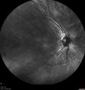
Recurrent MEWDS Left Eye - Teenage Girl450 viewsPatient presented in 2014 (first images) 2 months following vision loss left eye. Vision went from 20/60 - 20/20. OCT showed outer retinal irregularities which normalized. Then 3 years later in 2017 she had full blown MEWDS. She presented acutely and all the images are typical. Both episodes where in the same time of year, about 3 months after her flu shot for sports.May 05, 2017
|
|

Recurrent MEWDS Left Eye - Teenage Girl411 viewsPatient presented in 2014 (first images) 2 months following vision loss left eye. Vision went from 20/60 - 20/20. OCT showed outer retinal irregularities which normalized. Then 3 years later in 2017 she had full blown MEWDS. She presented acutely and all the images are typical. Both episodes where in the same time of year, about 3 months after her flu shot for sports.May 05, 2017
|
|

Recurrent MEWDS Left Eye - Teenage Girl441 viewsPatient presented in 2014 (first images) 2 months following vision loss left eye. Vision went from 20/60 - 20/20. OCT showed outer retinal irregularities which normalized. Then 3 years later in 2017 she had full blown MEWDS. She presented acutely and all the images are typical. Both episodes where in the same time of year, about 3 months after her flu shot for sports.May 05, 2017
|
|

Recurrent MEWDS Left Eye - Teenage Girl455 viewsPatient presented in 2014 (first images) 2 months following vision loss left eye. Vision went from 20/60 - 20/20. OCT showed outer retinal irregularities which normalized. Then 3 years later in 2017 she had full blown MEWDS. She presented acutely and all the images are typical. Both episodes where in the same time of year, about 3 months after her flu shot for sports.May 05, 2017
|
|

Recurrent MEWDS Left Eye - Teenage Girl503 viewsPatient presented in 2014 (first images) 2 months following vision loss left eye. Vision went from 20/60 - 20/20. OCT showed outer retinal irregularities which normalized. Then 3 years later in 2017 she had full blown MEWDS. She presented acutely and all the images are typical. Both episodes where in the same time of year, about 3 months after her flu shot for sports.May 05, 2017
|
|

Recurrent MEWDS Left Eye - Teenage Girl462 viewsPatient presented in 2014 (first images) 2 months following vision loss left eye. Vision went from 20/60 - 20/20. OCT showed outer retinal irregularities which normalized. Then 3 years later in 2017 she had full blown MEWDS. She presented acutely and all the images are typical. Both episodes where in the same time of year, about 3 months after her flu shot for sports.May 05, 2017
|
|

Recurrent MEWDS Left Eye - Teenage Girl478 viewsPatient presented in 2014 (first images) 2 months following vision loss left eye. Vision went from 20/60 - 20/20. OCT showed outer retinal irregularities which normalized. Then 3 years later in 2017 she had full blown MEWDS. She presented acutely and all the images are typical. Both episodes where in the same time of year, about 3 months after her flu shot for sports.May 05, 2017
|
|

Recurrent MEWDS Left Eye - Teenage Girl476 viewsPatient presented in 2014 (first images) 2 months following vision loss left eye. Vision went from 20/60 - 20/20. OCT showed outer retinal irregularities which normalized. Then 3 years later in 2017 she had full blown MEWDS. She presented acutely and all the images are typical. Both episodes where in the same time of year, about 3 months after her flu shot for sports.May 05, 2017
|
|
| 2409 files on 25 page(s) |
 |
|
|
|
|
13 | |
|
|
|
|


