Last additions - Posterior Uveitis - Chorioretinitis
|

Birdshot Chorioretinitis - Fresh Presentation488 viewsUltawidefield ICG shows best the multifocal lesions. 61 year old woman with floaters for less than 6 months. HLA-A29 PositiveJul 07, 2016
|
|

Birdshot Chorioretinitis - Fresh Presentation527 viewsUltawidefield ICG shows best the multifocal lesions. 61 year old woman with floaters for less than 6 months. HLA-A29 PositiveJul 07, 2016
|
|

476 viewsApr 21, 2016
|
|

467 viewsApr 21, 2016
|
|

399 viewsApr 21, 2016
|
|

425 viewsApr 21, 2016
|
|

396 viewsApr 21, 2016
|
|
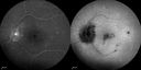
422 viewsApr 21, 2016
|
|
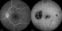
423 viewsApr 21, 2016
|
|

434 viewsApr 21, 2016
|
|

414 viewsApr 21, 2016
|
|

405 viewsApr 21, 2016
|
|

489 viewsApr 21, 2016
|
|
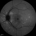
465 viewsApr 21, 2016
|
|
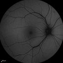
480 viewsApr 21, 2016
|
|

Syphillitic Optic Neuropathy and Maculopathy608 views46 year old woman with 2 weeks of vision loss OD. VA 20/200 OD; 20/20 OS. there are posterior vitreous cells on OCT in both eyes. ICG, FAF, and FA show an abnormal macula OD. VA improved to 20/30 within a month with treatment with PCN.Apr 18, 2016
|
|
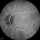
Syphillitic Optic Neuropathy and Maculopathy540 views46 year old woman with 2 weeks of vision loss OD. VA 20/200 OD; 20/20 OS. there are posterior vitreous cells on OCT in both eyes. ICG, FAF, and FA show an abnormal macula OD. VA improved to 20/30 within a month with treatment with PCN.Apr 18, 2016
|
|

Syphillitic Optic Neuropathy and Maculopathy553 views46 year old woman with 2 weeks of vision loss OD. VA 20/200 OD; 20/20 OS. there are posterior vitreous cells on OCT in both eyes. ICG, FAF, and FA show an abnormal macula OD. VA improved to 20/30 within a month with treatment with PCN.Apr 18, 2016
|
|

Syphillitic Optic Neuropathy and Maculopathy568 views46 year old woman with 2 weeks of vision loss OD. VA 20/200 OD; 20/20 OS. there are posterior vitreous cells on OCT in both eyes. ICG, FAF, and FA show an abnormal macula OD. VA improved to 20/30 within a month with treatment with PCN.Apr 18, 2016
|
|
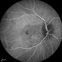
Syphillitic Optic Neuropathy and Maculopathy579 views46 year old woman with 2 weeks of vision loss OD. VA 20/200 OD; 20/20 OS. there are posterior vitreous cells on OCT in both eyes. ICG, FAF, and FA show an abnormal macula OD. VA improved to 20/30 within a month with treatment with PCN.Apr 18, 2016
|
|

Syphillitic Optic Neuropathy and Maculopathy589 views46 year old woman with 2 weeks of vision loss OD. VA 20/200 OD; 20/20 OS. there are posterior vitreous cells on OCT in both eyes. ICG, FAF, and FA show an abnormal macula OD. VA improved to 20/30 within a month with treatment with PCN.Apr 18, 2016
|
|
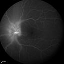
Syphillitic Optic Neuropathy and Maculopathy549 views46 year old woman with 2 weeks of vision loss OD. VA 20/200 OD; 20/20 OS. there are posterior vitreous cells on OCT in both eyes. ICG, FAF, and FA show an abnormal macula OD. VA improved to 20/30 within a month with treatment with PCN.Apr 18, 2016
|
|

Syphillitic Optic Neuropathy and Maculopathy528 views46 year old woman with 2 weeks of vision loss OD. VA 20/200 OD; 20/20 OS. there are posterior vitreous cells on OCT in both eyes. ICG, FAF, and FA show an abnormal macula OD. VA improved to 20/30 within a month with treatment with PCN.Apr 18, 2016
|
|

Syphillitic Optic Neuropathy and Maculopathy521 views46 year old woman with 2 weeks of vision loss OD. VA 20/200 OD; 20/20 OS. there are posterior vitreous cells on OCT in both eyes. ICG, FAF, and FA show an abnormal macula OD. VA improved to 20/30 within a month with treatment with PCN.Apr 18, 2016
|
|
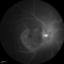
Syphillitic Optic Neuropathy and Maculopathy533 views46 year old woman with 2 weeks of vision loss OD. VA 20/200 OD; 20/20 OS. there are posterior vitreous cells on OCT in both eyes. ICG, FAF, and FA show an abnormal macula OD. VA improved to 20/30 within a month with treatment with PCN.Apr 18, 2016
|
|

Syphillitic Optic Neuropathy and Maculopathy549 views46 year old woman with 2 weeks of vision loss OD. VA 20/200 OD; 20/20 OS. there are posterior vitreous cells on OCT in both eyes. ICG, FAF, and FA show an abnormal macula OD. VA improved to 20/30 within a month with treatment with PCN.Apr 18, 2016
|
|

Syphillitic Optic Neuropathy and Maculopathy505 views46 year old woman with 2 weeks of vision loss OD. VA 20/200 OD; 20/20 OS. there are posterior vitreous cells on OCT in both eyes. ICG, FAF, and FA show an abnormal macula OD. VA improved to 20/30 within a month with treatment with PCN.Apr 18, 2016
|
|

Syphillitic Optic Neuropathy and Maculopathy523 views46 year old woman with 2 weeks of vision loss OD. VA 20/200 OD; 20/20 OS. there are posterior vitreous cells on OCT in both eyes. ICG, FAF, and FA show an abnormal macula OD. VA improved to 20/30 within a month with treatment with PCN.Apr 18, 2016
|
|

Syphillitic Optic Neuropathy and Maculopathy484 views46 year old woman with 2 weeks of vision loss OD. VA 20/200 OD; 20/20 OS. there are posterior vitreous cells on OCT in both eyes. ICG, FAF, and FA show an abnormal macula OD. VA improved to 20/30 within a month with treatment with PCN.Apr 18, 2016
|
|
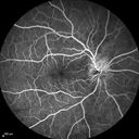
Syphillitic Optic Neuropathy and Maculopathy572 views46 year old woman with 2 weeks of vision loss OD. VA 20/200 OD; 20/20 OS. there are posterior vitreous cells on OCT in both eyes. ICG, FAF, and FA show an abnormal macula OD. VA improved to 20/30 within a month with treatment with PCN.Apr 18, 2016
|
|

Syphillitic Optic Neuropathy and Maculopathy553 views46 year old woman with 2 weeks of vision loss OD. VA 20/200 OD; 20/20 OS. there are posterior vitreous cells on OCT in both eyes. ICG, FAF, and FA show an abnormal macula OD. VA improved to 20/30 within a month with treatment with PCN.Apr 18, 2016
|
|

Syphillitic Optic Neuropathy and Maculopathy430 views46 year old woman with 2 weeks of vision loss OD. VA 20/200 OD; 20/20 OS. there are posterior vitreous cells on OCT in both eyes. ICG, FAF, and FA show an abnormal macula OD. VA improved to 20/30 within a month with treatment with PCN.Apr 18, 2016
|
|

Syphillitic Optic Neuropathy and Maculopathy490 views46 year old woman with 2 weeks of vision loss OD. VA 20/200 OD; 20/20 OS. there are posterior vitreous cells on OCT in both eyes. ICG, FAF, and FA show an abnormal macula OD. VA improved to 20/30 within a month with treatment with PCN.Apr 18, 2016
|
|

Syphillitic Optic Neuropathy and Maculopathy478 views46 year old woman with 2 weeks of vision loss OD. VA 20/200 OD; 20/20 OS. there are posterior vitreous cells on OCT in both eyes. ICG, FAF, and FA show an abnormal macula OD. VA improved to 20/30 within a month with treatment with PCN.Apr 18, 2016
|
|

Syphillitic Optic Neuropathy and Maculopathy515 views46 year old woman with 2 weeks of vision loss OD. VA 20/200 OD; 20/20 OS. there are posterior vitreous cells on OCT in both eyes. ICG, FAF, and FA show an abnormal macula OD. VA improved to 20/30 within a month with treatment with PCN.Apr 18, 2016
|
|

Syphillitic Optic Neuropathy and Maculopathy500 views46 year old woman with 2 weeks of vision loss OD. VA 20/200 OD; 20/20 OS. there are posterior vitreous cells on OCT in both eyes. ICG, FAF, and FA show an abnormal macula OD. VA improved to 20/30 within a month with treatment with PCN.Apr 18, 2016
|
|

Syphillitic Optic Neuropathy and Maculopathy548 views46 year old woman with 2 weeks of vision loss OD. VA 20/200 OD; 20/20 OS. there are posterior vitreous cells on OCT in both eyes. ICG, FAF, and FA show an abnormal macula OD. VA improved to 20/30 within a month with treatment with PCN.Apr 18, 2016
|
|

Syphillitic Optic Neuropathy and Maculopathy483 views46 year old woman with 2 weeks of vision loss OD. VA 20/200 OD; 20/20 OS. there are posterior vitreous cells on OCT in both eyes. ICG, FAF, and FA show an abnormal macula OD. VA improved to 20/30 within a month with treatment with PCN.Apr 18, 2016
|
|

Syphillitic Optic Neuropathy and Maculopathy501 views46 year old woman with 2 weeks of vision loss OD. VA 20/200 OD; 20/20 OS. there are posterior vitreous cells on OCT in both eyes. ICG, FAF, and FA show an abnormal macula OD. VA improved to 20/30 within a month with treatment with PCN.Apr 18, 2016
|
|

Syphillitic Optic Neuropathy and Maculopathy496 views46 year old woman with 2 weeks of vision loss OD. VA 20/200 OD; 20/20 OS. there are posterior vitreous cells on OCT in both eyes. ICG, FAF, and FA show an abnormal macula OD. VA improved to 20/30 within a month with treatment with PCN.Apr 18, 2016
|
|

Syphillitic Optic Neuropathy and Maculopathy555 views46 year old woman with 2 weeks of vision loss OD. VA 20/200 OD; 20/20 OS. there are posterior vitreous cells on OCT in both eyes. ICG, FAF, and FA show an abnormal macula OD. VA improved to 20/30 within a month with treatment with PCN.Apr 18, 2016
|
|
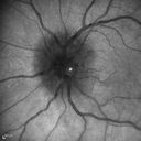
Syphillitic Optic Neuropathy and Maculopathy490 views46 year old woman with 2 weeks of vision loss OD. VA 20/200 OD; 20/20 OS. there are posterior vitreous cells on OCT in both eyes. ICG, FAF, and FA show an abnormal macula OD. VA improved to 20/30 within a month with treatment with PCN.Apr 18, 2016
|
|

Syphillitic Optic Neuropathy and Maculopathy520 views46 year old woman with 2 weeks of vision loss OD. VA 20/200 OD; 20/20 OS. there are posterior vitreous cells on OCT in both eyes. ICG, FAF, and FA show an abnormal macula OD. VA improved to 20/30 within a month with treatment with PCN.Apr 18, 2016
|
|

Syphillitic Optic Neuropathy and Maculopathy530 views46 year old woman with 2 weeks of vision loss OD. VA 20/200 OD; 20/20 OS. there are posterior vitreous cells on OCT in both eyes. ICG, FAF, and FA show an abnormal macula OD. VA improved to 20/30 within a month with treatment with PCN.Apr 18, 2016
|
|

Syphillitic Optic Neuropathy and Maculopathy528 views46 year old woman with 2 weeks of vision loss OD. VA 20/200 OD; 20/20 OS. there are posterior vitreous cells on OCT in both eyes. ICG, FAF, and FA show an abnormal macula OD. VA improved to 20/30 within a month with treatment with PCN.Apr 18, 2016
|
|

Syphillitic Optic Neuropathy and Maculopathy555 views46 year old woman with 2 weeks of vision loss OD. VA 20/200 OD; 20/20 OS. there are posterior vitreous cells on OCT in both eyes. ICG, FAF, and FA show an abnormal macula OD. VA improved to 20/30 within a month with treatment with PCN.Apr 18, 2016
|
|

Syphillitic Optic Neuropathy and Maculopathy610 views46 year old woman with 2 weeks of vision loss OD. VA 20/200 OD; 20/20 OS. there are posterior vitreous cells on OCT in both eyes. ICG, FAF, and FA show an abnormal macula OD. VA improved to 20/30 within a month with treatment with PCN.Apr 18, 2016
|
|

Syphillitic Optic Neuropathy and Maculopathy544 views46 year old woman with 2 weeks of vision loss OD. VA 20/200 OD; 20/20 OS. there are posterior vitreous cells on OCT in both eyes. ICG, FAF, and FA show an abnormal macula OD. VA improved to 20/30 within a month with treatment with PCN.Apr 18, 2016
|
|

Syphillitic Optic Neuropathy and Maculopathy568 views46 year old woman with 2 weeks of vision loss OD. VA 20/200 OD; 20/20 OS. there are posterior vitreous cells on OCT in both eyes. ICG, FAF, and FA show an abnormal macula OD. VA improved to 20/30 within a month with treatment with PCN.Apr 18, 2016
|
|

Syphillitic Optic Neuropathy and Maculopathy608 views46 year old woman with 2 weeks of vision loss OD. VA 20/200 OD; 20/20 OS. there are posterior vitreous cells on OCT in both eyes. ICG, FAF, and FA show an abnormal macula OD. VA improved to 20/30 within a month with treatment with PCN.Apr 18, 2016
|
|

Syphillitic Optic Neuropathy and Maculopathy658 views46 year old woman with 2 weeks of vision loss OD. VA 20/200 OD; 20/20 OS. there are posterior vitreous cells on OCT in both eyes. ICG, FAF, and FA show an abnormal macula OD. VA improved to 20/30 within a month with treatment with PCN.Apr 18, 2016
|
|

Acute Posterior Multifocal Placoid Pigment Epitheliopathy - 4 years after onset733 views4 years after presentation - vision 20/80 OD 20/16 OS and there is subretinal fibrosisMar 07, 2016
|
|
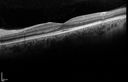
Acute Posterior Multifocal Placoid Pigment Epitheliopathy - 4 years after onset539 views4 years after presentation - vision 20/80 OD 20/16 OS and there is subretinal fibrosisMar 07, 2016
|
|
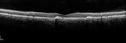
Acute Posterior Multifocal Placoid Pigment Epitheliopathy - 4 years after onset522 views4 years after presentation - vision 20/80 OD 20/16 OS and there is subretinal fibrosisMar 07, 2016
|
|

Acute Posterior Multifocal Placoid Pigment Epitheliopathy - 4 years after onset539 views4 years after presentation - vision 20/80 OD 20/16 OS and there is subretinal fibrosisMar 07, 2016
|
|

Acute Posterior Multifocal Placoid Pigment Epitheliopathy - 4 years after onset587 views4 years after presentation - vision 20/80 OD 20/16 OS and there is subretinal fibrosisMar 07, 2016
|
|

Acute Posterior Multifocal Placoid Pigment Epitheliopathy - 4 years after onset586 views4 years after presentation - vision 20/80 OD 20/16 OS and there is subretinal fibrosisMar 07, 2016
|
|

Acute Posterior Multifocal Placoid Pigment Epitheliopathy - 4 years after onset538 views4 years after presentation - vision 20/80 OD 20/16 OS and there is subretinal fibrosisMar 07, 2016
|
|

Acute Posterior Multifocal Placoid Pigment Epitheliopathy - 4 years after onset537 views4 years after presentation - vision 20/80 OD 20/16 OS and there is subretinal fibrosisMar 07, 2016
|
|

Acute Posterior Multifocal Placoid Pigment Epitheliopathy - 4 years after onset559 views4 years after presentation - vision 20/80 OD 20/16 OS and there is subretinal fibrosisMar 07, 2016
|
|

Acute Posterior Multifocal Placoid Pigment Epitheliopathy - 4 years after onset541 views4 years after presentation - vision 20/80 OD 20/16 OS and there is subretinal fibrosisMar 07, 2016
|
|

Acute Posterior Multifocal Placoid Pigment Epitheliopathy - 4 years after onset582 views4 years after presentation - vision 20/80 OD 20/16 OS and there is subretinal fibrosisMar 07, 2016
|
|

Acute Posterior Multifocal Placoid Pigment Epitheliopathy - 4 years after onset560 views4 years after presentation - vision 20/80 OD 20/16 OS and there is subretinal fibrosisMar 07, 2016
|
|

Acute Posterior Multifocal Placoid Pigment Epitheliopathy - 4 years after onset545 views4 years after presentation - vision 20/80 OD 20/16 OS and there is subretinal fibrosisMar 07, 2016
|
|

Acute Posterior Multifocal Placoid Pigment Epitheliopathy - 4 years after onset632 views4 years after presentation - vision 20/80 OD 20/16 OS and there is subretinal fibrosisMar 07, 2016
|
|

Acute Posterior Multifocal Placoid Pigment Epitheliopathy - 4 years after onset600 views4 years after presentation - vision 20/80 OD 20/16 OS and there is subretinal fibrosisMar 07, 2016
|
|
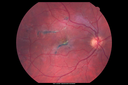
Acute Posterior Multifocal Placoid Pigment Epitheliopathy - 4 years after onset605 views4 years after presentation - vision 20/80 OD 20/16 OS and there is subretinal fibrosisMar 07, 2016
|
|
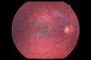
Acute Posterior Multifocal Placoid Pigment Epitheliopathy - 4 years after onset690 views4 years after presentation - vision 20/80 OD 20/16 OS and there is subretinal fibrosisMar 07, 2016
|
|

West Nile Virus Multifocal Choroiditis and diabetic retinopathy732 viewsMultifocal and clustered focal lesions are from west nile virus. The retinopathy is probably diabetic but could also be related to west nile virusNov 17, 2015
|
|

West Nile Virus Multifocal Choroiditis and diabetic retinopathy534 viewsMultifocal and clustered focal lesions are from west nile virus. The retinopathy is probably diabetic but could also be related to west nile virusNov 17, 2015
|
|

West Nile Virus Multifocal Choroiditis and diabetic retinopathy555 viewsMultifocal and clustered focal lesions are from west nile virus. The retinopathy is probably diabetic but could also be related to west nile virusNov 17, 2015
|
|
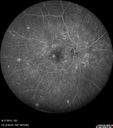
West Nile Virus Multifocal Choroiditis and diabetic retinopathy516 viewsMultifocal and clustered focal lesions are from west nile virus. The retinopathy is probably diabetic but could also be related to west nile virusNov 17, 2015
|
|

West Nile Virus Multifocal Choroiditis and diabetic retinopathy512 viewsMultifocal and clustered focal lesions are from west nile virus. The retinopathy is probably diabetic but could also be related to west nile virusNov 17, 2015
|
|

West Nile Virus Multifocal Choroiditis and diabetic retinopathy538 viewsMultifocal and clustered focal lesions are from west nile virus. The retinopathy is probably diabetic but could also be related to west nile virusNov 17, 2015
|
|

West Nile Virus Multifocal Choroiditis and diabetic retinopathy535 viewsMultifocal and clustered focal lesions are from west nile virus. The retinopathy is probably diabetic but could also be related to west nile virusNov 17, 2015
|
|

West Nile Virus Multifocal Choroiditis and diabetic retinopathy496 viewsMultifocal and clustered focal lesions are from west nile virus. The retinopathy is probably diabetic but could also be related to west nile virusNov 17, 2015
|
|

West Nile Virus Multifocal Choroiditis and diabetic retinopathy522 viewsMultifocal and clustered focal lesions are from west nile virus. The retinopathy is probably diabetic but could also be related to west nile virusNov 17, 2015
|
|

West Nile Virus Multifocal Choroiditis and diabetic retinopathy593 viewsMultifocal and clustered focal lesions are from west nile virus. The retinopathy is probably diabetic but could also be related to west nile virusNov 17, 2015
|
|

West Nile Virus Multifocal Choroiditis and diabetic retinopathy621 viewsMultifocal and clustered focal lesions are from west nile virus. The retinopathy is probably diabetic but could also be related to west nile virusNov 17, 2015
|
|

West Nile Virus Multifocal Choroiditis and diabetic retinopathy489 viewsMultifocal and clustered focal lesions are from west nile virus. The retinopathy is probably diabetic but could also be related to west nile virusNov 17, 2015
|
|

West Nile Virus Multifocal Choroiditis and diabetic retinopathy581 viewsMultifocal and clustered focal lesions are from west nile virus. The retinopathy is probably diabetic but could also be related to west nile virusNov 17, 2015
|
|

West Nile Virus Multifocal Choroiditis and diabetic retinopathy488 viewsMultifocal and clustered focal lesions are from west nile virus. The retinopathy is probably diabetic but could also be related to west nile virusNov 17, 2015
|
|
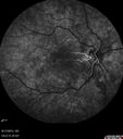
West Nile Virus Multifocal Choroiditis and diabetic retinopathy512 viewsMultifocal and clustered focal lesions are from west nile virus. The retinopathy is probably diabetic but could also be related to west nile virusNov 17, 2015
|
|

West Nile Virus Multifocal Choroiditis and diabetic retinopathy454 viewsMultifocal and clustered focal lesions are from west nile virus. The retinopathy is probably diabetic but could also be related to west nile virusNov 17, 2015
|
|

West Nile Virus Multifocal Choroiditis and diabetic retinopathy510 viewsMultifocal and clustered focal lesions are from west nile virus. The retinopathy is probably diabetic but could also be related to west nile virusNov 17, 2015
|
|

West Nile Virus Multifocal Choroiditis and diabetic retinopathy430 viewsMultifocal and clustered focal lesions are from west nile virus. The retinopathy is probably diabetic but could also be related to west nile virusNov 17, 2015
|
|

West Nile Virus Multifocal Choroiditis and diabetic retinopathy489 viewsMultifocal and clustered focal lesions are from west nile virus. The retinopathy is probably diabetic but could also be related to west nile virusNov 17, 2015
|
|

West Nile Virus Multifocal Choroiditis and diabetic retinopathy451 viewsMultifocal and clustered focal lesions are from west nile virus. The retinopathy is probably diabetic but could also be related to west nile virusNov 17, 2015
|
|

West Nile Virus Multifocal Choroiditis and diabetic retinopathy480 viewsMultifocal and clustered focal lesions are from west nile virus. The retinopathy is probably diabetic but could also be related to west nile virusNov 17, 2015
|
|

West Nile Virus Multifocal Choroiditis and diabetic retinopathy589 viewsMultifocal and clustered focal lesions are from west nile virus. The retinopathy is probably diabetic but could also be related to west nile virusNov 17, 2015
|
|

West Nile Virus Multifocal Choroiditis and diabetic retinopathy548 viewsMultifocal and clustered focal lesions are from west nile virus. The retinopathy is probably diabetic but could also be related to west nile virusNov 17, 2015
|
|

West Nile Virus Multifocal Choroiditis and diabetic retinopathy612 viewsMultifocal and clustered focal lesions are from west nile virus. The retinopathy is probably diabetic but could also be related to west nile virusNov 17, 2015
|
|

West Nile Virus Multifocal Choroiditis and diabetic retinopathy1080 viewsMultifocal and clustered focal lesions are from west nile virus. The retinopathy is probably diabetic but could also be related to west nile virusNov 17, 2015
|
|

West Nile Virus Multifocal Choroiditis and diabetic retinopathy786 viewsMultifocal and clustered focal lesions are from west nile virus. The retinopathy is probably diabetic but could also be related to west nile virusNov 17, 2015
|
|

West Nile Virus Multifocal Choroiditis and diabetic retinopathy689 viewsMultifocal and clustered focal lesions are from west nile virus. The retinopathy is probably diabetic but could also be related to west nile virusNov 17, 2015
|
|
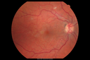
West Nile Virus Multifocal Choroiditis and diabetic retinopathy737 viewsMultifocal and clustered focal lesions are from west nile virus. The retinopathy is probably diabetic but could also be related to west nile virusNov 17, 2015
|
|

Retinal Atrophy 1 year following Streptococcus sanguinis endophthalmitis - Vision 5/200559 views79 year old woman with sleep apnea who sleeps with CPAP machineMay 29, 2015
|
|

Retinal Atrophy 1 year following Streptococcus sanguinis endophthalmitis - Vision 5/200529 views79 year old woman with sleep apnea who sleeps with CPAP machineMay 29, 2015
|
|

Retinal Atrophy 1 year following Streptococcus sanguinis endophthalmitis - Vision 5/200521 views79 year old woman with sleep apnea who sleeps with CPAP machineMay 29, 2015
|
|

Retinal Atrophy 1 year following Streptococcus sanguinis endophthalmitis - Vision 5/200533 views79 year old woman with sleep apnea who sleeps with CPAP machineMay 29, 2015
|
|
| 2409 files on 25 page(s) |
 |
|
|
|
|
15 | |
|
|
|
|


