Last additions - Posterior Uveitis - Chorioretinitis
|

Retinal Atrophy 1 year following Streptococcus sanguinis endophthalmitis - Vision 5/200521 views79 year old woman with sleep apnea who sleeps with CPAP machineMay 29, 2015
|
|

Retinal Atrophy 1 year following Streptococcus sanguinis endophthalmitis - Vision 5/200574 views79 year old woman with sleep apnea who sleeps with CPAP machineMay 29, 2015
|
|

Retinal Atrophy 1 year following Streptococcus sanguinis endophthalmitis - Vision 5/200503 views79 year old woman with sleep apnea who sleeps with CPAP machineMay 29, 2015
|
|

Retinal Atrophy 1 year following Streptococcus sanguinis endophthalmitis - Vision 5/200521 views79 year old woman with sleep apnea who sleeps with CPAP machineMay 29, 2015
|
|

Retinal Atrophy 1 year following Streptococcus sanguinis endophthalmitis - Vision 5/200600 views79 year old woman with sleep apnea who sleeps with CPAP machineMay 29, 2015
|
|

Retinal Atrophy 1 year following Streptococcus sanguinis endophthalmitis - Vision 5/200576 views79 year old woman with sleep apnea who sleeps with CPAP machineMay 29, 2015
|
|

Toxocariasis - Stalk from optic nerve to peripheral scar644 viewsMay 15, 2015
|
|
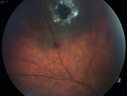
Toxocariasis - Stalk from optic nerve to peripheral scar650 viewsMay 15, 2015
|
|

Toxocariasis - Stalk from optic nerve to peripheral scar770 viewsMay 15, 2015
|
|

Toxocariasis - Stalk from optic nerve to peripheral scar653 viewsMay 15, 2015
|
|

Toxocariasis - Stalk from optic nerve to peripheral scar606 viewsMay 15, 2015
|
|

Unilateral Acute Idiopathic Maculopathy (UAIM) - VIDEO TAKES TIME TO LOAD - Raster Scan OCT547 viewsVision has improved back to 20/20 without any therapy. 1 month following vision lossFeb 05, 2015
|
|

Unilateral Acute Idiopathic Maculopathy (UAIM)614 viewsVision has improved back to 20/20 without any therapy. 1 month following vision lossFeb 05, 2015
|
|

Unilateral Acute Idiopathic Maculopathy (UAIM)735 viewsVision has improved back to 20/20 without any therapy. 1 month following vision lossFeb 05, 2015
|
|

Unilateral Acute Idiopathic Maculopathy (UAIM)899 views1 week later eye looks better (after initial scan) 41 year old with serous retinal detachment OS and sudden vision loss. He had a viral prodrome. Coxscackie IgG was high but no acute infection was documented. Vision improved in about 2 months without therapy. - Right eye has spot nasal to the optic nerveFeb 05, 2015
|
|
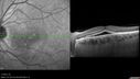
568 views1 week later eye looks better (after initial scan) 41 year old with serous retinal detachment OS and sudden vision loss. He had a viral prodrome. Coxscackie IgG was high but no acute infection was documented. Vision improved in about 2 months without therapy. - Right eye has spot nasal to the optic nerveFeb 05, 2015
|
|

Unilateral Acute Idiopathic Maculopathy (UAIM)605 views1 week later eye looks better (after initial scan) 41 year old with serous retinal detachment OS and sudden vision loss. He had a viral prodrome. Coxscackie IgG was high but no acute infection was documented. Vision improved in about 2 months without therapy. - Right eye has spot nasal to the optic nerveFeb 05, 2015
|
|

Unilateral Acute Idiopathic Maculopathy (UAIM)622 views41 year old with serous retinal detachment OS and sudden vision loss. He had a viral prodrome. Coxscackie IgG was high but no acute infection was documented. Vision improved in about 2 months without therapy. - Right eye has spot nasal to the optic nerveFeb 05, 2015
|
|

Unilateral Acute Idiopathic Maculopathy (UAIM)563 views41 year old with serous retinal detachment OS and sudden vision loss. He had a viral prodrome. Coxscackie IgG was high but no acute infection was documented. Vision improved in about 2 months without therapy. - Right eye has spot nasal to the optic nerveFeb 05, 2015
|
|

Unilateral Acute Idiopathic Maculopathy (UAIM)523 views41 year old with serous retinal detachment OS and sudden vision loss. He had a viral prodrome. Coxscackie IgG was high but no acute infection was documented. Vision improved in about 2 months without therapy. - Right eye has spot nasal to the optic nerveFeb 05, 2015
|
|

Unilateral Acute Idiopathic Maculopathy (UAIM)549 views41 year old with serous retinal detachment OS and sudden vision loss. He had a viral prodrome. Coxscackie IgG was high but no acute infection was documented. Vision improved in about 2 months without therapy. - Right eye has spot nasal to the optic nerveFeb 05, 2015
|
|

Unilateral Acute Idiopathic Maculopathy (UAIM)537 views41 year old with serous retinal detachment OS and sudden vision loss. He had a viral prodrome. Coxscackie IgG was high but no acute infection was documented. Vision improved in about 2 months without therapy. - Right eye has spot nasal to the optic nerveFeb 05, 2015
|
|

Unilateral Acute Idiopathic Maculopathy (UAIM)485 views41 year old with serous retinal detachment OS and sudden vision loss. He had a viral prodrome. Coxscackie IgG was high but no acute infection was documented. Vision improved in about 2 months without therapy. - Right eye has spot nasal to the optic nerveFeb 05, 2015
|
|

Unilateral Acute Idiopathic Maculopathy (UAIM)535 views41 year old with serous retinal detachment OS and sudden vision loss. He had a viral prodrome. Coxscackie IgG was high but no acute infection was documented. Vision improved in about 2 months without therapy. - Right eye has spot nasal to the optic nerveFeb 05, 2015
|
|

Unilateral Acute Idiopathic Maculopathy (UAIM)504 views41 year old with serous retinal detachment OS and sudden vision loss. He had a viral prodrome. Coxscackie IgG was high but no acute infection was documented. Vision improved in about 2 months without therapy. - Right eye has spot nasal to the optic nerveFeb 05, 2015
|
|

Unilateral Acute Idiopathic Maculopathy (UAIM)537 views41 year old with serous retinal detachment OS and sudden vision loss. He had a viral prodrome. Coxscackie IgG was high but no acute infection was documented. Vision improved in about 2 months without therapy. - Right eye has spot nasal to the optic nerveFeb 05, 2015
|
|

Unilateral Acute Idiopathic Maculopathy (UAIM)539 views41 year old with serous retinal detachment OS and sudden vision loss. He had a viral prodrome. Coxscackie IgG was high but no acute infection was documented. Vision improved in about 2 months without therapy. - Right eye has spot nasal to the optic nerveFeb 05, 2015
|
|

Unilateral Acute Idiopathic Maculopathy (UAIM)593 views41 year old with serous retinal detachment OS and sudden vision loss. He had a viral prodrome. Coxscackie IgG was high but no acute infection was documented. Vision improved in about 2 months without therapy. - Right eye has spot nasal to the optic nerveFeb 05, 2015
|
|

Unilateral Acute Idiopathic Maculopathy (UAIM)531 views41 year old with serous retinal detachment OS and sudden vision loss. He had a viral prodrome. Coxscackie IgG was high but no acute infection was documented. Vision improved in about 2 months without therapy. - Right eye has spot nasal to the optic nerveFeb 05, 2015
|
|

Unilateral Acute Idiopathic Maculopathy (UAIM)529 views41 year old with serous retinal detachment OS and sudden vision loss. He had a viral prodrome. Coxscackie IgG was high but no acute infection was documented. Vision improved in about 2 months without therapy. - Right eye has spot nasal to the optic nerveFeb 05, 2015
|
|

Unilateral Acute Idiopathic Maculopathy (UAIM)515 views41 year old with serous retinal detachment OS and sudden vision loss. He had a viral prodrome. Coxscackie IgG was high but no acute infection was documented. Vision improved in about 2 months without therapy. - Right eye has spot nasal to the optic nerveFeb 05, 2015
|
|
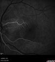
Unilateral Acute Idiopathic Maculopathy (UAIM)530 viewsCilioretinal artery fills earlyFeb 05, 2015
|
|

Unilateral Acute Idiopathic Maculopathy (UAIM)498 views41 year old with serous retinal detachment OS and sudden vision loss. He had a viral prodrome. Coxscackie IgG was high but no acute infection was documented. Vision improved in about 2 months without therapy. - Right eye has spot nasal to the optic nerveFeb 05, 2015
|
|

Unilateral Acute Idiopathic Maculopathy (UAIM)456 views41 year old with serous retinal detachment OS and sudden vision loss. He had a viral prodrome. Coxscackie IgG was high but no acute infection was documented. Vision improved in about 2 months without therapy. - Right eye has spot nasal to the optic nerveFeb 05, 2015
|
|
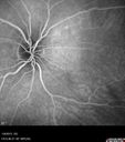
Unilateral Acute Idiopathic Maculopathy (UAIM)457 views41 year old with serous retinal detachment OS and sudden vision loss. He had a viral prodrome. Coxscackie IgG was high but no acute infection was documented. Vision improved in about 2 months without therapy. - Right eye has spot nasal to the optic nerveFeb 05, 2015
|
|

Unilateral Acute Idiopathic Maculopathy (UAIM)492 views41 year old with serous retinal detachment OS and sudden vision loss. He had a viral prodrome. Coxscackie IgG was high but no acute infection was documented. Vision improved in about 2 months without therapy. - Right eye has spot nasal to the optic nerveFeb 05, 2015
|
|

Unilateral Acute Idiopathic Maculopathy (UAIM)489 views41 year old with serous retinal detachment OS and sudden vision loss. He had a viral prodrome. Coxscackie IgG was high but no acute infection was documented. Vision improved in about 2 months without therapy. - Right eye has spot nasal to the optic nerveFeb 05, 2015
|
|

Unilateral Acute Idiopathic Maculopathy (UAIM)614 views41 year old with serous retinal detachment OS and sudden vision loss. He had a viral prodrome. Coxscackie IgG was high but no acute infection was documented. Vision improved in about 2 months without therapy. - Right eye has spot nasal to the optic nerveFeb 05, 2015
|
|
Unilateral Acute Idiopathic Maculopathy (UAIM) - VIDEO TAKES TIME TO LOAD - raster oct scan521 views41 year old with serous retinal detachment OS and sudden vision loss. He had a viral prodrome. Coxscackie IgG was high but no acute infection was documented. Vision improved in about 2 months without therapy.Feb 05, 2015
|
|

Unilateral Acute Idiopathic Maculopathy (UAIM)611 views41 year old with serous retinal detachment OS and sudden vision loss. He had a viral prodrome. Coxscackie IgG was high but no acute infection was documented. Vision improved in about 2 months without therapy.Feb 05, 2015
|
|

Unilateral Acute Idiopathic Maculopathy (UAIM)567 views41 year old with serous retinal detachment OS and sudden vision loss. He had a viral prodrome. Coxscackie IgG was high but no acute infection was documented. Vision improved in about 2 months without therapy.Feb 05, 2015
|
|

Unilateral Acute Idiopathic Maculopathy (UAIM)555 viewsFeb 05, 2015
|
|

Unilateral Acute Idiopathic Maculopathy (UAIM)616 viewsFeb 05, 2015
|
|

Unilateral Acute Idiopathic Maculopathy (UAIM)805 views41 year old with serous retinal detachment OS and sudden vision loss. He had a viral prodrome. Coxscackie IgG was high but no acute infection was documented. Vision improved in about 2 months without therapy.Feb 05, 2015
|
|

Unilateral Acute Idiopathic Maculopathy (UAIM)569 viewsFeb 05, 2015
|
|
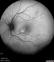
Unilateral Acute Idiopathic Maculopathy (UAIM)618 views41 year old with serous retinal detachment OS and sudden vision loss. He had a viral prodrome. Coxscackie IgG was high but no acute infection was documented. Vision improved in about 2 months without therapy.Feb 05, 2015
|
|
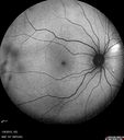
Unilateral Acute Idiopathic Maculopathy (UAIM)669 viewsFeb 05, 2015
|
|

Unilateral Acute Idiopathic Maculopathy (UAIM)590 viewsFeb 05, 2015
|
|

Unilateral Acute Idiopathic Maculopathy (UAIM)752 views41 year old with serous retinal detachment OS and sudden vision loss. He had a viral prodrome. Coxscackie IgG was high but no acute infection was documented. Vision improved in about 2 months without therapy.Feb 05, 2015
|
|

Unilateral Acute Idiopathic Maculopathy (UAIM)645 views41 year old with serous retinal detachment OS and sudden vision loss. He had a viral prodrome. Coxscackie IgG was high but no acute infection was documented. Vision improved in about 2 months without therapy.Feb 05, 2015
|
|

Unilateral Acute Idiopathic Maculopathy (UAIM)634 viewsNormal right eyeFeb 05, 2015
|
|

Unilateral Acute Idiopathic Maculopathy (UAIM)659 viewsNormal right eyeFeb 05, 2015
|
|

Acute Macular Neuroretinopathy - (AMN) - Spot in Vision OS - 40 Year old Female749 views4 months later spot is nearly gone and vision is back to normal (almost) - 20/20Feb 05, 2015
|
|

Acute Macular Neuroretinopathy - (AMN) - Spot in Vision OS - 40 Year old Female612 viewsVision improved 1 month laterFeb 05, 2015
|
|

Acute Macular Neuroretinopathy - (AMN) - Spot in Vision OS - 40 Year old Female732 viewsFeb 05, 2015
|
|

Acute Macular Neuroretinopathy - (AMN) - Spot in Vision OS - 40 Year old Female704 viewsFeb 05, 2015
|
|
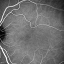
Acute Macular Neuroretinopathy - (AMN) - Spot in Vision OS - 40 Year old Female731 viewsICG angiogram is normalFeb 05, 2015
|
|

Acute Macular Neuroretinopathy - (AMN) - Spot in Vision OS - 40 Year old Female751 viewsICG angiogram is normalFeb 05, 2015
|
|
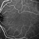
Acute Macular Neuroretinopathy - (AMN) - Spot in Vision OS - 40 Year old Female694 viewsICG angiogram is normalFeb 05, 2015
|
|

Acute Macular Neuroretinopathy - (AMN) - Spot in Vision OS - 40 Year old Female708 viewsICG angiogram is normalFeb 05, 2015
|
|

Acute Macular Neuroretinopathy - (AMN) - Spot in Vision OS - 40 Year old Female768 viewsICG angiogram is normalFeb 05, 2015
|
|

Acute Macular Neuroretinopathy - (AMN) - Spot in Vision OS - 40 Year old Female731 viewsICG angiogram is normalFeb 05, 2015
|
|

Acute Macular Neuroretinopathy - (AMN) - Spot in Vision OS - 40 Year old Female696 viewsICG angiogram is normalFeb 05, 2015
|
|
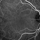
Acute Macular Neuroretinopathy - (AMN) - Spot in Vision OS - 40 Year old Female607 viewsFeb 05, 2015
|
|

Acute Macular Neuroretinopathy - (AMN) - Spot in Vision OS - 40 Year old Female586 viewsFluorescein Angiogram is normalFeb 05, 2015
|
|
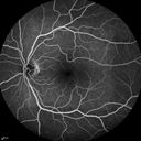
Acute Macular Neuroretinopathy - (AMN) - Spot in Vision OS - 40 Year old Female555 viewsFluorescein Angiogram is normalFeb 05, 2015
|
|

Acute Macular Neuroretinopathy - (AMN) - Spot in Vision OS - 40 Year old Female583 viewsFluorescein Angiogram is normalFeb 05, 2015
|
|

Acute Macular Neuroretinopathy - (AMN) - Spot in Vision OS - 40 Year old Female620 viewsFluorescein Angiogram is normalFeb 05, 2015
|
|

Acute Macular Neuroretinopathy - (AMN) - Spot in Vision OS - 40 Year old Female640 viewsFluorescein Angiogram is normalFeb 05, 2015
|
|

Acute Macular Neuroretinopathy - (AMN) - Spot in Vision OS - 40 Year old Female670 viewsFluorescein Angiogram is normalFeb 05, 2015
|
|
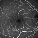
Acute Macular Neuroretinopathy - (AMN) - Spot in Vision OS - 40 Year old Female706 viewsFluorescein Angiogram is normalFeb 05, 2015
|
|

Acute Macular Neuroretinopathy - (AMN) - Spot in Vision OS - 40 Year old Female645 viewsFluorescein Angiogram is normalFeb 05, 2015
|
|

Acute Macular Neuroretinopathy - (AMN) - Spot in Vision OS - 40 Year old Female620 viewsFluorescein Angiogram is normalFeb 05, 2015
|
|

Acute Macular Neuroretinopathy - (AMN) - Spot in Vision OS - 40 Year old Female631 viewsFAF is normalFeb 05, 2015
|
|

Acute Macular Neuroretinopathy - (AMN) - Spot in Vision OS - 40 Year old Female664 viewsFAF is normalFeb 05, 2015
|
|

Acute Macular Neuroretinopathy - (AMN) - Spot in Vision OS - 40 Year old Female623 viewsIR shows gray spot nasal to the foveaFeb 05, 2015
|
|

Acute Macular Neuroretinopathy - (AMN) - Spot in Vision OS - 40 Year old Female633 viewsFeb 05, 2015
|
|
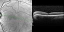
Acute Macular Neuroretinopathy - (AMN) - Spot in Vision OS - 40 Year old Female804 viewsSD OCT shows disruption of the outer retina nasal to the foveaFeb 05, 2015
|
|
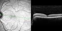
Acute Macular Neuroretinopathy - (AMN) - Spot in Vision OS - 40 Year old Female1046 viewsSD OCT shows disruption of the outer retina nasal to the foveaFeb 05, 2015
|
|

Acute Macular Neuroretinopathy - (AMN) - Spot in Vision OS - 40 Year old Female1000 viewsSD OCT shows disruption of the outer retina nasal to the foveaFeb 05, 2015
|
|
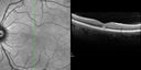
Acute Macular Neuroretinopathy - (AMN) - Spot in Vision OS - 40 Year old Female831 viewsSD OCT shows disruption of the outer retina nasal to the foveaFeb 05, 2015
|
|
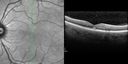
Acute Macular Neuroretinopathy - (AMN) - Spot in Vision OS - 40 Year old Female914 viewsSD OCT shows disruption of the outer retina nasal to the foveaFeb 05, 2015
|
|
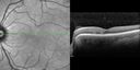
Acute Macular Neuroretinopathy - (AMN) - Spot in Vision OS - 40 Year old Female857 viewsSD OCT shows disruption of the outer retina nasal to the foveaFeb 05, 2015
|
|

Acute Macular Neuroretinopathy - (AMN) - Spot in Vision OS - 40 Year old Female757 viewsSD OCT shows disruption of the outer retina nasal to the foveaFeb 05, 2015
|
|

Acute Macular Neuroretinopathy - (AMN) - Spot in Vision OS - 40 Year old Female711 viewsFeb 05, 2015
|
|

Acute Macular Neuroretinopathy - (AMN) - Spot in Vision OS - 40 Year old Female671 viewsFeb 05, 2015
|
|

Acute Macular Neuroretinopathy - (AMN) - Spot in Vision OS - 40 Year old Female720 viewsFeb 05, 2015
|
|

Acute Macular Neuroretinopathy - (AMN) - Spot in Vision OS - 40 Year old Female722 viewsPhotos shows gray area nasal to the foveaFeb 05, 2015
|
|

Acute Macular Neuroretinopathy - (AMN) - Spot in Vision OS - 40 Year old Female781 viewsFeb 05, 2015
|
|

Acute Macular Neuroretinopathy - (AMN) - Spot in Vision OS - 40 Year old Female734 viewsFeb 05, 2015
|
|
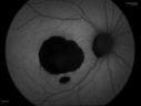
Macular Scar469 viewsFundus photography with Auto Fluorescent shows macular scar centrally, right eye. VA is 20/200 in the right eye. Aug 07, 2014
|
|

Macular Scar1845 viewsFundus photography with Auto Fluorescent shows macular scar centrally, right eye. VA is 20/200 in the right eye. Aug 07, 2014
|
|

Macular Star, Toxoplasmosis532 viewsYoung male with visual disturbance in the right eye. Fundus photo shows swelling around the optic nerve with macular star visible.Jun 03, 2014
|
|

Macular Star, Toxoplasmosis557 viewsYoung male with visual disturbance in the right eye. Fundus photo shows swelling around the optic nerve with macular star visible.Jun 03, 2014
|
|

Uveitis and Cystoid Macular Edema - Probably Sarcoidosis 851 views- Decreased CME after topical Pred Forte for one month QID Vision 20/40 OD, 20/25 OSMay 02, 2014
|
|
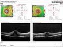
Uveitis and Cystoid Macular Edema - Probably Sarcoidosis - SD OCT Map805 viewsVA 20/63 OD 20/50 OS - 3 months vision loss - second episode by history of uveitisMay 02, 2014
|
|

Uveitis and Cystoid Macular Edema - Probably Sarcoidosis672 viewsVA 20/63 OD 20/50 OS - 3 months vision loss - second episode by history of uveitisMay 02, 2014
|
|

Uveitis and Cystoid Macular Edema - Probably Sarcoidosis734 viewsVA 20/63 OD 20/50 OS - 3 months vision loss - second episode by history of uveitisMay 02, 2014
|
|

Uveitis and Cystoid Macular Edema - Probably Sarcoidosis646 viewsVA 20/63 OD 20/50 OS - 3 months vision loss - second episode by history of uveitisMay 02, 2014
|
|

Uveitis and Cystoid Macular Edema - Probably Sarcoidosis641 viewsVA 20/63 OD 20/50 OS - 3 months vision loss - second episode by history of uveitisMay 02, 2014
|
|
| 2409 files on 25 page(s) |
 |
|
|
|
|
16 | |
|
|
|
|


