Last additions - Posterior Uveitis - Chorioretinitis
|
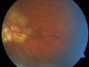
Serpiginouse Choroiditis (Chorioretinitis) - Acute Right Eye - Old Left Eye VA 20/25 OD , 20/50 OS865 views63-year-old woman has serpiginous choroiditis (date - March 2011). The right eye has not been previously involved, and then she noticed new onset floaters in the right eye for the last two weeks. Her vision in the right eye is hazy because of that.
VISUAL ACUITY: Vision OD is 20/25, OS is 20/50Apr 15, 2011
|
|

Serpiginouse Choroiditis (Chorioretinitis) - Acute Right Eye - Old Left Eye VA 20/25 OD , 20/50 OS642 views63-year-old woman has serpiginous choroiditis (date - March 2011). The right eye has not been previously involved, and then she noticed new onset floaters in the right eye for the last two weeks. Her vision in the right eye is hazy because of that.
VISUAL ACUITY: Vision OD is 20/25, OS is 20/50Apr 14, 2011
|
|

Serpiginouse Choroiditis (Chorioretinitis) - Acute Right Eye - Old Left Eye VA 20/25 OD , 20/50 OS663 views63-year-old woman has serpiginous choroiditis (date - March 2011). The right eye has not been previously involved, and then she noticed new onset floaters in the right eye for the last two weeks. Her vision in the right eye is hazy because of that.
VISUAL ACUITY: Vision OD is 20/25, OS is 20/50Apr 14, 2011
|
|
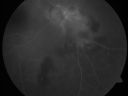
Serpiginouse Choroiditis (Chorioretinitis) - Acute Right Eye - Old Left Eye VA 20/25 OD , 20/50 OS594 views63-year-old woman has serpiginous choroiditis (date - March 2011). The right eye has not been previously involved, and then she noticed new onset floaters in the right eye for the last two weeks. Her vision in the right eye is hazy because of that.
VISUAL ACUITY: Vision OD is 20/25, OS is 20/50Apr 14, 2011
|
|
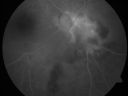
Serpiginouse Choroiditis (Chorioretinitis) - Acute Right Eye - Old Left Eye VA 20/25 OD , 20/50 OS570 views63-year-old woman has serpiginous choroiditis (date - March 2011). The right eye has not been previously involved, and then she noticed new onset floaters in the right eye for the last two weeks. Her vision in the right eye is hazy because of that.
VISUAL ACUITY: Vision OD is 20/25, OS is 20/50Apr 14, 2011
|
|
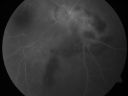
Serpiginouse Choroiditis (Chorioretinitis) - Acute Right Eye - Old Left Eye VA 20/25 OD , 20/50 OS642 views63-year-old woman has serpiginous choroiditis (date - March 2011). The right eye has not been previously involved, and then she noticed new onset floaters in the right eye for the last two weeks. Her vision in the right eye is hazy because of that.
VISUAL ACUITY: Vision OD is 20/25, OS is 20/50Apr 14, 2011
|
|

Serpiginouse Choroiditis (Chorioretinitis) - Acute Right Eye - Old Left Eye VA 20/25 OD , 20/50 OS673 views63-year-old woman has serpiginous choroiditis (date - March 2011). The right eye has not been previously involved, and then she noticed new onset floaters in the right eye for the last two weeks. Her vision in the right eye is hazy because of that.
VISUAL ACUITY: Vision OD is 20/25, OS is 20/50Apr 14, 2011
|
|

Serpiginouse Choroiditis (Chorioretinitis) - Acute Right Eye - Old Left Eye VA 20/25 OD , 20/50 OS860 views63-year-old woman has serpiginous choroiditis (date - March 2011). The right eye has not been previously involved, and then she noticed new onset floaters in the right eye for the last two weeks. Her vision in the right eye is hazy because of that.
VISUAL ACUITY: Vision OD is 20/25, OS is 20/50Apr 14, 2011
|
|

Serpiginouse Choroiditis (Chorioretinitis) - Acute Right Eye - Old Left Eye VA 20/25 OD , 20/50 OS678 views63-year-old woman has serpiginous choroiditis (date - March 2011). The right eye has not been previously involved, and then she noticed new onset floaters in the right eye for the last two weeks. Her vision in the right eye is hazy because of that.
VISUAL ACUITY: Vision OD is 20/25, OS is 20/50Apr 14, 2011
|
|

Serpiginouse Choroiditis (Chorioretinitis) - Acute Right Eye - Old Left Eye VA 20/25 OD , 20/50 OS661 views63-year-old woman has serpiginous choroiditis (date - March 2011). The right eye has not been previously involved, and then she noticed new onset floaters in the right eye for the last two weeks. Her vision in the right eye is hazy because of that.
VISUAL ACUITY: Vision OD is 20/25, OS is 20/50Apr 14, 2011
|
|

Serpiginouse Choroiditis (Chorioretinitis) - Acute Right Eye - Old Left Eye VA 20/25 OD , 20/50 OS650 views63-year-old woman has serpiginous choroiditis (date - March 2011). The right eye has not been previously involved, and then she noticed new onset floaters in the right eye for the last two weeks. Her vision in the right eye is hazy because of that.
VISUAL ACUITY: Vision OD is 20/25, OS is 20/50Apr 14, 2011
|
|

Serpiginouse Choroiditis (Chorioretinitis) - Acute Right Eye - Old Left Eye VA 20/25 OD , 20/50 OS679 views63-year-old woman has serpiginous choroiditis (date - March 2011). The right eye has not been previously involved, and then she noticed new onset floaters in the right eye for the last two weeks. Her vision in the right eye is hazy because of that.
VISUAL ACUITY: Vision OD is 20/25, OS is 20/50Apr 14, 2011
|
|

Serpiginouse Choroiditis (Chorioretinitis) - Acute Right Eye - Old Left Eye VA 20/25 OD , 20/50 OS670 views63-year-old woman has serpiginous choroiditis (date - March 2011). The right eye has not been previously involved, and then she noticed new onset floaters in the right eye for the last two weeks. Her vision in the right eye is hazy because of that.
VISUAL ACUITY: Vision OD is 20/25, OS is 20/50Apr 14, 2011
|
|

Serpiginouse Choroiditis (Chorioretinitis) - Acute Right Eye - Old Left Eye VA 20/25 OD , 20/50 OS776 views63-year-old woman has serpiginous choroiditis (date - March 2011). The right eye has not been previously involved, and then she noticed new onset floaters in the right eye for the last two weeks. Her vision in the right eye is hazy because of that.
VISUAL ACUITY: Vision OD is 20/25, OS is 20/50Apr 14, 2011
|
|

Serpiginouse Choroiditis (Chorioretinitis) - Acute Right Eye - Old Left Eye VA 20/25 OD , 20/50 OS938 views63-year-old woman has serpiginous choroiditis (date - March 2011). The right eye has not been previously involved, and then she noticed new onset floaters in the right eye for the last two weeks. Her vision in the right eye is hazy because of that.
VISUAL ACUITY: Vision OD is 20/25, OS is 20/50Apr 14, 2011
|
|

Serpiginouse Choroiditis (Chorioretinitis) - Acute Right Eye - Old Left Eye VA 20/25 OD , 20/50 OS958 views63-year-old woman has serpiginous choroiditis (date - March 2011). The right eye has not been previously involved, and then she noticed new onset floaters in the right eye for the last two weeks. Her vision in the right eye is hazy because of that.
VISUAL ACUITY: Vision OD is 20/25, OS is 20/50Apr 14, 2011
|
|

Serpiginouse Choroiditis (Chorioretinitis) - Acute Right Eye - Old Left Eye VA 20/25 OD , 20/50 OS958 views63-year-old woman has serpiginous choroiditis (date - March 2011). The right eye has not been previously involved, and then she noticed new onset floaters in the right eye for the last two weeks. Her vision in the right eye is hazy because of that.
VISUAL ACUITY: Vision OD is 20/25, OS is 20/50Apr 14, 2011
|
|

Serpiginouse Choroiditis (Chorioretinitis) - Acute Right Eye - Old Left Eye VA 20/25 OD , 20/50 OS840 views63-year-old woman has serpiginous choroiditis (date - March 2011). The right eye has not been previously involved, and then she noticed new onset floaters in the right eye for the last two weeks. Her vision in the right eye is hazy because of that.
VISUAL ACUITY: Vision OD is 20/25, OS is 20/50Apr 14, 2011
|
|

Serpiginouse Choroiditis (Chorioretinitis) - Acute Right Eye - Old Left Eye VA 20/25 OD , 20/50 OS1111 views63-year-old woman has serpiginous choroiditis (date - March 2011). The right eye has not been previously involved, and then she noticed new onset floaters in the right eye for the last two weeks. Her vision in the right eye is hazy because of that.
VISUAL ACUITY: Vision OD is 20/25, OS is 20/50Apr 14, 2011
|
|

Serpiginouse Choroiditis (Chorioretinitis) - Acute Right Eye - Old Left Eye VA 20/25 OD , 20/50 OS658 views63-year-old woman has serpiginous choroiditis (date - March 2011). The right eye has not been previously involved, and then she noticed new onset floaters in the right eye for the last two weeks. Her vision in the right eye is hazy because of that.
VISUAL ACUITY: Vision OD is 20/25, OS is 20/50Apr 14, 2011
|
|

Multiple Evanescent White Dot Syndrome - Atypical Yellow Foveal Spot Right Eye1255 views12-year-old had decreased vision in the left eye from reported amblyopia since around the year 2000. His right eye was doing fine until two days ago, when he had sudden severe vision loss. He sees a blurred spot in the middle of the vision in the right eye wherever he looks.
OD 20/60 with eccentric movement, OS 20/200. Pinhole 20/70. IOP: OD 14, OS 10.Dec 12, 2010
|
|

Multiple Evanescent White Dot Syndrome - Atypical Yellow Foveal Spot Right Eye1231 views12-year-old had decreased vision in the left eye from reported amblyopia since around the year 2000. His right eye was doing fine until two days ago, when he had sudden severe vision loss. He sees a blurred spot in the middle of the vision in the right eye wherever he looks.
OD 20/60 with eccentric movement, OS 20/200. Pinhole 20/70. IOP: OD 14, OS 10.Dec 12, 2010
|
|

Atypical Birdshot Chorioretinitis and Retinal Arterial Macroaneurysm Right Eye - HLA-B29 Positive871 views55-year-old woman has multifocal choroiditis in both eyes because she was HLA A-29 positive. She has presumptive birdshot chorioretinopathy. She has not noticed any vision changes. OD 20/40, OS 20/20.Dec 10, 2010
|
|
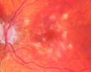
Atypical Birdshot Chorioretinitis and Retinal Arterial Macroaneurysm Right Eye - HLA-B29 Positive945 views55-year-old woman has multifocal choroiditis in both eyes because she was HLA A-29 positive. She has presumptive birdshot chorioretinopathy. She has not noticed any vision changes. OD 20/40, OS 20/20.Dec 10, 2010
|
|

Atypical Birdshot Chorioretinitis and Retinal Arterial Macroaneurysm Right Eye - HLA-B29 Positive1169 views55-year-old woman has multifocal choroiditis in both eyes because she was HLA A-29 positive. She has presumptive birdshot chorioretinopathy. She has not noticed any vision changes. OD 20/40, OS 20/20.Dec 10, 2010
|
|

Atypical Birdshot Chorioretinitis and Retinal Arterial Macroaneurysm Right Eye - HLA-B29 Positive841 views55-year-old woman has multifocal choroiditis in both eyes because she was HLA A-29 positive. She has presumptive birdshot chorioretinopathy. She has not noticed any vision changes. OD 20/40, OS 20/20.Dec 10, 2010
|
|
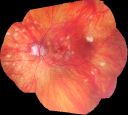
Atypical Birdshot Chorioretinitis and Retinal Arterial Macroaneurysm Right Eye - HLA-B29 Positive964 views55-year-old woman has multifocal choroiditis in both eyes because she was HLA A-29 positive. She has presumptive birdshot chorioretinopathy. She has not noticed any vision changes. OD 20/40, OS 20/20.Dec 10, 2010
|
|

Atypical Birdshot Chorioretinitis and Retinal Arterial Macroaneurysm Right Eye - HLA-B29 Positive872 views55-year-old woman has multifocal choroiditis in both eyes because she was HLA A-29 positive. She has presumptive birdshot chorioretinopathy. She has not noticed any vision changes. OD 20/40, OS 20/20.Dec 10, 2010
|
|

Atypical Birdshot Chorioretinitis and Retinal Arterial Macroaneurysm Right Eye - HLA-B29 Positive763 views55-year-old woman has multifocal choroiditis in both eyes because she was HLA A-29 positive. She has presumptive birdshot chorioretinopathy. She has not noticed any vision changes. OD 20/40, OS 20/20.Dec 10, 2010
|
|

Atypical Birdshot Chorioretinitis and Retinal Arterial Macroaneurysm Right Eye - HLA-B29 Positive933 views55-year-old woman has multifocal choroiditis in both eyes because she was HLA A-29 positive. She has presumptive birdshot chorioretinopathy. She has not noticed any vision changes. OD 20/40, OS 20/20..Dec 10, 2010
|
|

Atypical Birdshot Chorioretinitis and Retinal Arterial Macroaneurysm Right Eye - HLA-B29 Positive853 views55-year-old woman has multifocal choroiditis in both eyes because she was HLA A-29 positive. She has presumptive birdshot chorioretinopathy. She has not noticed any vision changes. OD 20/40, OS 20/20.Dec 10, 2010
|
|

Acute Zonal Occult Outer Retinopathy with Severe Peripheral Vision Loss Left Eye 569 views 58-year-old man developed a peripheral vision problem about 25 years ago. OD 20/30, OS 20/40.Dec 04, 2010
|
|

Acute Zonal Occult Outer Retinopathy with Severe Peripheral Vision Loss Left Eye 958 views 58-year-old man developed a peripheral vision problem about 25 years ago. OD 20/30, OS 20/40.Dec 04, 2010
|
|

Acute Zonal Occult Outer Retinopathy with Severe Peripheral Vision Loss Left Eye Post Treatment for CME723 views 58-year-old man developed a peripheral vision problem about 25 years ago. OD 20/30, OS 20/40.Dec 04, 2010
|
|
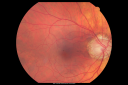
Acute Zonal Occult Outer Retinopathy with Severe Peripheral Vision Loss Left Eye 792 views 58-year-old man developed a peripheral vision problem about 25 years ago. OD 20/30, OS 20/40.Dec 04, 2010
|
|

Acute Zonal Occult Outer Retinopathy with Severe Peripheral Vision Loss Left Eye 653 views 58-year-old man developed a peripheral vision problem about 25 years ago. OD 20/30, OS 20/40.Dec 04, 2010
|
|

Acute Zonal Occult Outer Retinopathy with Severe Peripheral Vision Loss Left Eye 702 views 58-year-old man developed a peripheral vision problem about 25 years ago. OD 20/30, OS 20/40.Dec 04, 2010
|
|

Acute Zonal Occult Outer Retinopathy with Severe Peripheral Vision Loss Left Eye 586 views 58-year-old man developed a peripheral vision problem about 25 years ago. OD 20/30, OS 20/40.Dec 04, 2010
|
|

Acute Zonal Occult Outer Retinopathy with Severe Peripheral Vision Loss Left Eye 547 views 58-year-old man developed a peripheral vision problem about 25 years ago. OD 20/30, OS 20/40.Dec 04, 2010
|
|

Acute Zonal Occult Outer Retinopathy with Severe Peripheral Vision Loss Left Eye 583 views 58-year-old man developed a peripheral vision problem about 25 years ago. OD 20/30, OS 20/40.Dec 04, 2010
|
|
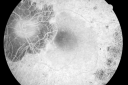
Acute Zonal Occult Outer Retinopathy with Severe Peripheral Vision Loss Left Eye 631 views 58-year-old man developed a peripheral vision problem about 25 years ago. OD 20/30, OS 20/40.Dec 04, 2010
|
|

Acute Zonal Occult Outer Retinopathy with Severe Peripheral Vision Loss Left Eye 662 views 58-year-old man developed a peripheral vision problem about 25 years ago. OD 20/30, OS 20/40.Dec 04, 2010
|
|

Acute Zonal Occult Outer Retinopathy with Severe Peripheral Vision Loss Left Eye 653 views 58-year-old man developed a peripheral vision problem about 25 years ago. OD 20/30, OS 20/40.Dec 04, 2010
|
|

Acute Zonal Occult Outer Retinopathy with Severe Peripheral Vision Loss Left Eye 617 views 58-year-old man developed a peripheral vision problem about 25 years ago. OD 20/30, OS 20/40.Dec 04, 2010
|
|

Acute Zonal Occult Outer Retinopathy with Severe Peripheral Vision Loss Left Eye 714 views 58-year-old man developed a peripheral vision problem about 25 years ago. OD 20/30, OS 20/40.Dec 04, 2010
|
|

Acute Zonal Occult Outer Retinopathy with Severe Peripheral Vision Loss Left Eye 735 views 58-year-old man developed a peripheral vision problem about 25 years ago. OD 20/30, OS 20/40.Dec 04, 2010
|
|

Acute Zonal Occult Outer Retinopathy with Severe Peripheral Vision Loss Left Eye 680 views 58-year-old man developed a peripheral vision problem about 25 years ago. OD 20/30, OS 20/40.Dec 04, 2010
|
|

Acute Zonal Occult Outer Retinopathy with Severe Peripheral Vision Loss Left Eye 687 views 58-year-old man developed a peripheral vision problem about 25 years ago. OD 20/30, OS 20/40.Dec 04, 2010
|
|

Acute Zonal Occult Outer Retinopathy with Severe Peripheral Vision Loss Left Eye 719 views 58-year-old man developed a peripheral vision problem about 25 years ago. OD 20/30, OS 20/40.Dec 04, 2010
|
|

Acute Zonal Occult Outer Retinopathy with Severe Peripheral Vision Loss Left Eye 630 views 58-year-old man developed a peripheral vision problem about 25 years ago. OD 20/30, OS 20/40.Dec 04, 2010
|
|

Acute Zonal Occult Outer Retinopathy with Severe Peripheral Vision Loss Left Eye Post Treatment for CME740 views 58-year-old man developed a peripheral vision problem about 25 years ago. OD 20/30, OS 20/40.Dec 04, 2010
|
|

Cytomegalovirus Retinitis - Partially Treated and AIDS retinopathy600 views44-year-old man a month agoe was diagnosed with HIV and he had pneumonia. He has had blurred vision for about a month, more in the left eye than the right eye. He has not yet seen anyone to start anti-AIDS therapy. He was given a medical doctor's name for a follow-up visit. He thinks his CD4 count is somewhere in the low 100s. Vision OU is 20/30Dec 03, 2010
|
|

Cytomegalovirus Retinitis - Partially Treated and AIDS retinopathy556 views44-year-old man a month agoe was diagnosed with HIV and he had pneumonia. He has had blurred vision for about a month, more in the left eye than the right eye. He has not yet seen anyone to start anti-AIDS therapy. He was given a medical doctor's name for a follow-up visit. He thinks his CD4 count is somewhere in the low 100s. Vision OU is 20/30Dec 03, 2010
|
|

Cytomegalovirus Retinitis - Partially Treated and AIDS retinopathy578 views44-year-old man a month agoe was diagnosed with HIV and he had pneumonia. He has had blurred vision for about a month, more in the left eye than the right eye. He has not yet seen anyone to start anti-AIDS therapy. He was given a medical doctor's name for a follow-up visit. He thinks his CD4 count is somewhere in the low 100s. Vision OU is 20/30Dec 03, 2010
|
|

Cytomegalovirus Retinitis - Partially Treated and AIDS retinopathy665 views44-year-old man a month agoe was diagnosed with HIV and he had pneumonia. He has had blurred vision for about a month, more in the left eye than the right eye. He has not yet seen anyone to start anti-AIDS therapy. He was given a medical doctor's name for a follow-up visit. He thinks his CD4 count is somewhere in the low 100s. Vision OU is 20/30Dec 03, 2010
|
|

Cytomegalovirus Retinitis - Partially Treated and AIDS retinopathy597 views44-year-old man a month agoe was diagnosed with HIV and he had pneumonia. He has had blurred vision for about a month, more in the left eye than the right eye. He has not yet seen anyone to start anti-AIDS therapy. He was given a medical doctor's name for a follow-up visit. He thinks his CD4 count is somewhere in the low 100s. Vision OU is 20/30Dec 03, 2010
|
|
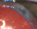
Propionibacterium acnes endophthalmitis with capsular plaque and uveitis1271 views61 year old man with inflammation after cataract surgery who ultimately needed removal of intraocular lens and capsule to quiet eye.Dec 02, 2010
|
|

Propionibacterium acnes endophthalmitis with capsular plaque and uveitis1470 views61 year old man with inflammation after cataract surgery who ultimately needed removal of intraocular lens and capsule to quiet eye.Dec 02, 2010
|
|
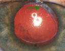
Propionibacterium acnes endophthalmitis with capsular plaque and uveitis1447 views61 year old man with inflammation after cataract surgery who ultimately needed removal of intraocular lens and capsule to quiet eye.Dec 02, 2010
|
|

Propionibacterium acnes endophthalmitis with capsular plaque and uveitis1121 views61 year old man with inflammation after cataract surgery who ultimately needed removal of intraocular lens and capsule to quiet eye.Dec 02, 2010
|
|

Propionibacterium acnes endophthalmitis with capsular plaque and uveitis828 views61 year old man with inflammation after cataract surgery who ultimately needed removal of intraocular lens and capsule to quiet eye.Dec 02, 2010
|
|

Multiple Evanescent White Dot Syndrome - Acute Idiopathic Blind Spot Enlargement725 views33-year-old woman one week ago she awoke and saw a red spots flashing out of her right eye. She notices them to the right side, which is the temporal visual field out of the right eye, then they changed to bursts of white lights without colors and then she notices straight ahead a couple spots. She has headaches in the back of her head. OD 20/20, OS 20/20Nov 30, 2010
|
|

Multiple Evanescent White Dot Syndrome - Acute Idiopathic Blind Spot Enlargement695 views33-year-old woman one week ago she awoke and saw a red spots flashing out of her right eye. She notices them to the right side, which is the temporal visual field out of the right eye, then they changed to bursts of white lights without colors and then she notices straight ahead a couple spots. She has headaches in the back of her head. OD 20/20, OS 20/20Nov 30, 2010
|
|

Multiple Evanescent White Dot Syndrome - Acute Idiopathic Blind Spot Enlargement780 views33-year-old woman one week ago she awoke and saw a red spots flashing out of her right eye. She notices them to the right side, which is the temporal visual field out of the right eye, then they changed to bursts of white lights without colors and then she notices straight ahead a couple spots. She has headaches in the back of her head. OD 20/20, OS 20/20Nov 30, 2010
|
|

Multiple Evanescent White Dot Syndrome - Acute Idiopathic Blind Spot Enlargement592 views33-year-old woman one week ago she awoke and saw a red spots flashing out of her right eye. She notices them to the right side, which is the temporal visual field out of the right eye, then they changed to bursts of white lights without colors and then she notices straight ahead a couple spots. She has headaches in the back of her head. OD 20/20, OS 20/20Nov 30, 2010
|
|
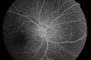
Multiple Evanescent White Dot Syndrome - Acute Idiopathic Blind Spot Enlargement618 views33-year-old woman one week ago she awoke and saw a red spots flashing out of her right eye. She notices them to the right side, which is the temporal visual field out of the right eye, then they changed to bursts of white lights without colors and then she notices straight ahead a couple spots. She has headaches in the back of her head. OD 20/20, OS 20/20Nov 30, 2010
|
|

Multiple Evanescent White Dot Syndrome - Acute Idiopathic Blind Spot Enlargement587 views33-year-old woman one week ago she awoke and saw a red spots flashing out of her right eye. She notices them to the right side, which is the temporal visual field out of the right eye, then they changed to bursts of white lights without colors and then she notices straight ahead a couple spots. She has headaches in the back of her head. OD 20/20, OS 20/20Nov 30, 2010
|
|

Multiple Evanescent White Dot Syndrome - Acute Idiopathic Blind Spot Enlargement618 views33-year-old woman one week ago she awoke and saw a red spots flashing out of her right eye. She notices them to the right side, which is the temporal visual field out of the right eye, then they changed to bursts of white lights without colors and then she notices straight ahead a couple spots. She has headaches in the back of her head. OD 20/20, OS 20/20Nov 30, 2010
|
|

Multiple Evanescent White Dot Syndrome - Acute Idiopathic Blind Spot Enlargement535 views33-year-old woman one week ago she awoke and saw a red spots flashing out of her right eye. She notices them to the right side, which is the temporal visual field out of the right eye, then they changed to bursts of white lights without colors and then she notices straight ahead a couple spots. She has headaches in the back of her head. OD 20/20, OS 20/20Nov 30, 2010
|
|

Multiple Evanescent White Dot Syndrome - Acute Idiopathic Blind Spot Enlargement520 views33-year-old woman one week ago she awoke and saw a red spots flashing out of her right eye. She notices them to the right side, which is the temporal visual field out of the right eye, then they changed to bursts of white lights without colors and then she notices straight ahead a couple spots. She has headaches in the back of her head. OD 20/20, OS 20/20Nov 30, 2010
|
|

Multiple Evanescent White Dot Syndrome - Acute Idiopathic Blind Spot Enlargement574 views33-year-old woman one week ago she awoke and saw a red spots flashing out of her right eye. She notices them to the right side, which is the temporal visual field out of the right eye, then they changed to bursts of white lights without colors and then she notices straight ahead a couple spots. She has headaches in the back of her head. OD 20/20, OS 20/20Nov 30, 2010
|
|

Multiple Evanescent White Dot Syndrome - Acute Idiopathic Blind Spot Enlargement644 views33-year-old woman one week ago she awoke and saw a red spots flashing out of her right eye. She notices them to the right side, which is the temporal visual field out of the right eye, then they changed to bursts of white lights without colors and then she notices straight ahead a couple spots. She has headaches in the back of her head. OD 20/20, OS 20/20Nov 30, 2010
|
|

Multiple Evanescent White Dot Syndrome - Acute Idiopathic Blind Spot Enlargement710 views33-year-old woman one week ago she awoke and saw a red spots flashing out of her right eye. She notices them to the right side, which is the temporal visual field out of the right eye, then they changed to bursts of white lights without colors and then she notices straight ahead a couple spots. She has headaches in the back of her head. OD 20/20, OS 20/20Nov 30, 2010
|
|
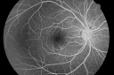
Multiple Evanescent White Dot Syndrome - Acute Idiopathic Blind Spot Enlargement694 views33-year-old woman one week ago she awoke and saw a red spots flashing out of her right eye. She notices them to the right side, which is the temporal visual field out of the right eye, then they changed to bursts of white lights without colors and then she notices straight ahead a couple spots. She has headaches in the back of her head. OD 20/20, OS 20/20Nov 30, 2010
|
|
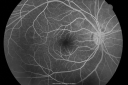
Multiple Evanescent White Dot Syndrome - Acute Idiopathic Blind Spot Enlargement693 views33-year-old woman one week ago she awoke and saw a red spots flashing out of her right eye. She notices them to the right side, which is the temporal visual field out of the right eye, then they changed to bursts of white lights without colors and then she notices straight ahead a couple spots. She has headaches in the back of her head. OD 20/20, OS 20/20Nov 30, 2010
|
|
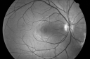
Multiple Evanescent White Dot Syndrome - Acute Idiopathic Blind Spot Enlargement714 views33-year-old woman one week ago she awoke and saw a red spots flashing out of her right eye. She notices them to the right side, which is the temporal visual field out of the right eye, then they changed to bursts of white lights without colors and then she notices straight ahead a couple spots. She has headaches in the back of her head. OD 20/20, OS 20/20Nov 30, 2010
|
|

Multiple Evanescent White Dot Syndrome - Acute Idiopathic Blind Spot Enlargement744 views33-year-old woman one week ago she awoke and saw a red spots flashing out of her right eye. She notices them to the right side, which is the temporal visual field out of the right eye, then they changed to bursts of white lights without colors and then she notices straight ahead a couple spots. She has headaches in the back of her head. OD 20/20, OS 20/20Nov 30, 2010
|
|

Multiple Evanescent White Dot Syndrome - Acute Idiopathic Blind Spot Enlargement825 views33-year-old woman one week ago she awoke and saw a red spots flashing out of her right eye. She notices them to the right side, which is the temporal visual field out of the right eye, then they changed to bursts of white lights without colors and then she notices straight ahead a couple spots. She has headaches in the back of her head. OD 20/20, OS 20/20Nov 30, 2010
|
|

Multiple Evanescent White Dot Syndrome - Acute Idiopathic Blind Spot Enlargement875 views33-year-old woman one week ago she awoke and saw a red spots flashing out of her right eye. She notices them to the right side, which is the temporal visual field out of the right eye, then they changed to bursts of white lights without colors and then she notices straight ahead a couple spots. She has headaches in the back of her head. OD 20/20, OS 20/20Nov 30, 2010
|
|

Multiple Evanescent White Dot Syndrome - Acute Idiopathic Blind Spot Enlargement996 views33-year-old woman one week ago she awoke and saw a red spots flashing out of her right eye. She notices them to the right side, which is the temporal visual field out of the right eye, then they changed to bursts of white lights without colors and then she notices straight ahead a couple spots. She has headaches in the back of her head. OD 20/20, OS 20/20Nov 30, 2010
|
|
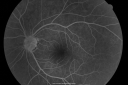
Multiple Evanescent White Dot Syndrome - Acute Idiopathic Blind Spot Enlargement871 views33-year-old woman one week ago she awoke and saw a red spots flashing out of her right eye. She notices them to the right side, which is the temporal visual field out of the right eye, then they changed to bursts of white lights without colors and then she notices straight ahead a couple spots. She has headaches in the back of her head. OD 20/20, OS 20/20Nov 30, 2010
|
|

Acute Zonal Occult Outer Retinopathy1002 views45-year-old man noticed a few years ago peripheral vision blurriness in the left eye and also some central vision loss. Previous to that, as far as he knows, the two eyes were okay.
VISUAL ACUITY: OD: 20/20; OS: 20/40. Nov 23, 2010
|
|

Acute Zonal Occult Outer Retinopathy836 views45-year-old man noticed a few years ago peripheral vision blurriness in the left eye and also some central vision loss. Previous to that, as far as he knows, the two eyes were okay.
VISUAL ACUITY: OD: 20/20; OS: 20/40. Nov 23, 2010
|
|

Acute Zonal Occult Outer Retinopathy810 views45-year-old man noticed a few years ago peripheral vision blurriness in the left eye and also some central vision loss. Previous to that, as far as he knows, the two eyes were okay.
VISUAL ACUITY: OD: 20/20; OS: 20/40. Nov 23, 2010
|
|

Acute Zonal Occult Outer Retinopathy902 views45-year-old man noticed a few years ago peripheral vision blurriness in the left eye and also some central vision loss. Previous to that, as far as he knows, the two eyes were okay.
VISUAL ACUITY: OD: 20/20; OS: 20/40. Nov 23, 2010
|
|

Acute Zonal Occult Outer Retinopathy863 views45-year-old man noticed a few years ago peripheral vision blurriness in the left eye and also some central vision loss. Previous to that, as far as he knows, the two eyes were okay.
VISUAL ACUITY: OD: 20/20; OS: 20/40. Nov 23, 2010
|
|

Acute Zonal Occult Outer Retinopathy863 views45-year-old man noticed a few years ago peripheral vision blurriness in the left eye and also some central vision loss. Previous to that, as far as he knows, the two eyes were okay.
VISUAL ACUITY: OD: 20/20; OS: 20/40. Nov 23, 2010
|
|

Acute Zonal Occult Outer Retinopathy749 views45-year-old man noticed a few years ago peripheral vision blurriness in the left eye and also some central vision loss. Previous to that, as far as he knows, the two eyes were okay.
VISUAL ACUITY: OD: 20/20; OS: 20/40. Nov 23, 2010
|
|

Acute Zonal Occult Outer Retinopathy715 views45-year-old man noticed a few years ago peripheral vision blurriness in the left eye and also some central vision loss. Previous to that, as far as he knows, the two eyes were okay.
VISUAL ACUITY: OD: 20/20; OS: 20/40. Nov 23, 2010
|
|

Acute Zonal Occult Outer Retinopathy794 views45-year-old man noticed a few years ago peripheral vision blurriness in the left eye and also some central vision loss. Previous to that, as far as he knows, the two eyes were okay.
VISUAL ACUITY: OD: 20/20; OS: 20/40. Nov 23, 2010
|
|

Acute Zonal Occult Outer Retinopathy791 views45-year-old man noticed a few years ago peripheral vision blurriness in the left eye and also some central vision loss. Previous to that, as far as he knows, the two eyes were okay.
VISUAL ACUITY: OD: 20/20; OS: 20/40. Nov 23, 2010
|
|

Acute Zonal Occult Outer Retinopathy915 views45-year-old man noticed a few years ago peripheral vision blurriness in the left eye and also some central vision loss. Previous to that, as far as he knows, the two eyes were okay.
VISUAL ACUITY: OD: 20/20; OS: 20/40. Nov 23, 2010
|
|

Acute Zonal Occult Outer Retinopathy1197 views45-year-old man noticed a few years ago peripheral vision blurriness in the left eye and also some central vision loss. Previous to that, as far as he knows, the two eyes were okay.
VISUAL ACUITY: OD: 20/20; OS: 20/40. Nov 23, 2010
|
|

Acute Zonal Occult Outer Retinopathy1079 views45-year-old man noticed a few years ago peripheral vision blurriness in the left eye and also some central vision loss. Previous to that, as far as he knows, the two eyes were okay.
VISUAL ACUITY: OD: 20/20; OS: 20/40. Nov 23, 2010
|
|

Acute Zonal Occult Outer Retinopathy1016 views45-year-old man noticed a few years ago peripheral vision blurriness in the left eye and also some central vision loss. Previous to that, as far as he knows, the two eyes were okay.
VISUAL ACUITY: OD: 20/20; OS: 20/40. Nov 23, 2010
|
|

Acute Zonal Occult Outer Retinopathy859 views45-year-old man noticed a few years ago peripheral vision blurriness in the left eye and also some central vision loss. Previous to that, as far as he knows, the two eyes were okay.
VISUAL ACUITY: OD: 20/20; OS: 20/40. Nov 23, 2010
|
|

Acute Zonal Occult Outer Retinopathy933 views45-year-old man noticed a few years ago peripheral vision blurriness in the left eye and also some central vision loss. Previous to that, as far as he knows, the two eyes were okay.
VISUAL ACUITY: OD: 20/20; OS: 20/40. Nov 23, 2010
|
|
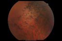
Acute Zonal Occult Outer Retinopathy1134 views45-year-old man noticed a few years ago peripheral vision blurriness in the left eye and also some central vision loss. Previous to that, as far as he knows, the two eyes were okay.
VISUAL ACUITY: OD: 20/20; OS: 20/40. Nov 23, 2010
|
|
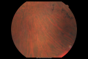
Acute Zonal Occult Outer Retinopathy903 views45-year-old man noticed a few years ago peripheral vision blurriness in the left eye and also some central vision loss. Previous to that, as far as he knows, the two eyes were okay.
VISUAL ACUITY: OD: 20/20; OS: 20/40. Nov 23, 2010
|
|

Acute Zonal Occult Outer Retinopathy879 views45-year-old man noticed a few years ago peripheral vision blurriness in the left eye and also some central vision loss. Previous to that, as far as he knows, the two eyes were okay.
VISUAL ACUITY: OD: 20/20; OS: 20/40. Nov 23, 2010
|
|
| 2412 files on 25 page(s) |
 |
|
|
|
|
|
|
|
23 | |
|


