Last additions - Posterior Uveitis - Chorioretinitis
|

Bilateral Diffuse Uveal Melanocytic Proliferation - BDUMP - Paraneoplastic Syndrome844 views80-year-old man vision loss for one year. He died about one year after these photos from Metastatic Poorly Differentiated Large Cell Carcinoma of unknown primary. He was a smoker. Nov 17, 2010
|
|

Bilateral Diffuse Uveal Melanocytic Proliferation - BDUMP - Paraneoplastic Syndrome2061 views80-year-old man vision loss for one year. He died about one year after these photos from Metastatic Poorly Differentiated Large Cell Carcinoma of unknown primary. He was a smoker. Nov 17, 2010
|
|

Bilateral Diffuse Uveal Melanocytic Proliferation - BDUMP - Paraneoplastic Syndrome1071 views80-year-old man vision loss for one year. He died about one year after these photos from Metastatic Poorly Differentiated Large Cell Carcinoma of unknown primary. He was a smoker. Nov 17, 2010
|
|

Bilateral Diffuse Uveal Melanocytic Proliferation - BDUMP - Paraneoplastic Syndrome1278 views80-year-old man vision loss for one year. He died about one year after these photos from Metastatic Poorly Differentiated Large Cell Carcinoma of unknown primary. He was a smoker. Nov 17, 2010
|
|

Bilateral Diffuse Uveal Melanocytic Proliferation - BDUMP - Paraneoplastic Syndrome1233 views80-year-old man vision loss for one year. He died about one year after these photos from Metastatic Poorly Differentiated Large Cell Carcinoma of unknown primary. He was a smoker. Nov 17, 2010
|
|

Bilateral Diffuse Uveal Melanocytic Proliferation - BDUMP - Paraneoplastic Syndrome1362 views80-year-old man vision loss for one year. He died about one year after these photos from Metastatic Poorly Differentiated Large Cell Carcinoma of unknown primary. He was a smoker. Nov 17, 2010
|
|

Fungal Endophthalmitis830 views65-year-old woman had chronic endophthalmitis in the left eye 1 day post-op vitrectomy.Nov 17, 2010
|
|

Fungal Endophthalmitis1020 views65-year-old woman had chronic endophthalmitis in the left eye 1 day post-op vitrectomy.Nov 17, 2010
|
|

Toxoplasmosis - Inner Retinal - Recurrent - Treated with Bactrim DS 718 views43-year-old man has macular toxoplasmosis. He has responded nicely to Bactrim OS: 20/60Nov 17, 2010
|
|

Toxoplasmosis - Inner Retinal - Recurrent - Treated with Bactrim DS 629 views43-year-old man has macular toxoplasmosis. He has responded nicely to Bactrim OS: 20/60Nov 17, 2010
|
|

Toxoplasmosis - Inner Retinal - Recurrent - Treated with Bactrim DS 679 views43-year-old man has macular toxoplasmosis. He has responded nicely to Bactrim OS: 20/60Nov 17, 2010
|
|

Toxoplasmosis - Inner Retinal - Recurrent - Treated with Bactrim DS 551 views43-year-old man has macular toxoplasmosis. He has responded nicely to Bactrim OS: 20/60Nov 17, 2010
|
|

Toxoplasmosis - Inner Retinal - Recurrent - Treated with Bactrim DS 587 views43-year-old man has macular toxoplasmosis. He has responded nicely to Bactrim OS: 20/60Nov 17, 2010
|
|
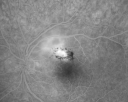
Toxoplasmosis - Inner Retinal - Recurrent - Treated with Bactrim DS 541 views43-year-old man has macular toxoplasmosis. He has responded nicely to Bactrim OS: 20/60Nov 17, 2010
|
|
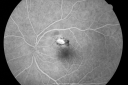
Toxoplasmosis - Inner Retinal - Recurrent - Treated with Bactrim DS 539 views43-year-old man has macular toxoplasmosis. He has responded nicely to Bactrim OS: 20/60Nov 17, 2010
|
|

Toxoplasmosis - Inner Retinal - Recurrent - Treated with Bactrim DS 585 views43-year-old man has macular toxoplasmosis. He has responded nicely to Bactrim OS: 20/60Nov 17, 2010
|
|

Toxoplasmosis - Inner Retinal - Recurrent - Treated with Bactrim DS 529 views43-year-old man has macular toxoplasmosis. He has responded nicely to Bactrim OS: 20/60Nov 17, 2010
|
|

Toxoplasmosis - Inner Retinal - Recurrent - Treated with Bactrim DS 546 views43-year-old man has macular toxoplasmosis. He has responded nicely to Bactrim OS: 20/60Nov 17, 2010
|
|

Toxoplasmosis - Inner Retinal - Recurrent - Treated with Bactrim DS 529 views43-year-old man has macular toxoplasmosis. He has responded nicely to Bactrim OS: 20/60Nov 17, 2010
|
|

Toxoplasmosis - Inner Retinal - Recurrent - Treated with Bactrim DS 583 views43-year-old man has macular toxoplasmosis. He has responded nicely to Bactrim OS: 20/60Nov 17, 2010
|
|

Toxoplasmosis - Inner Retinal - Recurrent - Treated with Bactrim DS 564 views43-year-old man has macular toxoplasmosis. He has responded nicely to Bactrim OS: 20/60Nov 17, 2010
|
|

Toxoplasmosis - Inner Retinal - Recurrent - Treated with Bactrim DS 735 views43-year-old man has macular toxoplasmosis. He has responded nicely to Bactrim OS: 20/60Nov 17, 2010
|
|

Toxoplasmosis - Inner Retinal - Recurrent - Treated with Bactrim DS 804 views43-year-old man has macular toxoplasmosis. He has responded nicely to Bactrim OS: 20/60Nov 17, 2010
|
|

Toxoplasmosis - Inner Retinal - Recurrent - Treated with Bactrim DS 633 views43-year-old man has macular toxoplasmosis. He has responded nicely to Bactrim OS: 20/60Nov 17, 2010
|
|

Toxoplasmosis - Inner Retinal - Recurrent - Treated with Bactrim DS 882 views43-year-old man has macular toxoplasmosis. He has responded nicely to Bactrim OS: 20/60Nov 17, 2010
|
|

Toxoplasmosis - Inner Retinal - Recurrent - Treated with Bactrim DS 730 views43-year-old man has macular toxoplasmosis. He has responded nicely to Bactrim OS: 20/60Nov 17, 2010
|
|

Serpiginous choroiditis682 viewsInactive scars in both eyesNov 14, 2010
|
|
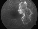
Serpiginous choroiditis577 viewsInactive scars in both eyesNov 14, 2010
|
|
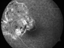
Serpiginous choroiditis585 viewsInactive scars in both eyesNov 14, 2010
|
|

Serpiginous choroiditis604 viewsInactive scars in both eyesNov 14, 2010
|
|

Serpiginous choroiditis653 viewsInactive scars in both eyesNov 14, 2010
|
|

Serpiginous choroiditis603 viewsInactive scars in both eyesNov 14, 2010
|
|

Serpiginous choroiditis571 viewsInactive scars in both eyesNov 14, 2010
|
|

Serpiginous choroiditis582 viewsInactive scars in both eyesNov 14, 2010
|
|

Serpiginous choroiditis577 viewsInactive scars in both eyesNov 14, 2010
|
|

Serpiginous choroiditis642 viewsInactive scars in both eyesNov 14, 2010
|
|

Serpiginous choroiditis628 viewsInactive scars in both eyesNov 14, 2010
|
|
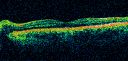
Multiple Evanescent White Dot Syndrome - Atypical Yellow Foveal Spot Right Eye619 views12-year-old had decreased vision in the left eye from reported amblyopia since around the year 2000. His right eye was doing fine until two days ago, when he had sudden severe vision loss. He sees a blurred spot in the middle of the vision in the right eye wherever he looks.
OD 20/60 with eccentric movement, OS 20/200. Pinhole 20/70. IOP: OD 14, OS 10.Nov 08, 2010
|
|

Multiple Evanescent White Dot Syndrome - Atypical Yellow Foveal Spot Right Eye620 views12-year-old had decreased vision in the left eye from reported amblyopia since around the year 2000. His right eye was doing fine until two days ago, when he had sudden severe vision loss. He sees a blurred spot in the middle of the vision in the right eye wherever he looks.
OD 20/60 with eccentric movement, OS 20/200. Pinhole 20/70. IOP: OD 14, OS 10.Nov 08, 2010
|
|

Multiple Evanescent White Dot Syndrome - Atypical Yellow Foveal Spot Right Eye701 views12-year-old had decreased vision in the left eye from reported amblyopia since around the year 2000. His right eye was doing fine until two days ago, when he had sudden severe vision loss. He sees a blurred spot in the middle of the vision in the right eye wherever he looks.
OD 20/60 with eccentric movement, OS 20/200. Pinhole 20/70. IOP: OD 14, OS 10.Nov 08, 2010
|
|
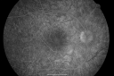
Multiple Evanescent White Dot Syndrome - Atypical Yellow Foveal Spot Right Eye564 views12-year-old had decreased vision in the left eye from reported amblyopia since around the year 2000. His right eye was doing fine until two days ago, when he had sudden severe vision loss. He sees a blurred spot in the middle of the vision in the right eye wherever he looks.
OD 20/60 with eccentric movement, OS 20/200. Pinhole 20/70. IOP: OD 14, OS 10.Nov 08, 2010
|
|

Multiple Evanescent White Dot Syndrome - Atypical Yellow Foveal Spot Right Eye568 views12-year-old had decreased vision in the left eye from reported amblyopia since around the year 2000. His right eye was doing fine until two days ago, when he had sudden severe vision loss. He sees a blurred spot in the middle of the vision in the right eye wherever he looks.
OD 20/60 with eccentric movement, OS 20/200. Pinhole 20/70. IOP: OD 14, OS 10.Nov 08, 2010
|
|

Multiple Evanescent White Dot Syndrome - Atypical Yellow Foveal Spot Right Eye518 views12-year-old had decreased vision in the left eye from reported amblyopia since around the year 2000. His right eye was doing fine until two days ago, when he had sudden severe vision loss. He sees a blurred spot in the middle of the vision in the right eye wherever he looks.
OD 20/60 with eccentric movement, OS 20/200. Pinhole 20/70. IOP: OD 14, OS 10.Nov 08, 2010
|
|

Multiple Evanescent White Dot Syndrome - Atypical Yellow Foveal Spot Right Eye566 views12-year-old had decreased vision in the left eye from reported amblyopia since around the year 2000. His right eye was doing fine until two days ago, when he had sudden severe vision loss. He sees a blurred spot in the middle of the vision in the right eye wherever he looks.
OD 20/60 with eccentric movement, OS 20/200. Pinhole 20/70. IOP: OD 14, OS 10.Nov 08, 2010
|
|

Multiple Evanescent White Dot Syndrome - Atypical Yellow Foveal Spot Right Eye467 views12-year-old had decreased vision in the left eye from reported amblyopia since around the year 2000. His right eye was doing fine until two days ago, when he had sudden severe vision loss. He sees a blurred spot in the middle of the vision in the right eye wherever he looks.
OD 20/60 with eccentric movement, OS 20/200. Pinhole 20/70. IOP: OD 14, OS 10.Nov 08, 2010
|
|
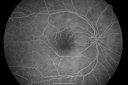
Multiple Evanescent White Dot Syndrome - Atypical Yellow Foveal Spot Right Eye508 views12-year-old had decreased vision in the left eye from reported amblyopia since around the year 2000. His right eye was doing fine until two days ago, when he had sudden severe vision loss. He sees a blurred spot in the middle of the vision in the right eye wherever he looks.
OD 20/60 with eccentric movement, OS 20/200. Pinhole 20/70. IOP: OD 14, OS 10.Nov 08, 2010
|
|

Multiple Evanescent White Dot Syndrome - Atypical Yellow Foveal Spot Right Eye509 views12-year-old had decreased vision in the left eye from reported amblyopia since around the year 2000. His right eye was doing fine until two days ago, when he had sudden severe vision loss. He sees a blurred spot in the middle of the vision in the right eye wherever he looks.
OD 20/60 with eccentric movement, OS 20/200. Pinhole 20/70. IOP: OD 14, OS 10.Nov 08, 2010
|
|

Multiple Evanescent White Dot Syndrome - Atypical Yellow Foveal Spot Right Eye534 views12-year-old had decreased vision in the left eye from reported amblyopia since around the year 2000. His right eye was doing fine until two days ago, when he had sudden severe vision loss. He sees a blurred spot in the middle of the vision in the right eye wherever he looks.
OD 20/60 with eccentric movement, OS 20/200. Pinhole 20/70. IOP: OD 14, OS 10.Nov 08, 2010
|
|

Multiple Evanescent White Dot Syndrome - Atypical Yellow Foveal Spot Right Eye591 views12-year-old had decreased vision in the left eye from reported amblyopia since around the year 2000. His right eye was doing fine until two days ago, when he had sudden severe vision loss. He sees a blurred spot in the middle of the vision in the right eye wherever he looks.
OD 20/60 with eccentric movement, OS 20/200. Pinhole 20/70. IOP: OD 14, OS 10.Nov 08, 2010
|
|

Multiple Evanescent White Dot Syndrome - Atypical Yellow Foveal Spot Right Eye618 views12-year-old had decreased vision in the left eye from reported amblyopia since around the year 2000. His right eye was doing fine until two days ago, when he had sudden severe vision loss. He sees a blurred spot in the middle of the vision in the right eye wherever he looks.
OD 20/60 with eccentric movement, OS 20/200. Pinhole 20/70. IOP: OD 14, OS 10.Nov 08, 2010
|
|

Multiple Evanescent White Dot Syndrome - Atypical Yellow Foveal Spot Right Eye932 views12-year-old had decreased vision in the left eye from reported amblyopia since around the year 2000. His right eye was doing fine until two days ago, when he had sudden severe vision loss. He sees a blurred spot in the middle of the vision in the right eye wherever he looks.
OD 20/60 with eccentric movement, OS 20/200. Pinhole 20/70. IOP: OD 14, OS 10.Nov 08, 2010
|
|

Multiple Evanescent White Dot Syndrome - Atypical Yellow Foveal Spot Right Eye825 views12-year-old had decreased vision in the left eye from reported amblyopia since around the year 2000. His right eye was doing fine until two days ago, when he had sudden severe vision loss. He sees a blurred spot in the middle of the vision in the right eye wherever he looks.
OD 20/60 with eccentric movement, OS 20/200. Pinhole 20/70. IOP: OD 14, OS 10.Nov 08, 2010
|
|

Multiple Evanescent White Dot Syndrome - Atypical Yellow Foveal Spot Right Eye907 views12-year-old had decreased vision in the left eye from reported amblyopia since around the year 2000. His right eye was doing fine until two days ago, when he had sudden severe vision loss. He sees a blurred spot in the middle of the vision in the right eye wherever he looks.
OD 20/60 with eccentric movement, OS 20/200. Pinhole 20/70. IOP: OD 14, OS 10.Nov 08, 2010
|
|

Multiple Evanescent White Dot Syndrome - Atypical Yellow Foveal Spot Right Eye930 views12-year-old had decreased vision in the left eye from reported amblyopia since around the year 2000. His right eye was doing fine until two days ago, when he had sudden severe vision loss. He sees a blurred spot in the middle of the vision in the right eye wherever he looks.
OD 20/60 with eccentric movement, OS 20/200. Pinhole 20/70. IOP: OD 14, OS 10.Nov 08, 2010
|
|

Multiple Evanescent White Dot Syndrome - Atypical Yellow Foveal Spot Right Eye540 views12-year-old had decreased vision in the left eye from reported amblyopia since around the year 2000. His right eye was doing fine until two days ago, when he had sudden severe vision loss. He sees a blurred spot in the middle of the vision in the right eye wherever he looks.
OD 20/60 with eccentric movement, OS 20/200. Pinhole 20/70. IOP: OD 14, OS 10.Nov 08, 2010
|
|

Birdshot Chorioretinitis Mild - Asymptomatic1030 views59-year-old man has birdshot chorioretinopathy in both eyes. Vision OD 20/25, OS 20/20. IOP: OD 16, OS 14. The right eye has a posterior chamber intraocular lens in good position. There is 1+ posterior capsular opacity. The left eye has 2+ nuclear sclerosis.
EXTENDED OPHTHALMOSCOPY:
OD: Vertical C/D ration is 0.8. There is patchy depigmented lesion in the nasal periphery. The retina is attached with peripheral laser.
OS: Vertical C/D ratio is 0.4. There are peripheral punched out lesions in the peripheryNov 03, 2010
|
|

Birdshot Chorioretinitis Mild - Asymptomatic768 views59-year-old man has birdshot chorioretinopathy in both eyes. Vision OD 20/25, OS 20/20. IOP: OD 16, OS 14. The right eye has a posterior chamber intraocular lens in good position. There is 1+ posterior capsular opacity. The left eye has 2+ nuclear sclerosis.
EXTENDED OPHTHALMOSCOPY:
OD: Vertical C/D ration is 0.8. There is patchy depigmented lesion in the nasal periphery. The retina is attached with peripheral laser.
OS: Vertical C/D ratio is 0.4. There are peripheral punched out lesions in the peripheryNov 03, 2010
|
|

Birdshot Chorioretinitis Mild - Asymptomatic920 views59-year-old man has birdshot chorioretinopathy in both eyes. Vision OD 20/25, OS 20/20. IOP: OD 16, OS 14. The right eye has a posterior chamber intraocular lens in good position. There is 1+ posterior capsular opacity. The left eye has 2+ nuclear sclerosis.
EXTENDED OPHTHALMOSCOPY:
OD: Vertical C/D ration is 0.8. There is patchy depigmented lesion in the nasal periphery. The retina is attached with peripheral laser.
OS: Vertical C/D ratio is 0.4. There are peripheral punched out lesions in the peripheryNov 03, 2010
|
|

Birdshot Chorioretinitis Mild - Asymptomatic1053 views59-year-old man has birdshot chorioretinopathy in both eyes. Vision OD 20/25, OS 20/20. IOP: OD 16, OS 14. The right eye has a posterior chamber intraocular lens in good position. There is 1+ posterior capsular opacity. The left eye has 2+ nuclear sclerosis.
EXTENDED OPHTHALMOSCOPY:
OD: Vertical C/D ration is 0.8. There is patchy depigmented lesion in the nasal periphery. The retina is attached with peripheral laser.
OS: Vertical C/D ratio is 0.4. There are peripheral punched out lesions in the peripheryNov 03, 2010
|
|

Birdshot Chorioretinitis Mild - Asymptomatic915 views59-year-old man has birdshot chorioretinopathy in both eyes. Vision OD 20/25, OS 20/20. IOP: OD 16, OS 14. The right eye has a posterior chamber intraocular lens in good position. There is 1+ posterior capsular opacity. The left eye has 2+ nuclear sclerosis.
EXTENDED OPHTHALMOSCOPY:
OD: Vertical C/D ration is 0.8. There is patchy depigmented lesion in the nasal periphery. The retina is attached with peripheral laser.
OS: Vertical C/D ratio is 0.4. There are peripheral punched out lesions in the peripheryNov 03, 2010
|
|

Birdshot Chorioretinitis Mild - Asymptomatic924 views59-year-old man has birdshot chorioretinopathy in both eyes. Vision OD 20/25, OS 20/20. IOP: OD 16, OS 14. The right eye has a posterior chamber intraocular lens in good position. There is 1+ posterior capsular opacity. The left eye has 2+ nuclear sclerosis.
EXTENDED OPHTHALMOSCOPY:
OD: Vertical C/D ration is 0.8. There is patchy depigmented lesion in the nasal periphery. The retina is attached with peripheral laser.
OS: Vertical C/D ratio is 0.4. There are peripheral punched out lesions in the peripheryNov 03, 2010
|
|
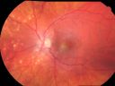
Multifocal Choroiditis and Subretinal Fibrosis - 32 yo Female Montage Initial Visit979 views32-year-old woman vision loss in the right eye associated with macular scarring and multifocal choroiditis in 1999 with new vision loss in left eye: OD 20/400, OS 20/50.
2 months post-rx with posterior subtenons kenalog and intravitreal avastin - va os 20/30 and lesion has retracted and organized. It never subsequently grew over 2 years follow-upNov 02, 2010
|
|

Multifocal Choroiditis and Subretinal Fibrosis - 32 yo Female Macula OS 2 months post rx1169 views32-year-old woman vision loss in the right eye associated with macular scarring and multifocal choroiditis in 1999 with new vision loss in left eye: OD 20/400, OS 20/50.
2 months post-rx with posterior subtenons kenalog and intravitreal avastin - va os 20/30 and lesion has retracted and organized. It never subsequently grew over 2 years follow-upNov 02, 2010
|
|

Multifocal Choroiditis and Subretinal Fibrosis - 32 yo Female Macula OS822 views32-year-old woman vision loss in the right eye associated with macular scarring and multifocal choroiditis in 1999 with new vision loss in left eye: OD 20/400, OS 20/50. Nov 02, 2010
|
|

Multifocal Choroiditis and Subretinal Fibrosis - 32 yo Female Multifocal and macular scar OD1286 views32-year-old woman vision loss in the right eye associated with macular scarring and multifocal choroiditis in 1999 with new vision loss in left eye: OD 20/400, OS 20/50. Nov 02, 2010
|
|

Multifocal Choroiditis and Subretinal Fibrosis - 32 yo Female Old Macular Scar OD1057 views32-year-old woman vision loss in the right eye associated with macular scarring and multifocal choroiditis in 1999 with new vision loss in left eye: OD 20/400, OS 20/50. Nov 02, 2010
|
|

Multifocal Choroiditis and Subretinal Fibrosis - 32 yo Female OCT map932 views32-year-old woman vision loss in the right eye associated with macular scarring and multifocal choroiditis in 1999 with new vision loss in left eye: OD 20/400, OS 20/50. Nov 02, 2010
|
|

Multifocal Choroiditis and Subretinal Fibrosis - 32 yo Female Line Scan New Lesion OS801 views32-year-old woman vision loss in the right eye associated with macular scarring and multifocal choroiditis in 1999 with new vision loss in left eye: OD 20/400, OS 20/50. Nov 02, 2010
|
|

Multifocal Choroiditis and Subretinal Fibrosis - 32 yo Female FA Old Scar OS758 views32-year-old woman vision loss in the right eye associated with macular scarring and multifocal choroiditis in 1999 with new vision loss in left eye: OD 20/400, OS 20/50. Nov 02, 2010
|
|

Multifocal Choroiditis and Subretinal Fibrosis - 32 yo Female FA New Lesion OS722 views32-year-old woman vision loss in the right eye associated with macular scarring and multifocal choroiditis in 1999 with new vision loss in left eye: OD 20/400, OS 20/50. Nov 02, 2010
|
|
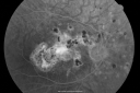
Multifocal Choroiditis and Subretinal Fibrosis - 32 yo Female Old Scar OD691 views32-year-old woman vision loss in the right eye associated with macular scarring and multifocal choroiditis in 1999 with new vision loss in left eye: OD 20/400, OS 20/50. Nov 02, 2010
|
|

Multifocal Choroiditis and Subretinal Fibrosis - 32 yo Female New Lesion OS672 views32-year-old woman vision loss in the right eye associated with macular scarring and multifocal choroiditis in 1999 with new vision loss in left eye: OD 20/400, OS 20/50. Nov 02, 2010
|
|
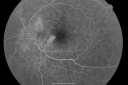
Multifocal Choroiditis and Subretinal Fibrosis - 32 yo Female New Lesion OS642 views32-year-old woman vision loss in the right eye associated with macular scarring and multifocal choroiditis in 1999 with new vision loss in left eye: OD 20/400, OS 20/50. Nov 02, 2010
|
|
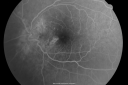
Multifocal Choroiditis and Subretinal Fibrosis - 32 yo Female New Lesion OS606 views32-year-old woman vision loss in the right eye associated with macular scarring and multifocal choroiditis in 1999 with new vision loss in left eye: OD 20/400, OS 20/50. Nov 02, 2010
|
|

Multifocal Choroiditis and Subretinal Fibrosis - 32 yo Female New Lesion OS730 views32-year-old woman vision loss in the right eye associated with macular scarring and multifocal choroiditis in 1999 with new vision loss in left eye: OD 20/400, OS 20/50. Nov 02, 2010
|
|
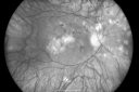
Multifocal Choroiditis and Subretinal Fibrosis - 32 yo Female Old Scar OD749 views32-year-old woman vision loss in the right eye associated with macular scarring and multifocal choroiditis in 1999 with new vision loss in left eye: OD 20/400, OS 20/50. Nov 02, 2010
|
|

Multifocal Choroiditis and Subretinal Fibrosis - 32 yo Female New Lesion OS861 views32-year-old woman vision loss in the right eye associated with macular scarring and multifocal choroiditis in 1999 with new vision loss in left eye: OD 20/400, OS 20/50. Nov 02, 2010
|
|

Multifocal Choroiditis and Subretinal Fibrosis - 32 yo Female Old Scar OD1017 views32-year-old woman vision loss in the right eye associated with macular scarring and multifocal choroiditis in 1999 with new vision loss in left eye: OD 20/400, OS 20/50. Nov 02, 2010
|
|

Multifocal Choroiditis and Subretinal Fibrosis - 32 yo Female New Lesion OS616 views32-year-old woman vision loss in the right eye associated with macular scarring and multifocal choroiditis in 1999 with new vision loss in left eye: OD 20/400, OS 20/50. Nov 02, 2010
|
|

Multifocal Choroiditis and Subretinal Fibrosis - 32 yo Female New Lesion OS681 views32-year-old woman vision loss in the right eye associated with macular scarring and multifocal choroiditis in 1999 with new vision loss in left eye: OD 20/400, OS 20/50. Nov 02, 2010
|
|

Multifocal Choroiditis and Subretinal Fibrosis - 32 yo Female Multifocal Scars - PIC Like786 views32-year-old woman vision loss in the right eye associated with macular scarring and multifocal choroiditis in 1999 with new vision loss in left eye: OD 20/400, OS 20/50. Nov 02, 2010
|
|
|
|
|
| 2381 files on 24 page(s) |
 |
|
|
|
|
|
|
|
|
24 |
|


