Last additions - Retina Vascular Occlusions
|

Cardiac Catheterization induced Cilioretinal Artery Occlusion - Paracentral Acute Middle Maculopathy457 views69 year old female with sudden vision loss on completing cardiac catheterization. The SD OCT shows a PAMM lesion and the lesion looks gray on color imaging. There is delayed filling of the cilioretinal artery.Mar 06, 2018
|
|

Cardiac Catheterization induced Cilioretinal Artery Occlusion - Paracentral Acute Middle Maculopathy494 views69 year old female with sudden vision loss on completing cardiac catheterization. The SD OCT shows a PAMM lesion and the lesion looks gray on color imaging. There is delayed filling of the cilioretinal artery.Mar 06, 2018
|
|

Cardiac Catheterization induced Cilioretinal Artery Occlusion - Paracentral Acute Middle Maculopathy448 views69 year old female with sudden vision loss on completing cardiac catheterization. The SD OCT shows a PAMM lesion and the lesion looks gray on color imaging. There is delayed filling of the cilioretinal artery.Mar 06, 2018
|
|

Cardiac Catheterization induced Cilioretinal Artery Occlusion - Paracentral Acute Middle Maculopathy451 views69 year old female with sudden vision loss on completing cardiac catheterization. The SD OCT shows a PAMM lesion and the lesion looks gray on color imaging. There is delayed filling of the cilioretinal artery.Mar 06, 2018
|
|

Cardiac Catheterization induced Cilioretinal Artery Occlusion - Paracentral Acute Middle Maculopathy464 views69 year old female with sudden vision loss on completing cardiac catheterization. The SD OCT shows a PAMM lesion and the lesion looks gray on color imaging. There is delayed filling of the cilioretinal artery.Mar 06, 2018
|
|

Cardiac Catheterization induced Cilioretinal Artery Occlusion - Paracentral Acute Middle Maculopathy468 views69 year old female with sudden vision loss on completing cardiac catheterization. The SD OCT shows a PAMM lesion and the lesion looks gray on color imaging. There is delayed filling of the cilioretinal artery.Mar 06, 2018
|
|

Cardiac Catheterization induced Cilioretinal Artery Occlusion - Paracentral Acute Middle Maculopathy587 views69 year old female with sudden vision loss on completing cardiac catheterization. The SD OCT shows a PAMM lesion and the lesion looks gray on color imaging. There is delayed filling of the cilioretinal artery.Mar 06, 2018
|
|

Cardiac Catheterization induced Cilioretinal Artery Occlusion - Paracentral Acute Middle Maculopathy445 views69 year old female with sudden vision loss on completing cardiac catheterization. The SD OCT shows a PAMM lesion and the lesion looks gray on color imaging. There is delayed filling of the cilioretinal artery.Mar 06, 2018
|
|

Cardiac Catheterization induced Cilioretinal Artery Occlusion - Paracentral Acute Middle Maculopathy510 views69 year old female with sudden vision loss on completing cardiac catheterization. The SD OCT shows a PAMM lesion and the lesion looks gray on color imaging. There is delayed filling of the cilioretinal artery.Mar 06, 2018
|
|

Cardiac Catheterization induced Cilioretinal Artery Occlusion - Paracentral Acute Middle Maculopathy445 views69 year old female with sudden vision loss on completing cardiac catheterization. The SD OCT shows a PAMM lesion and the lesion looks gray on color imaging. There is delayed filling of the cilioretinal artery.Mar 06, 2018
|
|

Cardiac Catheterization induced Cilioretinal Artery Occlusion - Paracentral Acute Middle Maculopathy514 views69 year old female with sudden vision loss on completing cardiac catheterization. The SD OCT shows a PAMM lesion and the lesion looks gray on color imaging. There is delayed filling of the cilioretinal artery.Mar 06, 2018
|
|

Cardiac Catheterization induced Cilioretinal Artery Occlusion - Paracentral Acute Middle Maculopathy492 views69 year old female with sudden vision loss on completing cardiac catheterization. The SD OCT shows a PAMM lesion and the lesion looks gray on color imaging. There is delayed filling of the cilioretinal artery.Mar 06, 2018
|
|

Cardiac Catheterization induced Cilioretinal Artery Occlusion - Paracentral Acute Middle Maculopathy475 views69 year old female with sudden vision loss on completing cardiac catheterization. The SD OCT shows a PAMM lesion and the lesion looks gray on color imaging. There is delayed filling of the cilioretinal artery.Mar 06, 2018
|
|

Cardiac Catheterization induced Cilioretinal Artery Occlusion - Paracentral Acute Middle Maculopathy483 views69 year old female with sudden vision loss on completing cardiac catheterization. The SD OCT shows a PAMM lesion and the lesion looks gray on color imaging. There is delayed filling of the cilioretinal artery.Mar 06, 2018
|
|
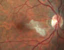
Cardiac Catheterization induced Cilioretinal Artery Occlusion - Paracentral Acute Middle Maculopathy525 views69 year old female with sudden vision loss on completing cardiac catheterization. The SD OCT shows a PAMM lesion and the lesion looks gray on color imaging. There is delayed filling of the cilioretinal artery.Mar 06, 2018
|
|

Vascular Remodeling Bilateral BRVO528 views85 year old female 20/25 OU with BRVO 7 years ago in both eyes. She has a change in vision. The FA shows extensive collaterallization and vascular remodeling in each macula.Jan 25, 2018
|
|

Vascular Remodeling Bilateral BRVO467 views85 year old female 20/25 OU with BRVO 7 years ago in both eyes. She has a change in vision. The FA shows extensive collaterallization and vascular remodeling in each macula.Jan 25, 2018
|
|

Vascular Remodeling Bilateral BRVO507 views85 year old female 20/25 OU with BRVO 7 years ago in both eyes. She has a change in vision. The FA shows extensive collaterallization and vascular remodeling in each macula.Jan 25, 2018
|
|

Vascular Remodeling Bilateral BRVO504 views85 year old female 20/25 OU with BRVO 7 years ago in both eyes. She has a change in vision. The FA shows extensive collaterallization and vascular remodeling in each macula.Jan 25, 2018
|
|

Vascular Remodeling Bilateral BRVO457 views85 year old female 20/25 OU with BRVO 7 years ago in both eyes. She has a change in vision. The FA shows extensive collaterallization and vascular remodeling in each macula.Jan 25, 2018
|
|

Vascular Remodeling Bilateral BRVO534 views85 year old female 20/25 OU with BRVO 7 years ago in both eyes. She has a change in vision. The FA shows extensive collaterallization and vascular remodeling in each macula.Jan 25, 2018
|
|

Vascular Remodeling Bilateral BRVO496 views85 year old female 20/25 OU with BRVO 7 years ago in both eyes. She has a change in vision. The FA shows extensive collaterallization and vascular remodeling in each macula.Jan 25, 2018
|
|

Vascular Remodeling Bilateral BRVO458 views85 year old female 20/25 OU with BRVO 7 years ago in both eyes. She has a change in vision. The FA shows extensive collaterallization and vascular remodeling in each macula.Jan 25, 2018
|
|

Vascular Remodeling Bilateral BRVO443 views85 year old female 20/25 OU with BRVO 7 years ago in both eyes. She has a change in vision. The FA shows extensive collaterallization and vascular remodeling in each macula.Jan 25, 2018
|
|

Vascular Remodeling Bilateral BRVO505 views85 year old female 20/25 OU with BRVO 7 years ago in both eyes. She has a change in vision. The FA shows extensive collaterallization and vascular remodeling in each macula.Jan 25, 2018
|
|

Holenhorst Plaque - Small and asymptomatic644 views72 year old woman with mild dry ARMD. VA 20/40 OU. Asymptomatic plaque noted in the right eye.Jan 18, 2018
|
|

Holenhorst Plaque - Small and asymptomatic517 views72 year old woman with mild dry ARMD. VA 20/40 OU. Asymptomatic plaque noted in the right eye.Jan 18, 2018
|
|

Intraluminal Plaque Retinal Vessel 640 views69 year old female referred with atypical plaque in the right retina. Her vision is normal. The plaque looks to be adjacent to the blood column visible on the images and the SC OCT shows it to be in the vessel wall. Jan 15, 2018
|
|

Intraluminal Plaque Retinal Vessel 497 views69 year old female referred with atypical plaque in the right retina. Her vision is normal. The plaque looks to be adjacent to the blood column visible on the images and the SC OCT shows it to be in the vessel wall. Jan 15, 2018
|
|

Intraluminal Plaque Retinal Vessel 487 views69 year old female referred with atypical plaque in the right retina. Her vision is normal. The plaque looks to be adjacent to the blood column visible on the images and the SC OCT shows it to be in the vessel wall. Jan 15, 2018
|
|

556 viewsJan 15, 2018
|
|

518 viewsJan 15, 2018
|
|
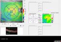
502 viewsJan 15, 2018
|
|

Disorganized Retinal Inner Layers (DRIL)1206 views72 year old man treated with Avastin for CRVO VA is still 20/200 with absent macular edema (Only 1 month post rx)Jan 11, 2018
|
|

Disorganized Retinal Inner Layers (DRIL)665 views72 year old man treated with Avastin for CRVO VA is still 20/200 with absent macular edema (Only 1 month post rx)Jan 11, 2018
|
|

Disorganized Retinal Inner Layers (DRIL)638 views72 year old man treated with Avastin for CRVO VA is still 20/200 with absent macular edema (Only 1 month post rx)Jan 11, 2018
|
|

Branch Retinal Vein Occlusion (BRVO) no macular edema - cotton wool spot557 views85 year old man with asymptomatic BRVO - his vision stayed great without needing any treatment.Sep 03, 2017
|
|

Branch Retinal Vein Occlusion (BRVO) no macular edema - cotton wool spot587 views85 year old man with asymptomatic BRVO - his vision stayed great without needing any treatment.Sep 03, 2017
|
|

Branch Retinal Vein Occlusion (BRVO) no macular edema - cotton wool spot481 views85 year old man with asymptomatic BRVO - his vision stayed great without needing any treatment.Sep 03, 2017
|
|

Branch Retinal Vein Occlusion (BRVO) no macular edema - cotton wool spot468 views85 year old man with asymptomatic BRVO - his vision stayed great without needing any treatment.Sep 03, 2017
|
|

Branch Retinal Vein Occlusion (BRVO) no macular edema - cotton wool spot555 views85 year old man with asymptomatic BRVO - his vision stayed great without needing any treatment.Sep 03, 2017
|
|

Branch Retinal Vein Occlusion (BRVO) no macular edema - cotton wool spot452 views85 year old man with asymptomatic BRVO - his vision stayed great without needing any treatment.Sep 03, 2017
|
|

Branch Retinal Vein Occlusion (BRVO) no macular edema - cotton wool spot487 views85 year old man with asymptomatic BRVO - his vision stayed great without needing any treatment.Sep 03, 2017
|
|

Branch Retinal Vein Occlusion (BRVO) no macular edema - cotton wool spot517 views85 year old man with asymptomatic BRVO - his vision stayed great without needing any treatment.Sep 03, 2017
|
|

Branch Retinal Vein Occlusion (BRVO) no macular edema - cotton wool spot478 views85 year old man with asymptomatic BRVO - his vision stayed great without needing any treatment.Sep 03, 2017
|
|

Branch Retinal Vein Occlusion (BRVO) no macular edema - cotton wool spot460 views85 year old man with asymptomatic BRVO - his vision stayed great without needing any treatment.Sep 03, 2017
|
|

Branch Retinal Vein Occlusion (BRVO) no macular edema - cotton wool spot469 views85 year old man with asymptomatic BRVO - his vision stayed great without needing any treatment.Sep 03, 2017
|
|

Branch Retinal Vein Occlusion (BRVO) no macular edema - cotton wool spot496 views85 year old man with asymptomatic BRVO - his vision stayed great without needing any treatment.Sep 03, 2017
|
|

Branch Retinal Vein Occlusion (BRVO) no macular edema - cotton wool spot584 views85 year old man with asymptomatic BRVO - his vision stayed great without needing any treatment.Sep 03, 2017
|
|

Branch Retinal Vein Occlusion (BRVO) no macular edema - cotton wool spot476 views85 year old man with asymptomatic BRVO - his vision stayed great without needing any treatment.Sep 03, 2017
|
|

Branch Retinal Vein Occlusion (BRVO) no macular edema - cotton wool spot529 views85 year old man with asymptomatic BRVO - his vision stayed great without needing any treatment.Sep 03, 2017
|
|

Branch Retinal Vein Occlusion (BRVO) no macular edema - cotton wool spot491 views85 year old man with asymptomatic BRVO - his vision stayed great without needing any treatment.Sep 03, 2017
|
|

Branch Retinal Vein Occlusion (BRVO) no macular edema - cotton wool spot477 views85 year old man with asymptomatic BRVO - his vision stayed great without needing any treatment.Sep 03, 2017
|
|

Branch Retinal Vein Occlusion (BRVO) no macular edema - cotton wool spot723 views85 year old man with asymptomatic BRVO - his vision stayed great without needing any treatment.Sep 03, 2017
|
|

Branch Retinal Vein Occlusion (BRVO) no macular edema - cotton wool spot495 views85 year old man with asymptomatic BRVO - his vision stayed great without needing any treatment.Sep 03, 2017
|
|

Branch Retinal Vein Occlusion (BRVO) no macular edema - cotton wool spot558 views85 year old man with asymptomatic BRVO - his vision stayed great without needing any treatment.Sep 03, 2017
|
|

Branch Retinal Vein Occlusion (BRVO) no macular edema - cotton wool spot763 views85 year old man with asymptomatic BRVO - his vision stayed great without needing any treatment.Sep 03, 2017
|
|

Branch Retinal Vein Occlusion (BRVO) no macular edema - cotton wool spot580 views85 year old man with asymptomatic BRVO - his vision stayed great without needing any treatment.Sep 03, 2017
|
|

Branch Retinal Vein Occlusion (BRVO) no macular edema - cotton wool spot588 views85 year old man with asymptomatic BRVO - his vision stayed great without needing any treatment.Sep 03, 2017
|
|
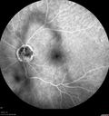
Branch Retinal Arterial Occlusion Right Eye - Atheromata - Both Eyes540 views83 year old woman with no symptoms - VA 20/40 in both eyes. Right eye has ST BRAO with silver wiring. Left eye has superotemporal atheromataJun 02, 2017
|
|

Branch Retinal Arterial Occlusion Right Eye - Atheromata - Both Eyes483 views83 year old woman with no symptoms - VA 20/40 in both eyes. Right eye has ST BRAO with silver wiring. Left eye has superotemporal atheromataJun 02, 2017
|
|
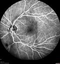
Branch Retinal Arterial Occlusion Right Eye - Atheromata - Both Eyes518 views83 year old woman with no symptoms - VA 20/40 in both eyes. Right eye has ST BRAO with silver wiring. Left eye has superotemporal atheromataJun 02, 2017
|
|

Branch Retinal Arterial Occlusion Right Eye - Atheromata - Both Eyes519 views83 year old woman with no symptoms - VA 20/40 in both eyes. Right eye has ST BRAO with silver wiring. Left eye has superotemporal atheromataJun 02, 2017
|
|

Branch Retinal Arterial Occlusion Right Eye - Atheromata - Both Eyes473 views83 year old woman with no symptoms - VA 20/40 in both eyes. Right eye has ST BRAO with silver wiring. Left eye has superotemporal atheromataJun 02, 2017
|
|

Branch Retinal Arterial Occlusion Right Eye - Atheromata - Both Eyes458 views83 year old woman with no symptoms - VA 20/40 in both eyes. Right eye has ST BRAO with silver wiring. Left eye has superotemporal atheromataJun 02, 2017
|
|

Branch Retinal Arterial Occlusion Right Eye - Atheromata - Both Eyes503 views83 year old woman with no symptoms - VA 20/40 in both eyes. Right eye has ST BRAO with silver wiring. Left eye has superotemporal atheromataJun 02, 2017
|
|

Branch Retinal Arterial Occlusion Right Eye - Atheromata - Both Eyes603 views83 year old woman with no symptoms - VA 20/40 in both eyes. Right eye has ST BRAO with silver wiring. Left eye has superotemporal atheromataJun 02, 2017
|
|

Branch Retinal Arterial Occlusion Right Eye - Atheromata - Both Eyes511 views83 year old woman with no symptoms - VA 20/40 in both eyes. Right eye has ST BRAO with silver wiring. Left eye has superotemporal atheromataJun 02, 2017
|
|

Branch Retinal Arterial Occlusion Right Eye - Atheromata - Both Eyes531 views83 year old woman with no symptoms - VA 20/40 in both eyes. Right eye has ST BRAO with silver wiring. Left eye has superotemporal atheromataJun 02, 2017
|
|

Branch Retinal Arterial Occlusion Right Eye - Atheromata - Both Eyes529 views83 year old woman with no symptoms - VA 20/40 in both eyes. Right eye has ST BRAO with silver wiring. Left eye has superotemporal atheromataJun 02, 2017
|
|

Branch Retinal Arterial Occlusion Right Eye - Atheromata - Both Eyes438 views83 year old woman with no symptoms - VA 20/40 in both eyes. Right eye has ST BRAO with silver wiring. Left eye has superotemporal atheromataJun 02, 2017
|
|

Branch Retinal Arterial Occlusion Right Eye - Atheromata - Both Eyes496 views83 year old woman with no symptoms - VA 20/40 in both eyes. Right eye has ST BRAO with silver wiring. Left eye has superotemporal atheromataJun 02, 2017
|
|

Branch Retinal Arterial Occlusion Right Eye - Atheromata - Both Eyes464 views83 year old woman with no symptoms - VA 20/40 in both eyes. Right eye has ST BRAO with silver wiring. Left eye has superotemporal atheromataJun 02, 2017
|
|

Branch Retinal Arterial Occlusion Right Eye - Atheromata - Both Eyes522 views83 year old woman with no symptoms - VA 20/40 in both eyes. Right eye has ST BRAO with silver wiring. Left eye has superotemporal atheromataJun 02, 2017
|
|

Branch Retinal Arterial Occlusion Right Eye - Atheromata - Both Eyes465 views83 year old woman with no symptoms - VA 20/40 in both eyes. Right eye has ST BRAO with silver wiring. Left eye has superotemporal atheromataJun 02, 2017
|
|

Branch Retinal Arterial Occlusion Right Eye - Atheromata - Both Eyes428 views83 year old woman with no symptoms - VA 20/40 in both eyes. Right eye has ST BRAO with silver wiring. Left eye has superotemporal atheromataJun 02, 2017
|
|

Branch Retinal Arterial Occlusion Right Eye - Atheromata - Both Eyes433 views83 year old woman with no symptoms - VA 20/40 in both eyes. Right eye has ST BRAO with silver wiring. Left eye has superotemporal atheromataJun 02, 2017
|
|

Branch Retinal Arterial Occlusion Right Eye - Atheromata - Both Eyes443 views83 year old woman with no symptoms - VA 20/40 in both eyes. Right eye has ST BRAO with silver wiring. Left eye has superotemporal atheromataJun 02, 2017
|
|

Branch Retinal Arterial Occlusion Right Eye - Atheromata - Both Eyes468 views83 year old woman with no symptoms - VA 20/40 in both eyes. Right eye has ST BRAO with silver wiring. Left eye has superotemporal atheromataJun 02, 2017
|
|

Branch Retinal Arterial Occlusion Right Eye - Atheromata - Both Eyes484 views83 year old woman with no symptoms - VA 20/40 in both eyes. Right eye has ST BRAO with silver wiring. Left eye has superotemporal atheromataJun 02, 2017
|
|

Branch Retinal Arterial Occlusion Right Eye - Atheromata - Both Eyes492 views83 year old woman with no symptoms - VA 20/40 in both eyes. Right eye has ST BRAO with silver wiring. Left eye has superotemporal atheromataJun 02, 2017
|
|

Branch Retinal Arterial Occlusion Right Eye - Atheromata - Both Eyes497 views83 year old woman with no symptoms - VA 20/40 in both eyes. Right eye has ST BRAO with silver wiring. Left eye has superotemporal atheromataJun 02, 2017
|
|

Branch Retinal Arterial Occlusion Right Eye - Atheromata - Both Eyes499 views83 year old woman with no symptoms - VA 20/40 in both eyes. Right eye has ST BRAO with silver wiring. Left eye has superotemporal atheromataJun 02, 2017
|
|

Branch Retinal Arterial Occlusion Right Eye - Atheromata - Both Eyes502 views83 year old woman with no symptoms - VA 20/40 in both eyes. Right eye has ST BRAO with silver wiring. Left eye has superotemporal atheromataJun 02, 2017
|
|

Branch Retinal Arterial Occlusion Right Eye - Atheromata - Both Eyes588 views83 year old woman with no symptoms - VA 20/40 in both eyes. Right eye has ST BRAO with silver wiring. Left eye has superotemporal atheromataJun 02, 2017
|
|

Branch Retinal Arterial Occlusion Right Eye - Atheromata - Both Eyes600 views83 year old woman with no symptoms - VA 20/40 in both eyes. Right eye has ST BRAO with silver wiring. Left eye has superotemporal atheromataJun 02, 2017
|
|

Branch Retinal Arterial Occlusion Right Eye - Atheromata - Both Eyes607 views83 year old woman with no symptoms - VA 20/40 in both eyes. Right eye has ST BRAO with silver wiring. Left eye has superotemporal atheromataJun 02, 2017
|
|

Branch Retinal Arterial Occlusion Right Eye - Atheromata - Both Eyes616 views83 year old woman with no symptoms - VA 20/40 in both eyes. Right eye has ST BRAO with silver wiring. Left eye has superotemporal atheromataJun 02, 2017
|
|

Branch Retinal Arterial Occlusion Right Eye - Atheromata - Both Eyes679 views83 year old woman with no symptoms - VA 20/40 in both eyes. Right eye has ST BRAO with silver wiring. Left eye has superotemporal atheromataJun 02, 2017
|
|

CRVO with Paracentral Acute Middle Maculopath - PAMM684 views56 year old woman with sudden vision loss, hemorrhages in all four quadrants. FA shows good retinal circulation. The OCT shows PAMM lesions in the affected eye. Vision did improve on second visit from 20/160 to 20/60 in about a monthMay 05, 2017
|
|

CRVO with Paracentral Acute Middle Maculopath - PAMM578 views56 year old woman with sudden vision loss, hemorrhages in all four quadrants. FA shows good retinal circulation. The OCT shows PAMM lesions in the affected eye. Vision did improve on second visit from 20/160 to 20/60 in about a monthMay 05, 2017
|
|

CRVO with Paracentral Acute Middle Maculopath - PAMM568 views56 year old woman with sudden vision loss, hemorrhages in all four quadrants. FA shows good retinal circulation. The OCT shows PAMM lesions in the affected eye. Vision did improve on second visit from 20/160 to 20/60 in about a monthMay 05, 2017
|
|

CRVO with Paracentral Acute Middle Maculopath - PAMM597 views56 year old woman with sudden vision loss, hemorrhages in all four quadrants. FA shows good retinal circulation. The OCT shows PAMM lesions in the affected eye. Vision did improve on second visit from 20/160 to 20/60 in about a monthMay 05, 2017
|
|

CRVO with Paracentral Acute Middle Maculopath - PAMM583 views56 year old woman with sudden vision loss, hemorrhages in all four quadrants. FA shows good retinal circulation. The OCT shows PAMM lesions in the affected eye. Vision did improve on second visit from 20/160 to 20/60 in about a monthMay 05, 2017
|
|

CRVO with Paracentral Acute Middle Maculopath - PAMM447 views56 year old woman with sudden vision loss, hemorrhages in all four quadrants. FA shows good retinal circulation. The OCT shows PAMM lesions in the affected eye. Vision did improve on second visit from 20/160 to 20/60 in about a monthMay 05, 2017
|
|

CRVO with Paracentral Acute Middle Maculopath - PAMM512 views56 year old woman with sudden vision loss, hemorrhages in all four quadrants. FA shows good retinal circulation. The OCT shows PAMM lesions in the affected eye. Vision did improve on second visit from 20/160 to 20/60 in about a monthMay 05, 2017
|
|

CRVO with Paracentral Acute Middle Maculopath - PAMM470 views56 year old woman with sudden vision loss, hemorrhages in all four quadrants. FA shows good retinal circulation. The OCT shows PAMM lesions in the affected eye. Vision did improve on second visit from 20/160 to 20/60 in about a monthMay 05, 2017
|
|

CRVO with Paracentral Acute Middle Maculopath - PAMM486 views56 year old woman with sudden vision loss, hemorrhages in all four quadrants. FA shows good retinal circulation. The OCT shows PAMM lesions in the affected eye. Vision did improve on second visit from 20/160 to 20/60 in about a monthMay 05, 2017
|
|

CRVO with Paracentral Acute Middle Maculopath - PAMM509 views56 year old woman with sudden vision loss, hemorrhages in all four quadrants. FA shows good retinal circulation. The OCT shows PAMM lesions in the affected eye. Vision did improve on second visit from 20/160 to 20/60 in about a monthMay 05, 2017
|
|

CRVO with Paracentral Acute Middle Maculopath - PAMM554 views56 year old woman with sudden vision loss, hemorrhages in all four quadrants. FA shows good retinal circulation. The OCT shows PAMM lesions in the affected eye. Vision did improve on second visit from 20/160 to 20/60 in about a monthMay 05, 2017
|
|
| 1798 files on 18 page(s) |
 |
|
|
|
|
10 | |
|
|
|
|


