Last additions - Retina Vascular Occlusions
|

Branch Retinal Vein Occlusion (BRVO) no macular edema - cotton wool spot726 views85 year old man with asymptomatic BRVO - his vision stayed great without needing any treatment.Sep 03, 2017
|
|

Branch Retinal Vein Occlusion (BRVO) no macular edema - cotton wool spot498 views85 year old man with asymptomatic BRVO - his vision stayed great without needing any treatment.Sep 03, 2017
|
|

Branch Retinal Vein Occlusion (BRVO) no macular edema - cotton wool spot562 views85 year old man with asymptomatic BRVO - his vision stayed great without needing any treatment.Sep 03, 2017
|
|

Branch Retinal Vein Occlusion (BRVO) no macular edema - cotton wool spot767 views85 year old man with asymptomatic BRVO - his vision stayed great without needing any treatment.Sep 03, 2017
|
|

Branch Retinal Vein Occlusion (BRVO) no macular edema - cotton wool spot583 views85 year old man with asymptomatic BRVO - his vision stayed great without needing any treatment.Sep 03, 2017
|
|

Branch Retinal Vein Occlusion (BRVO) no macular edema - cotton wool spot593 views85 year old man with asymptomatic BRVO - his vision stayed great without needing any treatment.Sep 03, 2017
|
|
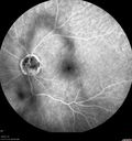
Branch Retinal Arterial Occlusion Right Eye - Atheromata - Both Eyes543 views83 year old woman with no symptoms - VA 20/40 in both eyes. Right eye has ST BRAO with silver wiring. Left eye has superotemporal atheromataJun 02, 2017
|
|

Branch Retinal Arterial Occlusion Right Eye - Atheromata - Both Eyes487 views83 year old woman with no symptoms - VA 20/40 in both eyes. Right eye has ST BRAO with silver wiring. Left eye has superotemporal atheromataJun 02, 2017
|
|
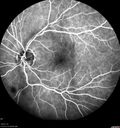
Branch Retinal Arterial Occlusion Right Eye - Atheromata - Both Eyes520 views83 year old woman with no symptoms - VA 20/40 in both eyes. Right eye has ST BRAO with silver wiring. Left eye has superotemporal atheromataJun 02, 2017
|
|

Branch Retinal Arterial Occlusion Right Eye - Atheromata - Both Eyes525 views83 year old woman with no symptoms - VA 20/40 in both eyes. Right eye has ST BRAO with silver wiring. Left eye has superotemporal atheromataJun 02, 2017
|
|

Branch Retinal Arterial Occlusion Right Eye - Atheromata - Both Eyes480 views83 year old woman with no symptoms - VA 20/40 in both eyes. Right eye has ST BRAO with silver wiring. Left eye has superotemporal atheromataJun 02, 2017
|
|

Branch Retinal Arterial Occlusion Right Eye - Atheromata - Both Eyes462 views83 year old woman with no symptoms - VA 20/40 in both eyes. Right eye has ST BRAO with silver wiring. Left eye has superotemporal atheromataJun 02, 2017
|
|

Branch Retinal Arterial Occlusion Right Eye - Atheromata - Both Eyes505 views83 year old woman with no symptoms - VA 20/40 in both eyes. Right eye has ST BRAO with silver wiring. Left eye has superotemporal atheromataJun 02, 2017
|
|

Branch Retinal Arterial Occlusion Right Eye - Atheromata - Both Eyes606 views83 year old woman with no symptoms - VA 20/40 in both eyes. Right eye has ST BRAO with silver wiring. Left eye has superotemporal atheromataJun 02, 2017
|
|

Branch Retinal Arterial Occlusion Right Eye - Atheromata - Both Eyes513 views83 year old woman with no symptoms - VA 20/40 in both eyes. Right eye has ST BRAO with silver wiring. Left eye has superotemporal atheromataJun 02, 2017
|
|

Branch Retinal Arterial Occlusion Right Eye - Atheromata - Both Eyes535 views83 year old woman with no symptoms - VA 20/40 in both eyes. Right eye has ST BRAO with silver wiring. Left eye has superotemporal atheromataJun 02, 2017
|
|

Branch Retinal Arterial Occlusion Right Eye - Atheromata - Both Eyes533 views83 year old woman with no symptoms - VA 20/40 in both eyes. Right eye has ST BRAO with silver wiring. Left eye has superotemporal atheromataJun 02, 2017
|
|

Branch Retinal Arterial Occlusion Right Eye - Atheromata - Both Eyes442 views83 year old woman with no symptoms - VA 20/40 in both eyes. Right eye has ST BRAO with silver wiring. Left eye has superotemporal atheromataJun 02, 2017
|
|

Branch Retinal Arterial Occlusion Right Eye - Atheromata - Both Eyes500 views83 year old woman with no symptoms - VA 20/40 in both eyes. Right eye has ST BRAO with silver wiring. Left eye has superotemporal atheromataJun 02, 2017
|
|

Branch Retinal Arterial Occlusion Right Eye - Atheromata - Both Eyes469 views83 year old woman with no symptoms - VA 20/40 in both eyes. Right eye has ST BRAO with silver wiring. Left eye has superotemporal atheromataJun 02, 2017
|
|

Branch Retinal Arterial Occlusion Right Eye - Atheromata - Both Eyes527 views83 year old woman with no symptoms - VA 20/40 in both eyes. Right eye has ST BRAO with silver wiring. Left eye has superotemporal atheromataJun 02, 2017
|
|

Branch Retinal Arterial Occlusion Right Eye - Atheromata - Both Eyes467 views83 year old woman with no symptoms - VA 20/40 in both eyes. Right eye has ST BRAO with silver wiring. Left eye has superotemporal atheromataJun 02, 2017
|
|

Branch Retinal Arterial Occlusion Right Eye - Atheromata - Both Eyes435 views83 year old woman with no symptoms - VA 20/40 in both eyes. Right eye has ST BRAO with silver wiring. Left eye has superotemporal atheromataJun 02, 2017
|
|

Branch Retinal Arterial Occlusion Right Eye - Atheromata - Both Eyes437 views83 year old woman with no symptoms - VA 20/40 in both eyes. Right eye has ST BRAO with silver wiring. Left eye has superotemporal atheromataJun 02, 2017
|
|

Branch Retinal Arterial Occlusion Right Eye - Atheromata - Both Eyes451 views83 year old woman with no symptoms - VA 20/40 in both eyes. Right eye has ST BRAO with silver wiring. Left eye has superotemporal atheromataJun 02, 2017
|
|

Branch Retinal Arterial Occlusion Right Eye - Atheromata - Both Eyes474 views83 year old woman with no symptoms - VA 20/40 in both eyes. Right eye has ST BRAO with silver wiring. Left eye has superotemporal atheromataJun 02, 2017
|
|

Branch Retinal Arterial Occlusion Right Eye - Atheromata - Both Eyes489 views83 year old woman with no symptoms - VA 20/40 in both eyes. Right eye has ST BRAO with silver wiring. Left eye has superotemporal atheromataJun 02, 2017
|
|

Branch Retinal Arterial Occlusion Right Eye - Atheromata - Both Eyes498 views83 year old woman with no symptoms - VA 20/40 in both eyes. Right eye has ST BRAO with silver wiring. Left eye has superotemporal atheromataJun 02, 2017
|
|

Branch Retinal Arterial Occlusion Right Eye - Atheromata - Both Eyes502 views83 year old woman with no symptoms - VA 20/40 in both eyes. Right eye has ST BRAO with silver wiring. Left eye has superotemporal atheromataJun 02, 2017
|
|

Branch Retinal Arterial Occlusion Right Eye - Atheromata - Both Eyes502 views83 year old woman with no symptoms - VA 20/40 in both eyes. Right eye has ST BRAO with silver wiring. Left eye has superotemporal atheromataJun 02, 2017
|
|

Branch Retinal Arterial Occlusion Right Eye - Atheromata - Both Eyes505 views83 year old woman with no symptoms - VA 20/40 in both eyes. Right eye has ST BRAO with silver wiring. Left eye has superotemporal atheromataJun 02, 2017
|
|

Branch Retinal Arterial Occlusion Right Eye - Atheromata - Both Eyes592 views83 year old woman with no symptoms - VA 20/40 in both eyes. Right eye has ST BRAO with silver wiring. Left eye has superotemporal atheromataJun 02, 2017
|
|

Branch Retinal Arterial Occlusion Right Eye - Atheromata - Both Eyes603 views83 year old woman with no symptoms - VA 20/40 in both eyes. Right eye has ST BRAO with silver wiring. Left eye has superotemporal atheromataJun 02, 2017
|
|

Branch Retinal Arterial Occlusion Right Eye - Atheromata - Both Eyes612 views83 year old woman with no symptoms - VA 20/40 in both eyes. Right eye has ST BRAO with silver wiring. Left eye has superotemporal atheromataJun 02, 2017
|
|

Branch Retinal Arterial Occlusion Right Eye - Atheromata - Both Eyes618 views83 year old woman with no symptoms - VA 20/40 in both eyes. Right eye has ST BRAO with silver wiring. Left eye has superotemporal atheromataJun 02, 2017
|
|

Branch Retinal Arterial Occlusion Right Eye - Atheromata - Both Eyes682 views83 year old woman with no symptoms - VA 20/40 in both eyes. Right eye has ST BRAO with silver wiring. Left eye has superotemporal atheromataJun 02, 2017
|
|

CRVO with Paracentral Acute Middle Maculopath - PAMM692 views56 year old woman with sudden vision loss, hemorrhages in all four quadrants. FA shows good retinal circulation. The OCT shows PAMM lesions in the affected eye. Vision did improve on second visit from 20/160 to 20/60 in about a monthMay 05, 2017
|
|

CRVO with Paracentral Acute Middle Maculopath - PAMM582 views56 year old woman with sudden vision loss, hemorrhages in all four quadrants. FA shows good retinal circulation. The OCT shows PAMM lesions in the affected eye. Vision did improve on second visit from 20/160 to 20/60 in about a monthMay 05, 2017
|
|

CRVO with Paracentral Acute Middle Maculopath - PAMM571 views56 year old woman with sudden vision loss, hemorrhages in all four quadrants. FA shows good retinal circulation. The OCT shows PAMM lesions in the affected eye. Vision did improve on second visit from 20/160 to 20/60 in about a monthMay 05, 2017
|
|

CRVO with Paracentral Acute Middle Maculopath - PAMM602 views56 year old woman with sudden vision loss, hemorrhages in all four quadrants. FA shows good retinal circulation. The OCT shows PAMM lesions in the affected eye. Vision did improve on second visit from 20/160 to 20/60 in about a monthMay 05, 2017
|
|

CRVO with Paracentral Acute Middle Maculopath - PAMM589 views56 year old woman with sudden vision loss, hemorrhages in all four quadrants. FA shows good retinal circulation. The OCT shows PAMM lesions in the affected eye. Vision did improve on second visit from 20/160 to 20/60 in about a monthMay 05, 2017
|
|

CRVO with Paracentral Acute Middle Maculopath - PAMM452 views56 year old woman with sudden vision loss, hemorrhages in all four quadrants. FA shows good retinal circulation. The OCT shows PAMM lesions in the affected eye. Vision did improve on second visit from 20/160 to 20/60 in about a monthMay 05, 2017
|
|

CRVO with Paracentral Acute Middle Maculopath - PAMM514 views56 year old woman with sudden vision loss, hemorrhages in all four quadrants. FA shows good retinal circulation. The OCT shows PAMM lesions in the affected eye. Vision did improve on second visit from 20/160 to 20/60 in about a monthMay 05, 2017
|
|

CRVO with Paracentral Acute Middle Maculopath - PAMM472 views56 year old woman with sudden vision loss, hemorrhages in all four quadrants. FA shows good retinal circulation. The OCT shows PAMM lesions in the affected eye. Vision did improve on second visit from 20/160 to 20/60 in about a monthMay 05, 2017
|
|

CRVO with Paracentral Acute Middle Maculopath - PAMM491 views56 year old woman with sudden vision loss, hemorrhages in all four quadrants. FA shows good retinal circulation. The OCT shows PAMM lesions in the affected eye. Vision did improve on second visit from 20/160 to 20/60 in about a monthMay 05, 2017
|
|

CRVO with Paracentral Acute Middle Maculopath - PAMM513 views56 year old woman with sudden vision loss, hemorrhages in all four quadrants. FA shows good retinal circulation. The OCT shows PAMM lesions in the affected eye. Vision did improve on second visit from 20/160 to 20/60 in about a monthMay 05, 2017
|
|

CRVO with Paracentral Acute Middle Maculopath - PAMM559 views56 year old woman with sudden vision loss, hemorrhages in all four quadrants. FA shows good retinal circulation. The OCT shows PAMM lesions in the affected eye. Vision did improve on second visit from 20/160 to 20/60 in about a monthMay 05, 2017
|
|

CRVO with Paracentral Acute Middle Maculopath - PAMM503 views56 year old woman with sudden vision loss, hemorrhages in all four quadrants. FA shows good retinal circulation. The OCT shows PAMM lesions in the affected eye. Vision did improve on second visit from 20/160 to 20/60 in about a monthMay 05, 2017
|
|

CRVO with Paracentral Acute Middle Maculopath - PAMM516 views56 year old woman with sudden vision loss, hemorrhages in all four quadrants. FA shows good retinal circulation. The OCT shows PAMM lesions in the affected eye. Vision did improve on second visit from 20/160 to 20/60 in about a monthMay 05, 2017
|
|
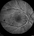
CRVO with Paracentral Acute Middle Maculopath - PAMM645 views56 year old woman with sudden vision loss, hemorrhages in all four quadrants. FA shows good retinal circulation. The OCT shows PAMM lesions in the affected eye. Vision did improve on second visit from 20/160 to 20/60 in about a monthMay 05, 2017
|
|

CRVO with Paracentral Acute Middle Maculopath - PAMM547 views56 year old woman with sudden vision loss, hemorrhages in all four quadrants. FA shows good retinal circulation. The OCT shows PAMM lesions in the affected eye. Vision did improve on second visit from 20/160 to 20/60 in about a monthMay 05, 2017
|
|

CRVO with Paracentral Acute Middle Maculopath - PAMM591 views56 year old woman with sudden vision loss, hemorrhages in all four quadrants. FA shows good retinal circulation. The OCT shows PAMM lesions in the affected eye. Vision did improve on second visit from 20/160 to 20/60 in about a monthMay 05, 2017
|
|

CRVO with Paracentral Acute Middle Maculopath - PAMM609 views56 year old woman with sudden vision loss, hemorrhages in all four quadrants. FA shows good retinal circulation. The OCT shows PAMM lesions in the affected eye. Vision did improve on second visit from 20/160 to 20/60 in about a monthMay 05, 2017
|
|

CRVO with Paracentral Acute Middle Maculopath - PAMM569 views56 year old woman with sudden vision loss, hemorrhages in all four quadrants. FA shows good retinal circulation. The OCT shows PAMM lesions in the affected eye. Vision did improve on second visit from 20/160 to 20/60 in about a monthMay 05, 2017
|
|

CRVO with Paracentral Acute Middle Maculopath - PAMM764 views56 year old woman with sudden vision loss, hemorrhages in all four quadrants. FA shows good retinal circulation. The OCT shows PAMM lesions in the affected eye. Vision did improve on second visit from 20/160 to 20/60 in about a monthMay 05, 2017
|
|

CRVO with Paracentral Acute Middle Maculopath - PAMM508 views56 year old woman with sudden vision loss, hemorrhages in all four quadrants. FA shows good retinal circulation. The OCT shows PAMM lesions in the affected eye. Vision did improve on second visit from 20/160 to 20/60 in about a monthMay 05, 2017
|
|

CRVO with Paracentral Acute Middle Maculopath - PAMM521 views56 year old woman with sudden vision loss, hemorrhages in all four quadrants. FA shows good retinal circulation. The OCT shows PAMM lesions in the affected eye. Vision did improve on second visit from 20/160 to 20/60 in about a monthMay 05, 2017
|
|

CRVO with Paracentral Acute Middle Maculopath - PAMM546 views56 year old woman with sudden vision loss, hemorrhages in all four quadrants. FA shows good retinal circulation. The OCT shows PAMM lesions in the affected eye. Vision did improve on second visit from 20/160 to 20/60 in about a monthMay 05, 2017
|
|

CRVO with Paracentral Acute Middle Maculopath - PAMM482 views56 year old woman with sudden vision loss, hemorrhages in all four quadrants. FA shows good retinal circulation. The OCT shows PAMM lesions in the affected eye. Vision did improve on second visit from 20/160 to 20/60 in about a monthMay 05, 2017
|
|

CRVO with Paracentral Acute Middle Maculopath - PAMM492 views56 year old woman with sudden vision loss, hemorrhages in all four quadrants. FA shows good retinal circulation. The OCT shows PAMM lesions in the affected eye. Vision did improve on second visit from 20/160 to 20/60 in about a monthMay 05, 2017
|
|

CRVO with Paracentral Acute Middle Maculopath - PAMM453 views56 year old woman with sudden vision loss, hemorrhages in all four quadrants. FA shows good retinal circulation. The OCT shows PAMM lesions in the affected eye. Vision did improve on second visit from 20/160 to 20/60 in about a monthMay 05, 2017
|
|

CRVO with Paracentral Acute Middle Maculopath - PAMM435 views56 year old woman with sudden vision loss, hemorrhages in all four quadrants. FA shows good retinal circulation. The OCT shows PAMM lesions in the affected eye. Vision did improve on second visit from 20/160 to 20/60 in about a monthMay 05, 2017
|
|

CRVO with Paracentral Acute Middle Maculopath - PAMM540 views56 year old woman with sudden vision loss, hemorrhages in all four quadrants. FA shows good retinal circulation. The OCT shows PAMM lesions in the affected eye. Vision did improve on second visit from 20/160 to 20/60 in about a monthMay 05, 2017
|
|

CRVO with Paracentral Acute Middle Maculopath - PAMM522 views56 year old woman with sudden vision loss, hemorrhages in all four quadrants. FA shows good retinal circulation. The OCT shows PAMM lesions in the affected eye. Vision did improve on second visit from 20/160 to 20/60 in about a monthMay 05, 2017
|
|

CRVO with Paracentral Acute Middle Maculopath - PAMM748 views56 year old woman with sudden vision loss, hemorrhages in all four quadrants. FA shows good retinal circulation. The OCT shows PAMM lesions in the affected eye. Vision did improve on second visit from 20/160 to 20/60 in about a monthMay 05, 2017
|
|

Hemicentral Vein Occlusion Left Eye544 viewsVision improved from 20/100 - 2 to 20/32 with 5 monthly Avastin treatments.Mar 06, 2017
|
|

Hemicentral Vein Occlusion Left Eye491 viewsVision improved from 20/100 - 2 to 20/32 with 5 monthly Avastin treatments.Mar 06, 2017
|
|

Hemicentral Vein Occlusion Left Eye512 viewsVision improved from 20/100 - 2 to 20/32 with 5 monthly Avastin treatments.Mar 06, 2017
|
|

Hemicentral Vein Occlusion Left Eye533 viewsVision improved from 20/100 - 2 to 20/32 with 5 monthly Avastin treatments.Mar 06, 2017
|
|

Hemicentral Vein Occlusion Left Eye457 viewsVision improved from 20/100 - 2 to 20/32 with 5 monthly Avastin treatments.Mar 06, 2017
|
|

Hemicentral Vein Occlusion Left Eye509 viewsVision improved from 20/100 - 2 to 20/32 with 5 monthly Avastin treatments.Mar 06, 2017
|
|

Hemicentral Vein Occlusion Left Eye527 viewsVision improved from 20/100 - 2 to 20/32 with 5 monthly Avastin treatments.Mar 06, 2017
|
|

Hemicentral Vein Occlusion Left Eye538 viewsVision improved from 20/100 - 2 to 20/32 with 5 monthly Avastin treatments.Mar 06, 2017
|
|

Hemicentral Vein Occlusion Left Eye571 viewsVision improved from 20/100 - 2 to 20/32 with 5 monthly Avastin treatments.Mar 06, 2017
|
|

Hemicentral Vein Occlusion Left Eye585 viewsVision improved from 20/100 - 2 to 20/32 with 5 monthly Avastin treatments.Mar 06, 2017
|
|

Hemicentral Vein Occlusion Left Eye587 viewsVision improved from 20/100 - 2 to 20/32 with 5 monthly Avastin treatments.Mar 06, 2017
|
|

Hemicentral Vein Occlusion Left Eye581 viewsVision improved from 20/100 - 2 to 20/32 with 5 monthly Avastin treatments.Mar 06, 2017
|
|

Hemicentral Vein Occlusion Left Eye585 viewsVision improved from 20/100 - 2 to 20/32 with 5 monthly Avastin treatments.Mar 06, 2017
|
|

Hemicentral Vein Occlusion Left Eye538 viewsVision improved from 20/100 - 2 to 20/32 with 5 monthly Avastin treatments.Mar 06, 2017
|
|

Hemicentral Vein Occlusion Left Eye508 viewsVision improved from 20/100 - 2 to 20/32 with 5 monthly Avastin treatments.Mar 06, 2017
|
|

Hemicentral Vein Occlusion Left Eye473 viewsVision improved from 20/100 - 2 to 20/32 with 5 monthly Avastin treatments.Mar 06, 2017
|
|

Hemicentral Vein Occlusion Left Eye510 viewsVision improved from 20/100 - 2 to 20/32 with 5 monthly Avastin treatments.Mar 06, 2017
|
|

Hemicentral Vein Occlusion Left Eye552 viewsVision improved from 20/100 - 2 to 20/32 with 5 monthly Avastin treatments.Mar 06, 2017
|
|

Hemicentral Vein Occlusion Left Eye586 viewsVision improved from 20/100 - 2 to 20/32 with 5 monthly Avastin treatments.Mar 06, 2017
|
|

Hemicentral Vein Occlusion Left Eye623 viewsVision improved from 20/100 - 2 to 20/32 with 5 monthly Avastin treatments.Mar 06, 2017
|
|

Bilateral Branch Retinal Artery Occlusion - Intraarterial (hollenhorst) plaques visible503 views67 year old with acute visual field loss in each eye. Vision is 20/25 right eye 20/32 left eye. Images show intra-arterial plaques in each eye. The right eye on the optic nerve and the left eye in a branch retinal artery.Feb 06, 2017
|
|

Bilateral Branch Retinal Artery Occlusion - Intraarterial (hollenhorst) plaques visible532 views67 year old with acute visual field loss in each eye. Vision is 20/25 right eye 20/32 left eye. Images show intra-arterial plaques in each eye. The right eye on the optic nerve and the left eye in a branch retinal artery.Feb 06, 2017
|
|

Bilateral Branch Retinal Artery Occlusion - Intraarterial (hollenhorst) plaques visible465 views67 year old with acute visual field loss in each eye. Vision is 20/25 right eye 20/32 left eye. Images show intra-arterial plaques in each eye. The right eye on the optic nerve and the left eye in a branch retinal artery.Feb 06, 2017
|
|

Bilateral Branch Retinal Artery Occlusion - Intraarterial (hollenhorst) plaques visible595 views67 year old with acute visual field loss in each eye. Vision is 20/25 right eye 20/32 left eye. Images show intra-arterial plaques in each eye. The right eye on the optic nerve and the left eye in a branch retinal artery.Feb 06, 2017
|
|

Bilateral Branch Retinal Artery Occlusion - Intraarterial (hollenhorst) plaques visible508 views67 year old with acute visual field loss in each eye. Vision is 20/25 right eye 20/32 left eye. Images show intra-arterial plaques in each eye. The right eye on the optic nerve and the left eye in a branch retinal artery.Feb 06, 2017
|
|

Bilateral Branch Retinal Artery Occlusion - Intraarterial (hollenhorst) plaques visible478 views67 year old with acute visual field loss in each eye. Vision is 20/25 right eye 20/32 left eye. Images show intra-arterial plaques in each eye. The right eye on the optic nerve and the left eye in a branch retinal artery.Feb 06, 2017
|
|

Bilateral Branch Retinal Artery Occlusion - Intraarterial (hollenhorst) plaques visible537 views67 year old with acute visual field loss in each eye. Vision is 20/25 right eye 20/32 left eye. Images show intra-arterial plaques in each eye. The right eye on the optic nerve and the left eye in a branch retinal artery.Feb 06, 2017
|
|

Bilateral Branch Retinal Artery Occlusion - Intraarterial (hollenhorst) plaques visible447 views67 year old with acute visual field loss in each eye. Vision is 20/25 right eye 20/32 left eye. Images show intra-arterial plaques in each eye. The right eye on the optic nerve and the left eye in a branch retinal artery.Feb 06, 2017
|
|

Bilateral Branch Retinal Artery Occlusion - Intraarterial (hollenhorst) plaques visible520 views67 year old with acute visual field loss in each eye. Vision is 20/25 right eye 20/32 left eye. Images show intra-arterial plaques in each eye. The right eye on the optic nerve and the left eye in a branch retinal artery.Feb 06, 2017
|
|

Bilateral Branch Retinal Artery Occlusion - Intraarterial (hollenhorst) plaques visible462 views67 year old with acute visual field loss in each eye. Vision is 20/25 right eye 20/32 left eye. Images show intra-arterial plaques in each eye. The right eye on the optic nerve and the left eye in a branch retinal artery.Feb 06, 2017
|
|

Bilateral Branch Retinal Artery Occlusion - Intraarterial (hollenhorst) plaques visible514 views67 year old with acute visual field loss in each eye. Vision is 20/25 right eye 20/32 left eye. Images show intra-arterial plaques in each eye. The right eye on the optic nerve and the left eye in a branch retinal artery.Feb 06, 2017
|
|

Bilateral Branch Retinal Artery Occlusion - Intraarterial (hollenhorst) plaques visible546 views67 year old with acute visual field loss in each eye. Vision is 20/25 right eye 20/32 left eye. Images show intra-arterial plaques in each eye. The right eye on the optic nerve and the left eye in a branch retinal artery.Feb 06, 2017
|
|

Bilateral Branch Retinal Artery Occlusion - Intraarterial (hollenhorst) plaques visible514 views67 year old with acute visual field loss in each eye. Vision is 20/25 right eye 20/32 left eye. Images show intra-arterial plaques in each eye. The right eye on the optic nerve and the left eye in a branch retinal artery.Feb 06, 2017
|
|

Bilateral Branch Retinal Artery Occlusion - Intraarterial (hollenhorst) plaques visible493 views67 year old with acute visual field loss in each eye. Vision is 20/25 right eye 20/32 left eye. Images show intra-arterial plaques in each eye. The right eye on the optic nerve and the left eye in a branch retinal artery.Feb 06, 2017
|
|

Bilateral Branch Retinal Artery Occlusion - Intraarterial (hollenhorst) plaques visible508 views67 year old with acute visual field loss in each eye. Vision is 20/25 right eye 20/32 left eye. Images show intra-arterial plaques in each eye. The right eye on the optic nerve and the left eye in a branch retinal artery.Feb 06, 2017
|
|
| 1845 files on 19 page(s) |
 |
|
|
|
|
11 | |
|
|
|
|


